. 2013 Dec 5;122(24):3940-50.
doi: 10.1182/blood-2013-06-506865. Epub 2013 Oct 24.

IL-21 signalling via STAT3 primes human naive B cells to respond to IL-2 to enhance their differentiation into plasmablasts

Affiliations

IL-21 signalling via STAT3 primes human naive B cells to respond to IL-2 to enhance their differentiation into plasmablasts

Lucinda J Berglund et al. Blood. .

Abstract

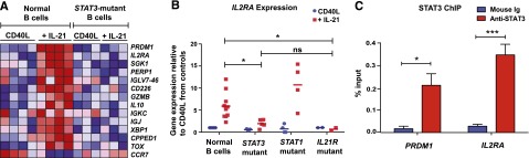
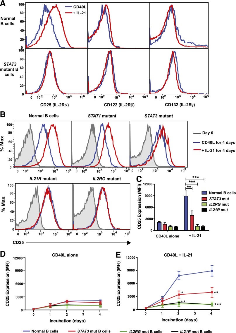
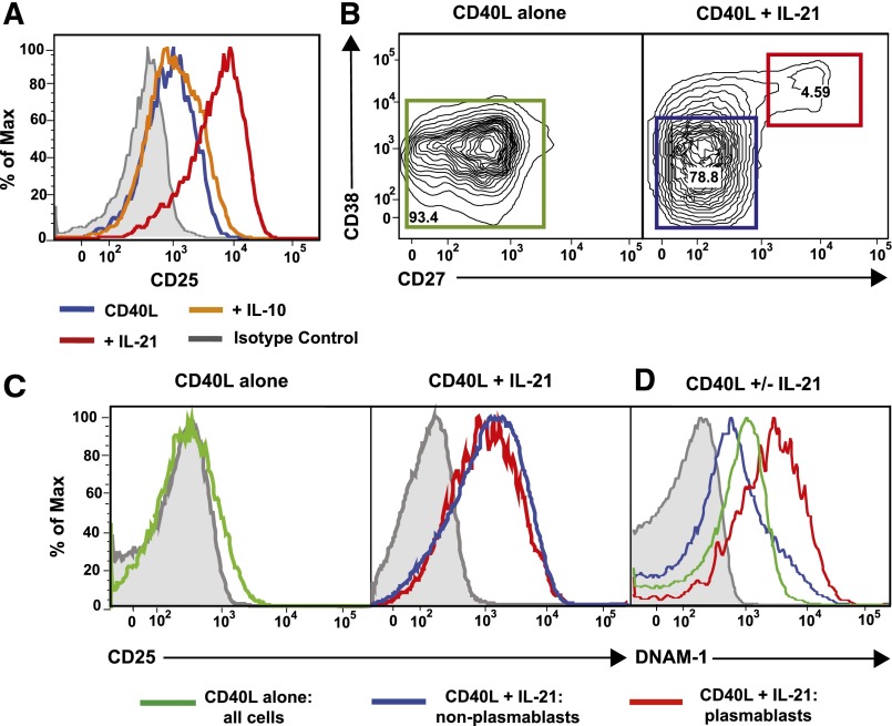
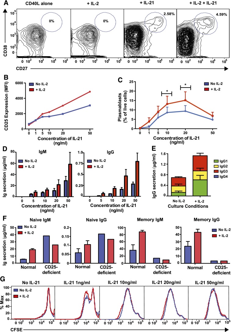
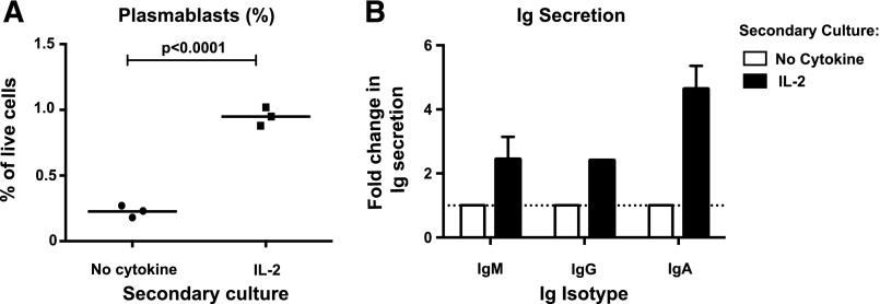
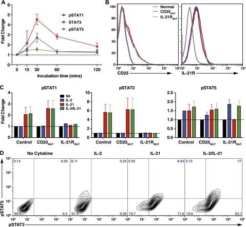
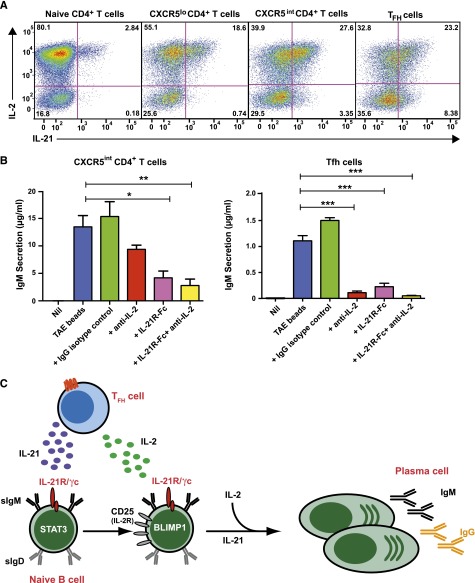
B-cell responses are guided by the integration of signals through the B-cell receptor (BCR), CD40, and cytokine receptors. The common γ chain (γc)-binding cytokine interleukin (IL)-21 drives humoral immune responses via STAT3-dependent induction of transcription factors required for plasma cell generation. We investigated additional mechanisms by which IL-21/STAT3 signaling modulates human B-cell responses by studying patients with STAT3 mutations. IL-21 strongly induced CD25 (IL-2Rα) in normal, but not STAT3-deficient, CD40L-stimulated naïve B cells. Chromatin immunoprecipitation confirmed IL2RA as a direct target of STAT3. IL-21-induced CD25 expression was also impaired on B cells from patients with IL2RG or IL21R mutations, confirming a requirement for intact IL-21R signaling in this process. IL-2 increased plasmablast generation and immunoglobulin secretion from normal, but not CD25-deficient, naïve B cells stimulated with CD40L/IL-21. IL-2 and IL-21 were produced by T follicular helper cells, and neutralizing both cytokines abolished the B-cell helper capacity of these cells. Our results demonstrate that IL-21, via STAT3, sensitizes B cells to the stimulatory effects of IL-2. Thus, IL-2 may play an adjunctive role in IL-21-induced B-cell differentiation. Lack of this secondary effect of IL-21 may amplify the humoral immunodeficiency in patients with mutations in STAT3, IL2RG, or IL21R due to impaired responsiveness to IL-21.


Figures

Figure 1
Figure 1
IL-21 directly induces IL2RA in normal, but not STAT3MUT, naïve B cells. (A) Naïve B cells were purified from the peripheral blood of normal donors (n = 4) and STAT3-deficient AD-HIES patients (n = 3) and then cultured with CD40L alone (“CD40L”) or together with IL-21 (“+IL-21”). RNA was extracted after 4 days and microarrays performed using Affymetrix Human Gene 1.0 ST Arrays. Genes with marked differences in expression between normal and STAT3-mutant (STAT3MUT) cells are shown. (B) Naïve B cells from normal donors (n = 10) or patients with loss-of-function mutations in STAT3 (n = 6), STAT1 (n = 4), or IL21R (n = 2) were cultured with CD40L alone (blue) or together with IL-21 (red). RNA was extracted after 5 days and used to determine expression of IL2RA by quantitative polymerase chain reaction. Results show expression levels relative to B cells cultured with CD40L alone. Each symbol represents an individual experiment using cells from a different donor or patient; the horizontal line represents the mean, *P < .05. (C) Chromatin immunoprecipitation (ChIP) was performed on normal LCLs using mouse Ig or anti-STAT3 Ab. Immunoprecipitated chromatin was assessed for the presence of PRDM1 and IL2RA Results are expressed relative to gene expression in the input DNA and represent the mean ± SEM from 3 separate experiments using different LCLs. *P < .05, ***P < .005.
Figure 2
Figure 2
IL-21–induced expression of CD25 is impaired on STAT3MUT naïve B cells. (A-C) Naive B cells from normal donors or patients with loss-of-function mutations in STAT3, STAT1, IL2RG, or IL21R were cultured with CD40L alone (blue) or in combination with IL-21 (red). (A) Expression of IL-2Rα (CD25), IL-2Rβ (CD122), and IL-2Rγ (CD132, γc) on normal and STAT3MUT B cells or of (B-C) IL-2Rα (CD25) on normal and STAT1MUT, STAT3MUT, IL-21RMUT, or IL2RγMUT B cells was determined after 5 days (or on day 0 for B). The histogram plots in A and B are representative of experiments performed on naïve B cells isolated from 6 normal donors, 6 STAT3-deficient patients, 1 STAT1-deficient patient, 2 IL-2Rγ–deficient patients, and 3 IL-21R–deficient patients. The summary graph in C depicts the mean ± SEM of CD25 expression on cultured B cells from the indicated numbers of patients. (D-E) Naïve B cells from normal donors (blue, n = 7), STAT3-deficient AD-HIES patients (red, n = 6), IL-2Rγ–deficient X-SCID patients (green, n = 2), or IL-21R–deficient patients (black, n = 3) were cultured with CD40L alone (D) or in the presence of IL-21 (E). The mean fluorescence intensity (MFI) of CD25 expression was determined at the indicated times. Results represent mean ± SEM for the indicated number of controls and patients. *P < .005, **P < .001, ***P < .0001.
Figure 3
Figure 3
IL-21, but not IL-10, induces CD25 expression on all activated naïve B cells. (A) Naïve B cells from healthy donors were cultured for 5 days with CD40L alone (blue histogram) or in the presence of IL-10 (orange histogram) or IL-21 (red histogram). Expression of CD25 was determined after 5 days. (B-D) Normal naïve B cells were cultured for 5 days with CD40L alone or together with IL-21 for 5 days. (B) Differential expression of CD27 and CD38 delineates IL-21–induced plasmablasts (CD38hiCD27hi; red gate) and nonplasmablasts (CD38loCD27lo; blue gate). Expression of CD25 (C) or DNAM-1 (D) was determined on all B cells in cultures stimulated with CD40L alone or on nonplasmablasts and plasmablasts present in cultures of CD40L/IL-21–stimulated B cells. Gray histograms represent isotype controls. Numbers represent percentage of live cells within each gate.
Figure 4
Figure 4
IL-21induced CD25 expression enables naïve B cells to respond to IL-2 with enhanced plasmablast generation and Ig secretion. Naïve B cells were sorted from normal spleens and then cultured with CD40L alone in the presence or absence of IL-2 and/or IL-21. The proportion of plasmablasts (CD27hiCD38hi) (A,C) and expression of CD25 (B) were determined by flow cytometry after 5 days. The data depicted are representative of 4 (A) or 3 (B) separate experiments using naïve B cells isolated from different donor spleens. C represents the mean ± SEM from 4 separate experiments; *P < .05, comparing cultures with and without IL-2. Secretion of IgM and IgG (D) or IgG subclasses (E) was determined after 10 to 12 days by enzyme-linked immunosorbent assay. Statistical analysis using 2-way ANOVA with Bonferroni post-test analysis confirmed a statistically significant difference between cultures with or without IL-2 (P < .005 for both IgM and IgG) and between different concentrations of IL-21 (P < .005 for IgM; P < .001 for IgG). Data in D show mean ± SEM from 3 independent experiments on 3 different donor spleens, each performed in triplicate. Data in E show mean ± SEM from a single experiment performed in triplicate but is representative of 2 independent experiments performed on different normal donor spleens. (F) Naïve and memory B cells were isolated from a normal donor and a CD25-deficient patient and then cultured with CD40L/IL-21 alone or together with IL-2. Secretion of IgM and IgG was determined after 10 days. Values represent the mean ± SEM of triplicate cultures for normal B cells and the mean of single cultures for CD25-deficient B cells. (G) Carboxyfluorescein diacetate succinimidyl ester profiles of splenic naïve B cells from normal donors cultured for 5 days with CD40L alone or with varying concentrations of IL-21 in the absence or presence of IL-2. Results are representative of 4 independent experiments using different normal donor spleens.
Figure 5
Figure 5
Initial upregulation of CD25 by IL-21 licenses IL-2 to maintain plasmablast generation in the absence of IL-21. Naive B cells from normal human spleens were initially cultured with CD40L together with IL-21. After 3 days, cells were harvested, washed, and recultured either in media alone or with IL-2. (A) After a further 3 days of culture, the proportion of plasmablasts (CD27hiCD38hi) was determined by flow cytometry. Each symbol corresponds to an individual experiment that used naïve B cells from a different normal donor spleen; the horizontal line represents the mean. (B) Secretion of IgM, IgG, and IgA was determined after 7 days of secondary culture in media alone or with IL-2 and Ig secretion was determined by enzyme-linked immunosorbent assay. Results are expressed as fold-change in Ig secretion relative to the “no cytokine” secondary culture (set to equal 1.0). The results for IgM represent mean ± SEM (n = 3); IgG secretion was detected in only 1 of 3 experiments; IgA was detected in 2 of 3 experiments.
Figure 6
Figure 6
Effect of IL-2 and IL-21 on STAT phosphorylation in human B-cell lines. (A) LCLs from normal donors were incubated for varying times in the absence or presence of IL-21. Phosphorylation of STAT1, STAT3, and STAT5 was then determined by intracellular staining and flow cytometry. Data are depicted as fold change in mean fluorescence intensity of pSTAT in the presence of IL-21 over media alone. Results represent mean ± SEM of experiments using LCLs from 6 different donors. (B) Expression of CD25 and IL-21R was determined on LCLs derived from different normal donors (red histogram) or CD25-deficient (blue histogram) or IL21R-deficient (gray histogram) patients (n = 2/group). (C-D) Normal, CD25-deficient (CD25MUT), and IL-21R–deficient (IL-21RMUT) LCLs were incubated in the absence (Nil) or presence of IL-2, IL-21, or IL-2/IL-21. Expression of pSTAT1, pSTAT3, and pSTAT5 was determined after 30 minutes. Results in C are expressed as fold change above pSTAT expression in cells cultured with no cytokine and represent mean ± SEM of experiments using LCLs from 2 different donors or patients. D shows representative contour plots demonstrating coexpression of pSTAT3 and pSTAT5 in LCLs cultured in the absence (no cytokine) or presence of IL-2, IL-21, or IL-2/IL-21.
Figure 7
Figure 7
IL-2 and IL-21 produced by Tfh cells cooperate to induce Ig secretion by co-cultured naïve B cells. (A) Naïve, CXCR5lo, CXCR5intermediate, and CXCR5hi Tfh CD4+ T cells were sort-purified from human tonsils and then stimulated in vitro with TAE beads. After 5 days, expression of IL-2 and IL-21 in these different populations following restimulation with PMA/ionomycin was determined. The values represent the proportions of cytokine-expressing cells. (B) CD4+CXCR5intermediate T cells and CXCR5hi Tfh cells were sorted from tonsil and co-cultured with autologous naïve B cells either in the absence (Nil) or presence of TAE beads (blue). Endogenous IL-2 and/or IL-21 were neutralized by the addition of anti-IL-2 mAb (red), IL-21R-Fc (pink), or anti-IL-2 mAb plus IL-21R-Fc (yellow), respectively. An isotype control mAb (green) was also included. IgM secretion was measured after 9 days. Results represent 2 independent experiments performed using cells from different donor tonsils. *P < .05; **P < .01; ***P < .0001. (C) IL-21, secreted from Tfh cells, promotes B-cell maturation by inducing Blimp-1. IL-21 also enhances CD25 expression on naïve B cells, sensitizing them to the effects of IL-2, which is also secreted by Tfh cells. IL-2 then enhances the effects of IL-21 on B cells. IL-21 and IL-2 thus work cooperatively to induce plasma cell development and Ig secretion.

References

    1. Tangye SG, Tarlinton DM. Memory B cells: effectors of long-lived immune responses. Eur J Immunol. 2009;39(8):2065–2075. - PubMed
    1. Goodnow CC, Vinuesa CG, Randall KL, Mackay F, Brink R. Control systems and decision making for antibody production. Nat Immunol. 2010;11(8):681–688. - PubMed
    1. Victora GD, Nussenzweig MC. Germinal centers. Annu Rev Immunol. 2012;30:429–457. - PubMed
    1. Crotty S. Follicular helper CD4 T cells (TFH). Annu Rev Immunol. 2011;29:621–663. - PubMed
    1. Ma CS, Deenick EK, Batten M, Tangye SG. The origins, function, and regulation of T follicular helper cells. J Exp Med. 2012;209(7):1241–1253. - PMC - PubMed

Publication types

MeSH terms

Associated data