

Congenital lumbar hernia with lumbocostovertebral syndrome: a case report and review of the literature
- PMID: 24159401
- PMCID: PMC3789277
- DOI: 10.1155/2013/532910
Congenital lumbar hernia with lumbocostovertebral syndrome: a case report and review of the literature
Abstract
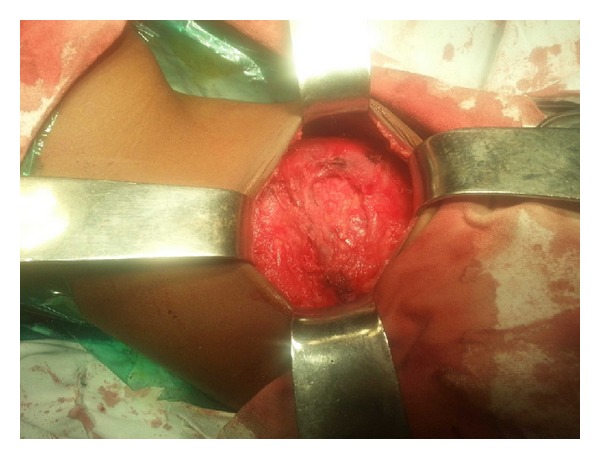
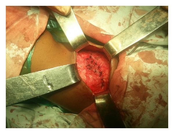
Introduction. Congenital lumbar hernia is one of the rare types of hernias. Anomalies of the ribs, spine, and muscles which constitute the lumbocostovertebral syndrome in association with congenital lumbar hernia make it the rarest of entities. In addition, a multitude of other organ systems may be involved. Case Report. A case of congenital lumbar hernia associated with lumbocostovertebral syndrome is presented in view of its rarity and diagnostic and therapeutic challenges. Discussion. Anatomical background of congenital lumbar hernia associated with various other anomalies especially of the musculoskeletal structures is discussed. All cases of congenital lumbar hernia should be investigated for other congenital anomalies. Both open and laparoscopic approaches have been described for surgical treatment. Conclusion. Open surgical intervention is the mainstay of treatment taking into consideration the technical challenges posed by distorted anatomy due to the associated congenital anomalies.
Figures


Similar articles
-
Laparoscopic lumbar hernia repair in a child with lumbocostovertebral syndrome.J Laparoendosc Adv Surg Tech A. 2010 Feb;20(1):97-8. doi: 10.1089/lap.2008.0278. J Laparoendosc Adv Surg Tech A. 2010. PMID: 19645602
-
Congenital lumbar hernia associated with the lumbocostovertebral syndrome: two cases.Eur J Pediatr Surg. 1997 Apr;7(2):122-4. doi: 10.1055/s-2008-1071071. Eur J Pediatr Surg. 1997. PMID: 9165264
-
A different type of congenital lumbar hernia associated with the lumbocostovertebral syndrome.J Pediatr Surg. 2008 Jan;43(1):e21-3. doi: 10.1016/j.jpedsurg.2007.08.065. J Pediatr Surg. 2008. PMID: 18206440
-
Intercostal variant of lumbar hernia in lumbocostovertebral syndrome: our experience with 6 cases.J Pediatr Surg. 2011 Oct;46(10):1974-7. doi: 10.1016/j.jpedsurg.2011.05.010. J Pediatr Surg. 2011. PMID: 22008337 Review.
-
[Lumbar hernia. Case report and literature review].Cir Cir. 2007 Sep-Oct;75(5):381-4. Cir Cir. 2007. PMID: 18158886 Review. Spanish.
Cited by
-
Open Approach to Primary Lumbar Hernia Repair: A Lucid Option.Case Rep Surg. 2017;2017:5839491. doi: 10.1155/2017/5839491. Epub 2017 Oct 17. Case Rep Surg. 2017. PMID: 29312793 Free PMC article.
-
Primary Lumbar Hernia, Review and Proposals for a Standardized Treatment.J Abdom Wall Surg. 2023 Dec 6;2:11754. doi: 10.3389/jaws.2023.11754. eCollection 2023. J Abdom Wall Surg. 2023. PMID: 38312404 Free PMC article.
-
Management of Lumbar Hernia Secondary to Retroperitoneal Abscess Drainage.Cureus. 2025 Apr 29;17(4):e83220. doi: 10.7759/cureus.83220. eCollection 2025 Apr. Cureus. 2025. PMID: 40309509 Free PMC article.
References
-
- Wakhlu A, Wakhlu AK. Congenital lumbar hernia. Pediatric Surgery International. 2000;16(1-2):146–148. - PubMed
-
- Pelaez Mata DJ, Alvarez Munoz V, Fernandez Jimenez I, Garcia Crespo JM, Teixidor de Otto JL. Congenital Lumbar hernia. Cirugia Pediatrica. 1998;11(3):126–128. - PubMed
-
- Stamatiou D, Skandalakis JE, Skandalakis LJ, Mirilas P. Lumbar hernia: surgical anatomy, embryology, and technique of repair. American Surgeon. 2009;75(3):202–207. - PubMed
-
- Akçora B, Temiz A, Babayiğit C. A different type of congenital lumbar hernia associated with the lumbocostovertebral syndrome. Journal of Pediatric Surgery. 2008;43(1):e21–e23. - PubMed
-
- Singh G, Ahuja S, Kumar R, et al. Posterior spinal dysraphism with lumbocostovertebral syndrome. British Journal of Neurosurgery. 2010;24(2):216–218. - PubMed
LinkOut - more resources
Full Text Sources
Other Literature Sources
