In vivo Noninvasive Small Animal Molecular Imaging
- PMID: 24159487
- PMCID: PMC3738683
- DOI: 10.1016/j.phrp.2012.02.002
In vivo Noninvasive Small Animal Molecular Imaging
Abstract
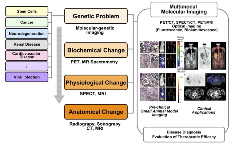
The remarkable efforts that are made on molecular imaging technologies demonstrate its potential importance and range of applications. The generation of disease-specific animal models, and the developments of target-specific probes and genetically encoded reporters are another important component. Continued improvements in the instrumentation, the identification of novel targets and genes, and the availability of improved imaging probes should be made. Multimodal imaging probes should provide easier transitions between laboratory studies, including small animal studies and clinical applications. Here, we reviewed basic strategies of noninvasive in vivo imaging methods in small animals to introducing the concept of molecular imaging.
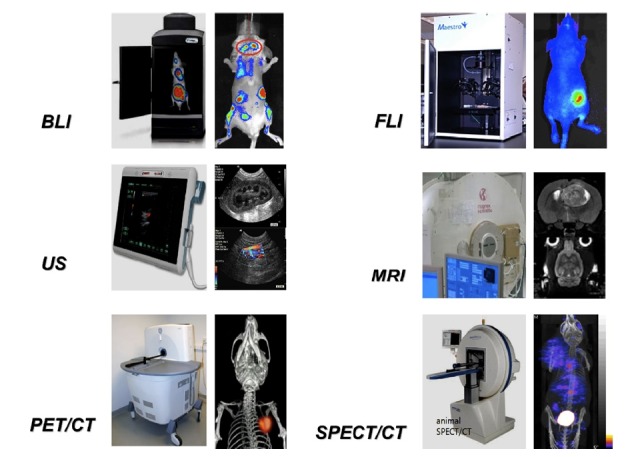
Keywords: animal imaging; computed tomography; magnetic resonance imaging; nuclear imaging; optical imaging; ultrasonography.
Figures
References
-
- Blasberg RG, Gelovani-Tjuvajev J. In vivo molecular-genetic Imaging. J Cell Biochem. 2002;(Suppl. 39):172–83. - PubMed
-
- Tjuvajev JG, Stockhammer G, Desai R, et al. Imaging the expression of transfected genes in vivo. Cancer Res. 1995 Dec 15;55(24):6126–32. - PubMed
-
- Brown RS, Leung JY, Fisher SJ, et al. Intratumoral distribution of tritiated-FDG in breast carcinoma: correlation between Glut-1 expression and FDG uptake. J Nucl Med. 1996 Jun;37(6):1042–7. - PubMed
Publication types
LinkOut - more resources
Full Text Sources
Other Literature Sources