. 2013 Jul 6:3:1-7.
doi: 10.1016/j.nicl.2013.06.017. eCollection 2013.

Multi-delay multi-parametric arterial spin-labeled perfusion MRI in acute ischemic stroke - Comparison with dynamic susceptibility contrast enhanced perfusion imaging

Affiliations

Multi-delay multi-parametric arterial spin-labeled perfusion MRI in acute ischemic stroke - Comparison with dynamic susceptibility contrast enhanced perfusion imaging

Danny J J Wang et al. Neuroimage Clin. .

Abstract

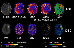
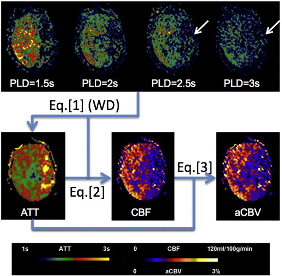
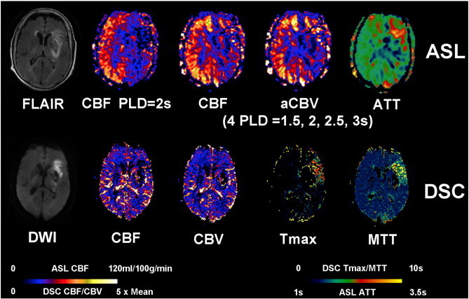
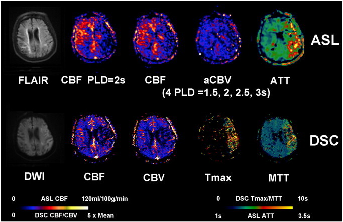
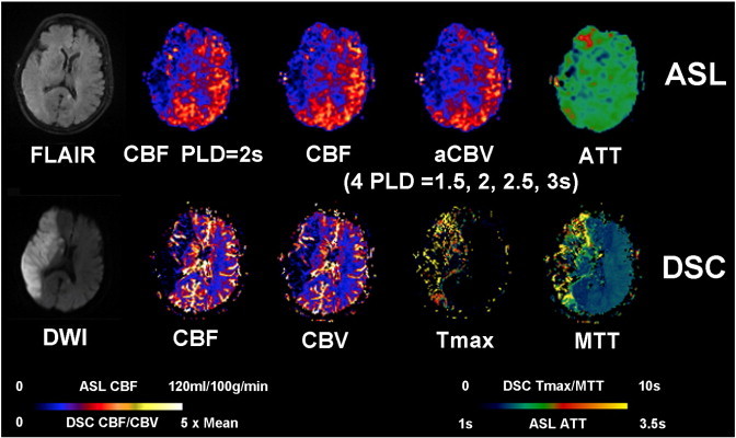
The purpose of the present study was to present a multi-delay multi-parametric pseudo-continuous arterial spin labeling (pCASL) protocol with background suppressed 3D GRASE (gradient and spin echo) readout for perfusion imaging in acute ischemic stroke. PCASL data at 4 post-labeling delay times (PLD = 1.5, 2, 2.5, 3 s) were acquired within 4.5 min in 24 patients (mean age 79.7 ± 11.4 years; 11 men) with acute middle cerebral artery (MCA) stroke who also underwent dynamic susceptibility contrast (DSC) enhanced perfusion imaging. Arterial transit times (ATT) were estimated through the calculation of weighted delays across the 4 PLDs, which were included in the calculation of cerebral blood flow (CBF) and arterial cerebral blood volume (CBV). Mean perfusion parameters derived using pCASL and DSC were measured within MCA territories and infarct regions identified on diffusion weighted MRI. The results showed highly significant correlations between pCASL and DSC CBF measurements (r > = 0.70, p < = 0.0001) and moderately significant correlations between pCASL and DSC CBV measurements (r > = 0.45, p < = 0.027) in both MCA territories and infarct regions. ASL ATT showed correlations with DSC time to the maximum of tissue residual function (Tmax)(r = 0.66, p = 0.0005) and mean transit time (MTT)(r = 0.59, p = 0.0023) in leptomeningeal MCA territories. The present study demonstrated the feasibility for noninvasive multi-parametric perfusion imaging using ASL for acute stroke imaging.

Keywords: Acute stroke; Arterial spin labeling (ASL); Dynamic susceptibility contrast (DSC); Ischemia; Multi-delay; Multi-parametric; Perfusion MRI.


Figures

None
Graphical abstract
Fig. 1
Fig. 1
Diagram of post-processing steps for a representative 4-delay pCASL dataset collected in a 61-year old female patient (case #15) with left MCA stroke. Labeled signal only appears in affected region when the PLD reaches 2.5 s (arrows), leading to prolonged ATT in the left MCA territory. CBF at each PLD is calculated by substituting ATT into Eq. (2) followed by averaging to generate the mean CBF map. The product of mean CBF and ATT provides aCBV map.
Fig. 2
Fig. 2
Representative AIS case with aligned FLAIR, DWI, multiparametric ASL and DSC PWI. An 89-year-old female (case #11) with a history of atrial fibrillation and hypertension was admitted to ER after being found down unconsciously and was found to have a left MCA stroke with a Baseline NIHSS of 20. The patient was scanned 8.1 h after the onset before receiving any endovascular treatment. Restricted diffusion and FLAIR hyperintensities were seen in the left basal ganglia, internal capsule and left frontal lobe. ASL shows decreased CBF, decreased CBV and prolonged ATT in the left MCA region. DSC Tmax and MTT were prolonged in the left frontal region with mildly decreased CBF and CBV.
Fig. 3
Fig. 3
A 94-year-old male (case #13) with a unclear past medical history presented with aphasia, right-sided numbness and weakness and was found to have a left MCA stroke with a Baseline NIHSS of 24. IV tPA was given before the patient was scanned 1.6 h after the onset. Acute restricted diffusion is present throughout the left parietal, temporal, and occipital lobes, as well as the left caudate head and body. ASL shows decreased CBF, decreased CBV and prolonged ATT in the left MCA region. DSC Tmax and MTT were prolonged in the left MCA region with decreased CBF and CBV.
Fig. 4
Fig. 4
An 85-year-old female (case #14) with a past medical history significant for paroxysmal atrial fibrillation, presented with aphasia, right-sided weakness and a baseline NIHSS of 19. IV tPA was given before the patient was scanned 2.5 h after the onset. Restricted diffusion is present throughout the left MCA territory with T2/FLAIR hyperintensity. ASL shows decreased CBF, decreased CBV and prolonged ATT in the left MCA region. DSC-Tmax and MTT were prolonged in the left MCA region with mildly decreased DSC-CBF and CBV.
Fig. 5
Fig. 5
An 88-year-old female (case #22) with history of hypertension presented with left-sided numbness and weakness, gait disturbance, limb ataxia, and altered mental status. The baseline NIHSS on admission was 18. Clot retrieval was performed before the patient was scanned 22.5 h after the onset. There is a large area of restricted diffusion involving the entire right MCA territory with T2/FLAIR hyperintensity. ASL shows decreased CBF and CBV in the right MCA region. However, ATT is not or only slightly prolonged in right MCA territory. DSC-Tmax and MTT were prolonged in the right MCA region with decreased DSC-CBF and CBV.

References

    1. Bokkers R.P., Hernandez D.A., Merino J.G., Mirasol R.V., van Osch M.J., Hendrikse J., Warach S., Latour L.L. Whole-brain arterial spin labeling perfusion MRI in patients with acute stroke. Stroke; A Journal of Cerebral Circulation. 2012;43:1290–1294. - PMC - PubMed
    1. Chalela J.A., Alsop D.C., Gonzalez-Atavalez J.B., Maldjian J.A., Kasner S.E., Detre J.A. Magnetic resonance perfusion imaging in acute ischemic stroke using continuous arterial spin labeling. Stroke. 2000;31:680–687. - PubMed
    1. Chen J., Licht D.J., Smith S.E., Agner S.C., Mason S., Wang S., Silvestre D.W., Detre J.A., Zimmerman R.A., Ichord R.N., Wang J. Arterial spin labeling perfusion MRI in pediatric arterial ischemic stroke: initial experiences. Journal of Magnetic Resonance Imaging. 2009;29:282–290. - PMC - PubMed
    1. Dai W., Robson P.M., Shankaranarayanan A., Alsop D.C. Reduced resolution transit delay prescan for quantitative continuous arterial spin labeling perfusion imaging. Magnetic Resonance in Medicine: Official Journal of the Society of Magnetic Resonance in Medicine/Society of Magnetic Resonance in Medicine. 2012;67:1252–1265. - PMC - PubMed
    1. Detre J.A., Rao H., Wang D.J., Chen Y.F., Wang Z. Applications of arterial spin labeled MRI in the brain. Journal of Magnetic Resonance Imaging: JMRI. 2012;35:1026–1037. - PMC - PubMed