

Clinical application of fluorescence imaging of liver cancer using indocyanine green
- PMID: 24159568
- PMCID: PMC3747548
- DOI: 10.1159/000339017
Clinical application of fluorescence imaging of liver cancer using indocyanine green
Abstract
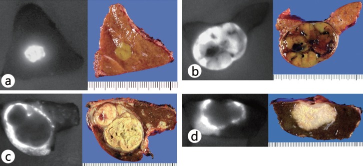
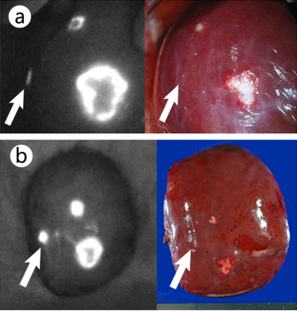
Recently, fluorescence imaging using indocyanine green (ICG) has been applied to hepatobiliary surgery, not only to visualize the bile ducts, but also to identify liver cancer during surgery. In this technique, ICG is administered intravenously at a dose of 0.5 mg/kg body weight for routine liver function testing before surgery. Intraoperatively, liver cancer can be readily identified by fluorescence imaging on the liver surface before resection and on the cut surface of the resected specimen. This is achieved by visualizing fluorescence from the area of impaired bile excretion in hepatocellular cancer tissue and in the liver parenchyma surrounding metastatic liver cancers. Liver cancer navigation surgery, first developed in Japan, is also possible, and it represents one of the few fluorescence imaging techniques for cancer that have reached the stage of clinical application; with further developments in basic research, fluorescence imaging is expected to become an indispensable technique for the diagnosis and treatment of liver cancer.
Keywords: Fluorescence imaging; Hepatocellular carcinoma; Indocyanine green; Metastatic liver cancer; Navigation surgery.
Figures
References
-
- Ishizawa T, Fukushima N, Shibahara J, et al. Real-time identification of liver cancers by using indocyanine green fluorescent imaging. Cancer. 2009;115:2491–2504. - PubMed
-
- Gotoh K, Yamada T, Ishikawa O, et al. A novel image-guided surgery of hepatocellular carcinoma by indocyanine green fluorescence imaging navigation. J Surg Oncol. 2009;100:75–79. - PubMed
-
- Stummer W, Stocker S, Wagner S, et al. Intraoperative detection of malignant gliomas by 5-aminolevulinic acid-induced porphyrin fluorescence. Neurosurgery. 1998;42:518–525. - PubMed
-
- Kriegmair M, Stepp H, Steinbach P, et al. Fluorescence cystoscopy following intravesical instillation of 5-aminolevulinic acid: a new procedure with high sensitivity for detection of hardly visible urothelial neoplasias. Urol Int. 1995;55:190–196. - PubMed
-
- Morton CA, Brown SB, Collins S, et al. Guidelines for topical photodynamic therapy: report of a workshop of the British Photodermatology Group. Br J Dermatol. 2002;146:552–567. - PubMed
Publication types
LinkOut - more resources
Full Text Sources
Other Literature Sources
