. 2014 Jan;120(1):73-86.
doi: 10.3171/2013.9.JNS122074. Epub 2013 Oct 25.

Stromal cell-derived factor-1 promoted angiogenesis and inflammatory cell infiltration in aneurysm walls

Affiliations

Stromal cell-derived factor-1 promoted angiogenesis and inflammatory cell infiltration in aneurysm walls

Brian L Hoh et al. J Neurosurg. 2014 Jan.

Abstract

Object: A small percentage of cerebral aneurysms rupture, but when they do, the effects are devastating. Current management of unruptured aneurysms consists of surgery, endovascular treatment, or watchful waiting. If the biology of how aneurysms grow and rupture were better known, a novel drug could be developed to prevent unruptured aneurysms from rupturing. Ruptured cerebral aneurysms are characterized by inflammation-mediated wall remodeling. The authors studied the role of stromal cell-derived factor-1 (SDF-1) in inflammation-mediated wall remodeling in cerebral aneurysms.

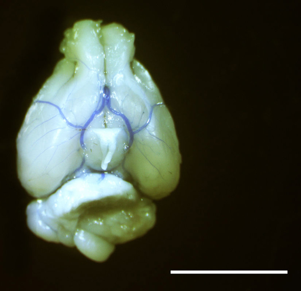
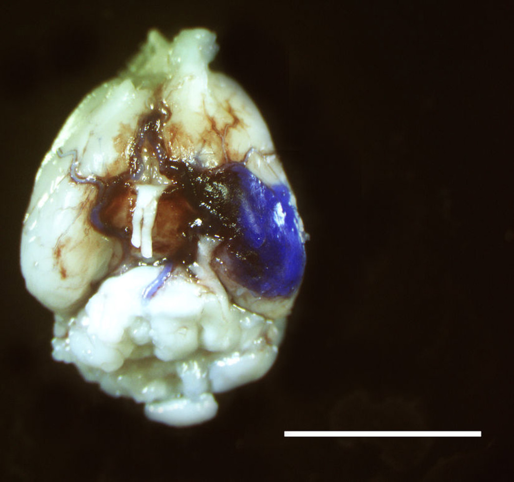
Methods: Human aneurysms, murine carotid artery aneurysms, and murine intracranial aneurysms were studied using immunohistochemistry. Flow cytometry analysis was performed on blood from mice developing carotid or intracranial aneurysms. The effect of SDF-1 on endothelial cells and macrophages was studied by chemotaxis cell migration assay and capillary tube formation assay. Anti-SDF-1 blocking antibody was given to mice and compared with control (vehicle)-administered mice for its effects on the walls of carotid aneurysms and the development of intracranial aneurysms.

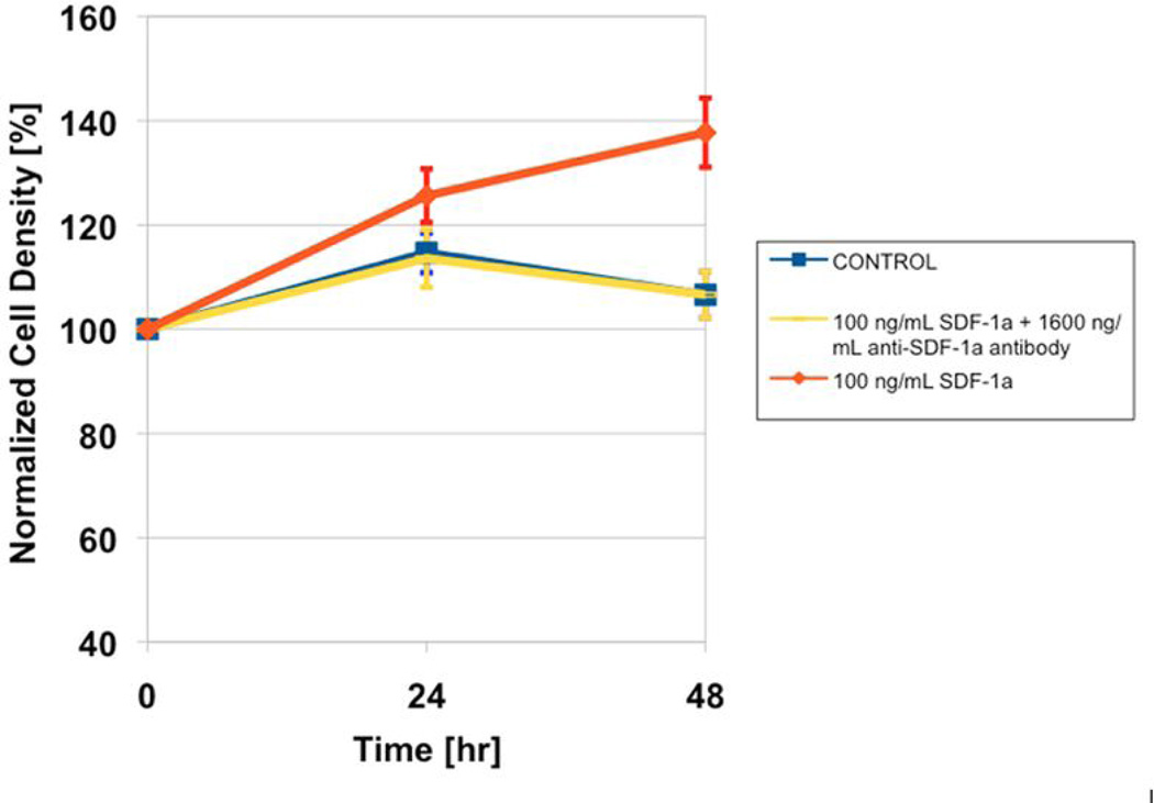
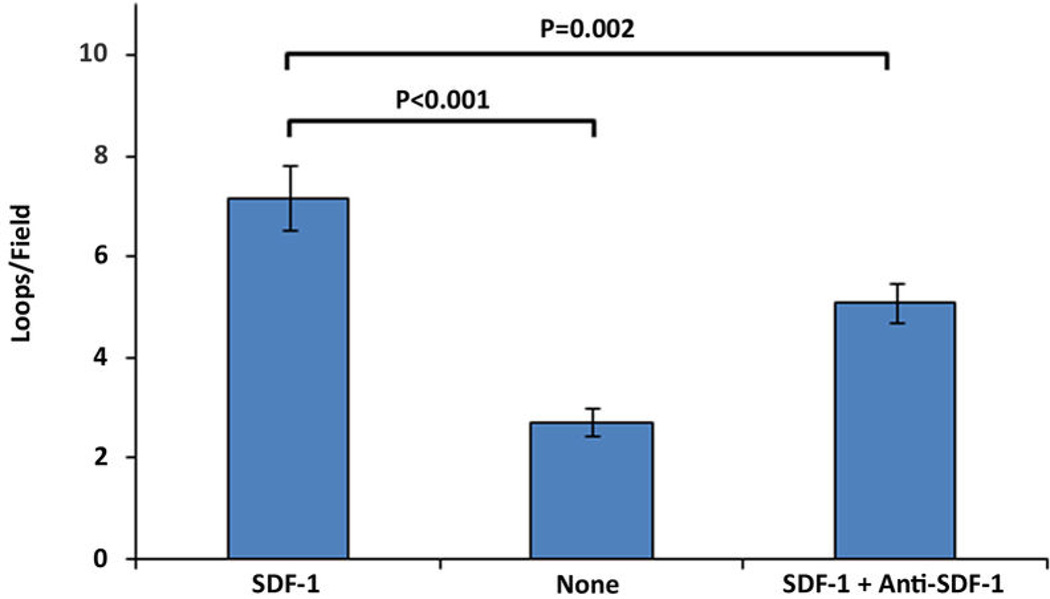
Results: Human aneurysms, murine carotid aneurysms, and murine intracranial aneurysms all expressed SDF-1, and mice with developing carotid or intracranial aneurysms had increased progenitor cells expressing CXCR4, the receptor for SDF-1 (p < 0.01 and p < 0.001, respectively). Human aneurysms and murine carotid aneurysms had endothelial cells, macrophages, and capillaries in the walls of the aneurysms, and the presence of capillaries in the walls of human aneurysms was associated with the presence of macrophages (p = 0.01). Stromal cell-derived factor-1 promoted endothelial cell and macrophage migration (p < 0.01 for each), and promoted capillary tube formation (p < 0.001). When mice were given anti-SDF-1 blocking antibody, there was a significant reduction in endothelial cells (p < 0.05), capillaries (p < 0.05), and cell proliferation (p < 0.05) in the aneurysm wall. Mice given anti-SDF-1 blocking antibody developed significantly fewer intracranial aneurysms (33% vs 89% in mice given control immunoglobulin G, respectively; p < 0.05).

Conclusions: These data suggest SDF-1 is associated with angiogenesis and inflammatory cell migration and proliferation in the walls of aneurysms, and may have a role in the development of intracranial aneurysms.


Conflict of interest statement

CONFLICTS OF INTEREST/DISCLOSURES

None

Figures

Figure 1
Figure 1
Angiogenesis Occurs in the Walls of Aneurysms. A, Human aneurysms contain endothelial cells (CD31+) and angiogenesis (capillary formation) within the media of the aneurysm walls. Representative section is depicted here at 20×. Scale bar is 200µm. Red: CD31+; blue: DAPI (4’,6-diamidino-2-phenylindole). B, Elastase-induced murine carotid aneurysms contain endothelial cells (MECA-32+) and angiogenesis (capillary formation) within the media of the aneurysm walls, but not in normal control murine carotid arteries. Representative section is depicted here at 20×. Scale bar is 200µm. Green: MECA-32+; blue: DAPI.
Figure 1
Figure 1
Angiogenesis Occurs in the Walls of Aneurysms. A, Human aneurysms contain endothelial cells (CD31+) and angiogenesis (capillary formation) within the media of the aneurysm walls. Representative section is depicted here at 20×. Scale bar is 200µm. Red: CD31+; blue: DAPI (4’,6-diamidino-2-phenylindole). B, Elastase-induced murine carotid aneurysms contain endothelial cells (MECA-32+) and angiogenesis (capillary formation) within the media of the aneurysm walls, but not in normal control murine carotid arteries. Representative section is depicted here at 20×. Scale bar is 200µm. Green: MECA-32+; blue: DAPI.
Figure 2
Figure 2
Angiogenesis is Associated with Inflammatory Cell Invasion in the Walls of Aneurysms. Human aneurysms contain abundant monocytes and macrophages (CD68+), and hematopoietic-derived inflammatory cells (CD45+) within the media of the walls of the aneurysms. Representative sections are depicted here at 40×. Scale bar is 100µm. −Red: CD68+ left panel, CD45+ right panel; blue: DAPI.
Figure 3
Figure 3
SDF-1 is Expressed in Aneurysms. A, Human aneurysms express SDF-1. Representative sections are depicted here at 60×. Scale bar is 50µm. Red: SDF-1+; blue: DAPI. B, Elastase-induced murine carotid aneurysms express SDF-1 and its receptor, CXCR4; but normal control murine carotid arteries do not. Representative sections are depicted here at 10× and 60×. Scale bar is 50µm for left top and left bottom panels, and 200µm for right top and right bottom panels. Green: SDF-1+ left panel, CXCR4+ right panel; blue: DAPI. C, Murine intracranial aneurysms express SDF-1, but normal murine circle of Willis do not. Representative sections are depicted here at 40× and 60×. Scale bar is 50µm for left panel, and 400µm for right panel. Red: SDF-1+; blue: DAPI.
Figure 3
Figure 3
SDF-1 is Expressed in Aneurysms. A, Human aneurysms express SDF-1. Representative sections are depicted here at 60×. Scale bar is 50µm. Red: SDF-1+; blue: DAPI. B, Elastase-induced murine carotid aneurysms express SDF-1 and its receptor, CXCR4; but normal control murine carotid arteries do not. Representative sections are depicted here at 10× and 60×. Scale bar is 50µm for left top and left bottom panels, and 200µm for right top and right bottom panels. Green: SDF-1+ left panel, CXCR4+ right panel; blue: DAPI. C, Murine intracranial aneurysms express SDF-1, but normal murine circle of Willis do not. Representative sections are depicted here at 40× and 60×. Scale bar is 50µm for left panel, and 400µm for right panel. Red: SDF-1+; blue: DAPI.
Figure 4
Figure 4
Aneurysm Formation Activates Circulation of Progenitor Cells Expressing CXCR4, the Receptor for SDF-1. A, Mice developing carotid aneurysms have increased circulation of progenitor cells (stem cell antigen-1 positive (SCA-1+)) expressing CXCR4, the receptor for SDF-1, compared to sham-operated mice. When anti-SDF-1 blocking antibody was given to mice, there were significantly less progenitor cells (SCA-1+) expressing CXCR4, than mice given IgG control. B, Mice developing intracranial aneurysms have increased circulation of progenitor cells (SCA-1+) expressing CXCR4, compared to sham-operated mice. C, Murine intracranial aneurysms demonstrate invasion of hematopoietic-derived inflammatory cells (CD45+), whereas none are found in normal circle of Willis. Representative sections are depicted here at 10× and 60×. Scale bar is 100µm for left panel and 400µm for right panel. Red: CD45+; blue: DAPI.
Figure 4
Figure 4
Aneurysm Formation Activates Circulation of Progenitor Cells Expressing CXCR4, the Receptor for SDF-1. A, Mice developing carotid aneurysms have increased circulation of progenitor cells (stem cell antigen-1 positive (SCA-1+)) expressing CXCR4, the receptor for SDF-1, compared to sham-operated mice. When anti-SDF-1 blocking antibody was given to mice, there were significantly less progenitor cells (SCA-1+) expressing CXCR4, than mice given IgG control. B, Mice developing intracranial aneurysms have increased circulation of progenitor cells (SCA-1+) expressing CXCR4, compared to sham-operated mice. C, Murine intracranial aneurysms demonstrate invasion of hematopoietic-derived inflammatory cells (CD45+), whereas none are found in normal circle of Willis. Representative sections are depicted here at 10× and 60×. Scale bar is 100µm for left panel and 400µm for right panel. Red: CD45+; blue: DAPI.
Figure 5
Figure 5
SDF-1 Promotes Angiogenesis (by Promoting Endothelial Cell Migration, Endothelial Cell Proliferation, and Tube Formation) and Inflammatory Cell Migration. A, In transwell cell migration assays, there is significant migration of endothelial cells in chambers containing SDF-1 compared to chambers containing SDF-1 and anti-SDF-1 blocking antibody, or neither. B, In endothelial cell proliferation assays, there was no significant difference between cells exposed to 100 ng/mL SDF-1 compared to control and anti-SDF-1 blocked groups at 24 hrs (p=0.18) by ANOVA. However, at 48 hrs, cells exposed to 100 ng/mL SDF-1 showed 31% higher proliferation than both the control (p=0.0012) and the anti-SDF-1 blocked group (p=0.0012) by ANOVA with Tukey’s pairwise comparison. The control and the anti-SDF-1 blocked group were not found to be significantly different at 48 hrs by the same analysis. C, In capillary tube formation assays, there is significantly greater complete loop networks formed by endothelial cells cultured with SDF-1 compared to SDF-1 and anti-SDF-1 blocking antibody, or neither. D, There is significantly greater macrophage migration in chambers containing SDF-1 compared to SDF-1 and anti-SDF-1 blocking antibody, or neither.
Figure 5
Figure 5
SDF-1 Promotes Angiogenesis (by Promoting Endothelial Cell Migration, Endothelial Cell Proliferation, and Tube Formation) and Inflammatory Cell Migration. A, In transwell cell migration assays, there is significant migration of endothelial cells in chambers containing SDF-1 compared to chambers containing SDF-1 and anti-SDF-1 blocking antibody, or neither. B, In endothelial cell proliferation assays, there was no significant difference between cells exposed to 100 ng/mL SDF-1 compared to control and anti-SDF-1 blocked groups at 24 hrs (p=0.18) by ANOVA. However, at 48 hrs, cells exposed to 100 ng/mL SDF-1 showed 31% higher proliferation than both the control (p=0.0012) and the anti-SDF-1 blocked group (p=0.0012) by ANOVA with Tukey’s pairwise comparison. The control and the anti-SDF-1 blocked group were not found to be significantly different at 48 hrs by the same analysis. C, In capillary tube formation assays, there is significantly greater complete loop networks formed by endothelial cells cultured with SDF-1 compared to SDF-1 and anti-SDF-1 blocking antibody, or neither. D, There is significantly greater macrophage migration in chambers containing SDF-1 compared to SDF-1 and anti-SDF-1 blocking antibody, or neither.
Figure 6
Figure 6
Blocking SDF-1 Inhibits Angiogenesis and Cell Proliferation in the Walls of Aneurysms. A, Carotid aneurysms from mice given anti-SDF-1 blocking antibody have significantly less endothelial cells in the walls of the aneurysms, compared to mice given Ig control. Representative sections are depicted here at 10× and 60×. Scale bar is 400µm for left top panel, and 50µm for right top panel. Red: MECA-32+; blue: DAPI. B, Carotid aneurysms from mice given anti-SDF-1 blocking antibody have significantly less number of formed capillaries in the walls of the aneurysms, compared to mice given Ig control. C, Carotid aneurysms from mice given anti-SDF-1 blocking antibody have significantly less cell proliferation, compared to mice given Ig control. Representative sections are depicted here at 20× and 60×. Scale bar is 400µm for left top panel, and 50µm for right top panel. Red: Ki-67+; blue: DAPI.
Figure 6
Figure 6
Blocking SDF-1 Inhibits Angiogenesis and Cell Proliferation in the Walls of Aneurysms. A, Carotid aneurysms from mice given anti-SDF-1 blocking antibody have significantly less endothelial cells in the walls of the aneurysms, compared to mice given Ig control. Representative sections are depicted here at 10× and 60×. Scale bar is 400µm for left top panel, and 50µm for right top panel. Red: MECA-32+; blue: DAPI. B, Carotid aneurysms from mice given anti-SDF-1 blocking antibody have significantly less number of formed capillaries in the walls of the aneurysms, compared to mice given Ig control. C, Carotid aneurysms from mice given anti-SDF-1 blocking antibody have significantly less cell proliferation, compared to mice given Ig control. Representative sections are depicted here at 20× and 60×. Scale bar is 400µm for left top panel, and 50µm for right top panel. Red: Ki-67+; blue: DAPI.
Figure 7
Figure 7
Blocking SDF-1 is Associated with Reduced Intracranial Aneurysm Formation A, C57BL6 mice given anti-SDF-1 blocking antibody (n=10 mice) developed significantly fewer intracranial aneurysms than C57BL6 mice given IgG control (n=10): 33% versus 89% respectively (P<0.05)(1 mouse from each group died perioperatively due to anesthetic issues). B, Representative example of intracranial aneurysm in mouse given IgG control. Scale bar is 5mm. C, Representative example of no intracranial aneurysm seen in mouse given anti-SDF-1 blocking antibody. Scale bar is 5mm. D, Representative example of ruptured intracranial aneurysm seen in mouse given IgG control. Scale bar is 5mm.
Figure 7
Figure 7
Blocking SDF-1 is Associated with Reduced Intracranial Aneurysm Formation A, C57BL6 mice given anti-SDF-1 blocking antibody (n=10 mice) developed significantly fewer intracranial aneurysms than C57BL6 mice given IgG control (n=10): 33% versus 89% respectively (P<0.05)(1 mouse from each group died perioperatively due to anesthetic issues). B, Representative example of intracranial aneurysm in mouse given IgG control. Scale bar is 5mm. C, Representative example of no intracranial aneurysm seen in mouse given anti-SDF-1 blocking antibody. Scale bar is 5mm. D, Representative example of ruptured intracranial aneurysm seen in mouse given IgG control. Scale bar is 5mm.

Comment in

  • Aneurysm wall inflammation.
    Macdonald RL. Macdonald RL. J Neurosurg. 2014 Jan;120(1):70-2. doi: 10.3171/2013.5.JNS13824. Epub 2013 Oct 25. J Neurosurg. 2014. PMID: 24160480 No abstract available.

References

    1. Atkinson JL, Okazaki H, Sundt TM, Jr., Nichols DA, Rufenacht DA. Intracranial cerebrovascular vasa vasorum associated with atherosclerosis and large thick-walled aneurysms. Surg Neurol. 1991;36:365–369. - PubMed
    1. Bavinzski G, Talazoglu V, Killer M, Richling B, Gruber A, Gross CE, Plenk H., Jr. Gross and microscopic histopathologic findings in aneurysms of the human brain treated with Guglielmi detachable coils. J Neurosurg. 1999;91:284–293. - PubMed
    1. Campi A, Ramzi N, Molyneux AJ, Summers PE, Kerr RS, Sneade M, Yarnold JA, Rischmiller J, Byrne JV. Retreatment of ruptured cerebral aneurysms in patients randomized by coiling or clipping in the International Subarachnoid Aneurysm Trial (ISAT) Stroke. 2007;38:1538–1544. - PubMed
    1. Ceradini DJ, Kulkarni AR, Callaghan MJ, Tepper OM, Bastidas N, Kleinman ME, Capla JM, Galiano RD, Levine JP, Gurtner GC. Progenitor cell trafficking is regulated by hypoxic gradients through HIF-1 induction of SDF-1. Nat Med. 2004;10:858–864. - PubMed
    1. Chen T, Bai H, Shao Y, Arzigian M, Janzen V, Attar E, Xie Y, Scadden DT, Wang ZZ. Stromal cell-derived factor-1/CXCR4 signaling modifies the capillary-like organization of human embryonic stem cell-derived endothelium in vitro. Stem Cells. 2007;25:392–401. - PubMed

Publication types

MeSH terms

Substances