Review
. 2013 Nov;33(6):508-30.
doi: 10.1016/j.semnephrol.2013.08.003.

Atypical hemolytic uremic syndrome

Affiliations
Review

Atypical hemolytic uremic syndrome

David Kavanagh et al. Semin Nephrol. 2013 Nov.

Abstract

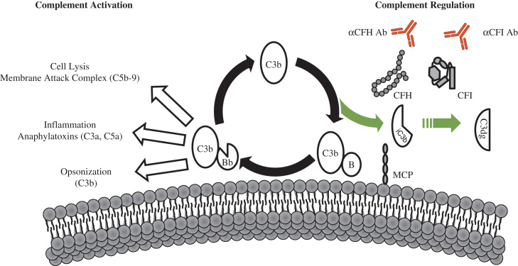
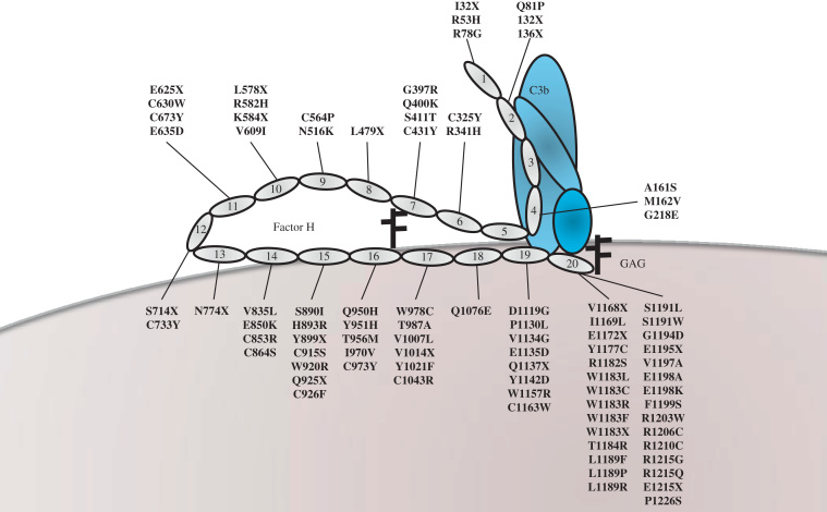
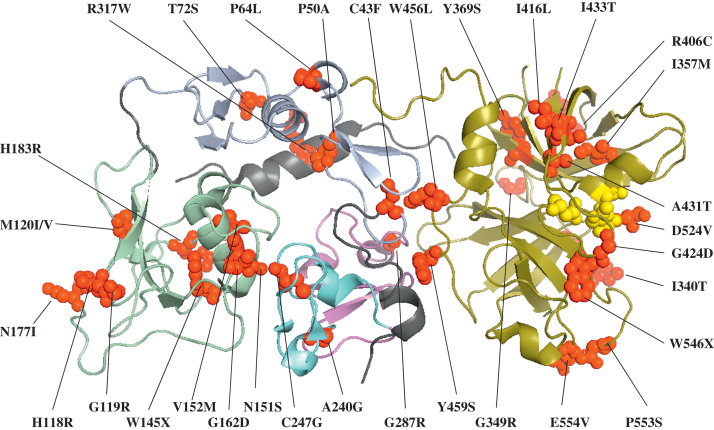
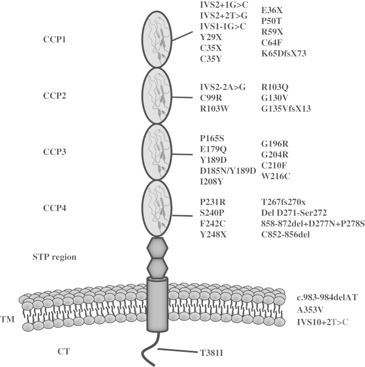
Hemolytic uremic syndrome (HUS) is a triad of microangiopathic hemolytic anemia, thrombocytopenia, and acute renal failure. The atypical form of HUS is a disease characterized by complement overactivation. Inherited defects in complement genes and acquired autoantibodies against complement regulatory proteins have been described. Incomplete penetrance of mutations in all predisposing genes is reported, suggesting that a precipitating event or trigger is required to unmask the complement regulatory deficiency. The underlying genetic defect predicts the prognosis both in native kidneys and after renal transplantation. The successful trials of the complement inhibitor eculizumab in the treatment of atypical HUS will revolutionize disease management.

Keywords: Complement; eculizumab; factor H; factor I; hemolytic uremic syndrome; membrane cofactor protein; thrombomodulin; transplantation.


Figures

Figure 1
Figure 1
Complement activation and regulation. The AP is a positive amplification loop. C3b interacts with factor B (B), which then is cleaved by factor D to form the AP C3 convertase (C3bBb). This enzyme complex is attached to the target covalently via C3b while Bb is the catalytic serine protease subunit. C3 is the substrate for this convertase, thus creating a powerful feedback loop. Unchecked, this leads to activation of the terminal complement pathway with generation of the effector molecules, the anaphylatoxin C5a, and the membrane attack complex (C5b-9). To protect host cells from bystander damage the AP is down-regulated by complement regulators including CFH, CFI, and MCP. Activating mutations in C3 and CFB and loss-of-function mutations in CFH, CFI, and MCP, in addition to autoantibodies to CFH and CFI, result in overactivation of the AP with resultant aHUS.
Figure 2
Figure 2
CFH and aHUS-associated mutations. CFH is composed of 20 CCP modules. The N-terminal modules (CCP1-4) bind to C3b and act as a cofactor for the CFI-mediated cleavage to the inactive iC3b. The C-terminal modules (CCP19 and 20) bind to C3b and glycosaminoglycans on host cells to mediate cell surface protection. Genetic variants described in aHUS cluster in CCPs 19 and 20, but can be seen throughout the molecule. Functional analysis of aHUS-associated variants has focused predominantly on the C-terminal variants (Table 3).
Figure 3
Figure 3
Location of aHUS-associated mutations within the crystal structure of factor I (protein database identification code: 2XRC). Factor I is a heterodimer consisting of a noncatalytic heavy chain linked by a disulfide bond to a catalytic light chain. The domain structure of CFI is shown with the heavy chain comprising the FIMAC domain, light blue; SRCR domain, pale green; LDLr1, cyan; and LDLr2, magenta; and the light chain or serine protease domain, deep olive. aHUS-associated genetic variants are shown as red spheres. Yellow spheres mark the catalytic triad of the serine protease domain. Functional analysis of aHUS-associated CFI variants are described in Table 4.
Figure 4
Figure 4
Mutations in MCP associated with aHUS. MCP is a transmembrane glycoprotein. It consists of 4 CCPs. Following the CCPs is an alternatively spliced region, rich in serine, threonine, and proline (STP region). The STP region is followed by a group of 12 amino acids of unknown function, a hydrophobic domain, a charged transmembrane anchor, and the alternatively spliced cytoplasmic tail (CT). Mutations associated with aHUS are clustered in the four extracellular CCPs of the molecule. Functional analysis of aHUS-associated MCP variants are described in Table 5.
Figure 5
Figure 5
Location of aHUS-associated mutations within the crystal structure of C3 (protein database identification code: 2A73). The structure of C3 is represented with the domains highlighted: MG1, green; MG2, blue; MG3, violet; MG4, olive; MG5, pink; MG6, orange; ANA, yellow; α’NT, grey; MG7, lime; CUB, light blue; TED, wheat; MG8, purple; and C345C, black. Genetic variants (red spheres) cluster around the MG2 and TED domains. Functional analysis of C3 mutations in aHUS has been performed in only a few cases (Table 6).

Similar articles

Cited by

References

    1. Kavanagh D., Richards A., Atkinson J. Complement regulatory genes and hemolytic uremic syndromes. Annu Rev Med. 2008;59:293–309. - PubMed
    1. Besbas N., Karpman D., Landau D., Loirat C., Proesmans W., Remuzzi G. A classification of hemolytic uremic syndrome and thrombotic thrombocytopenic purpura and related disorders. Kidney Int. 2006;70:423–431. - PubMed
    1. Constantinescu A.R., Bitzan M., Weiss L.S., Christen E., Kaplan B.S., Cnaan A. Non-enteropathic hemolytic uremic syndrome: causes and short-term course. Am J Kidney Dis. 2004;43:976–982. - PubMed
    1. Fogo A., Kashgarian M. Elsevier Science; Amsterdam: 2005. Diagnostic atlas of renal pathology.
    1. Ricklin D., Hajishengallis G., Yang K., Lambris J.D. Complement: a key system for immune surveillance and homeostasis. Nat Immunol. 2010;11:785–797. - PMC - PubMed

Publication types

MeSH terms