Comparative Study
. 2014 Jan;73(1):10-22.
doi: 10.1016/j.jdermsci.2013.09.001. Epub 2013 Sep 11.

Induction of a chloracne phenotype in an epidermal equivalent model by 2,3,7,8-tetrachlorodibenzo-p-dioxin (TCDD) is dependent on aryl hydrocarbon receptor activation and is not reproduced by aryl hydrocarbon receptor knock down

Affiliations
Comparative Study

Induction of a chloracne phenotype in an epidermal equivalent model by 2,3,7,8-tetrachlorodibenzo-p-dioxin (TCDD) is dependent on aryl hydrocarbon receptor activation and is not reproduced by aryl hydrocarbon receptor knock down

Alison R Forrester et al. J Dermatol Sci. 2014 Jan.

Abstract

Background: 2,3,7,8-Tetrachlorodibenzo-p-dioxin (TCDD) is a potent activator of the aryl hydrocarbon receptor (AhR) and causes chloracne in humans. The pathogenesis and role of AhR in chloracne remains incompletely understood.

Objective: To elucidate the mechanisms contributing to the development of the chloracne-like phenotype in a human epidermal equivalent model and identify potential biomarkers.

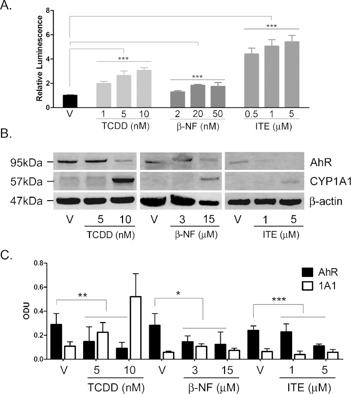
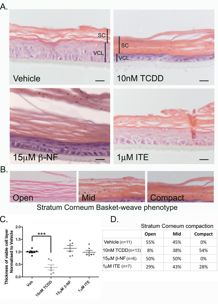
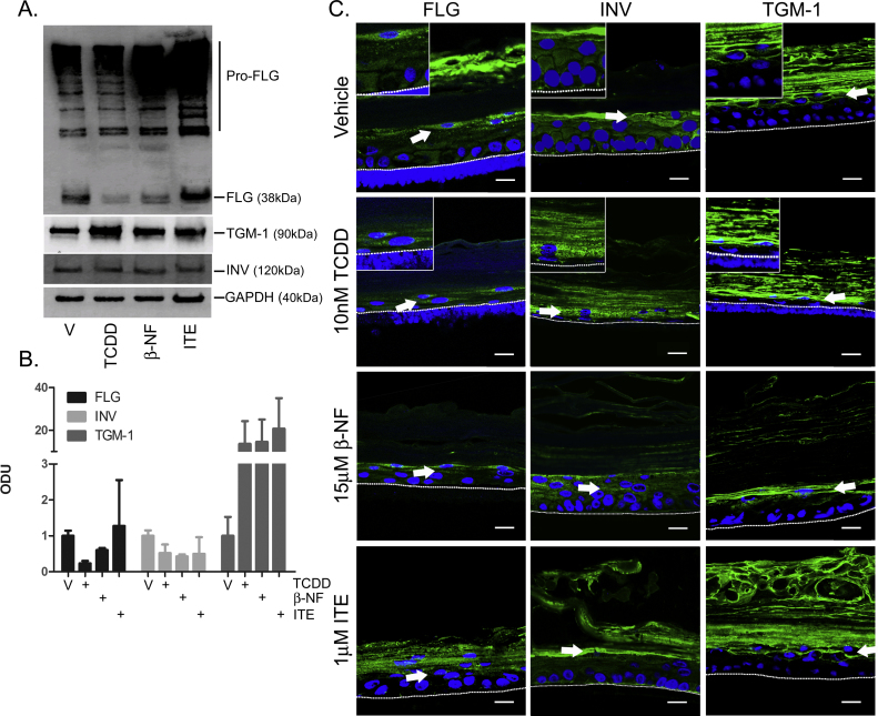
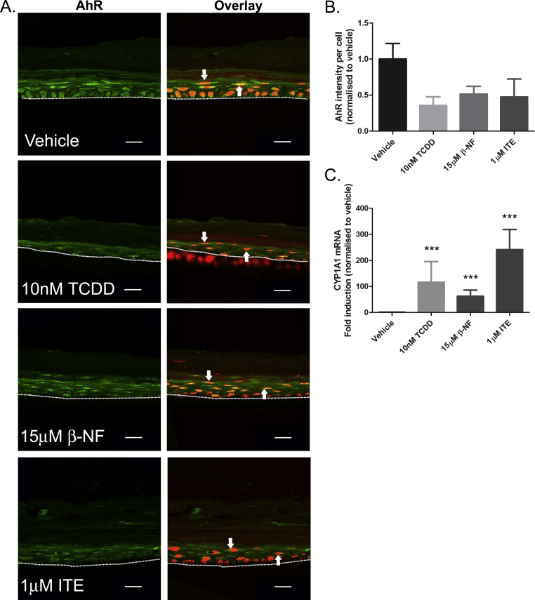
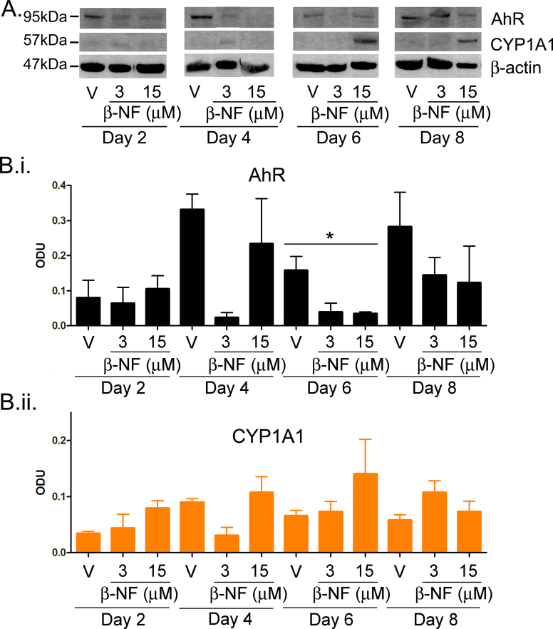
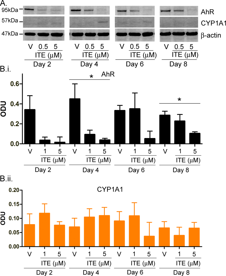
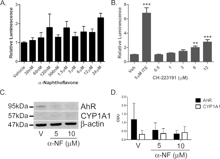
Methods: Using primary normal human epidermal keratinocytes (NHEK), we studied AhR activation by XRE-luciferase, AhR degradation and CYP1A1 induction. We treated epidermal equivalents with high affinity TCDD or two non-chloracnegens: β-naphthoflavone (β-NF) and 2-(1'H-indole-3'-carbonyl)-thiazole-4-carboxylic acid methyl ester (ITE). Using Western blotting and immunochemistry for filaggrin (FLG), involucrin (INV) and transglutaminase-1 (TGM-1), we compared the effects of the ligands on keratinocyte differentiation and development of the chloracne-like phenotype by H&E.

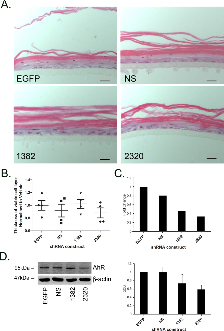
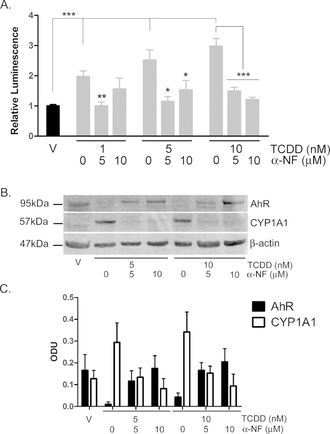
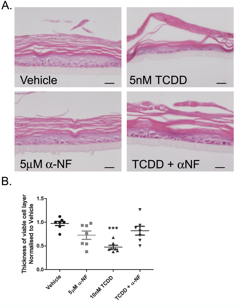
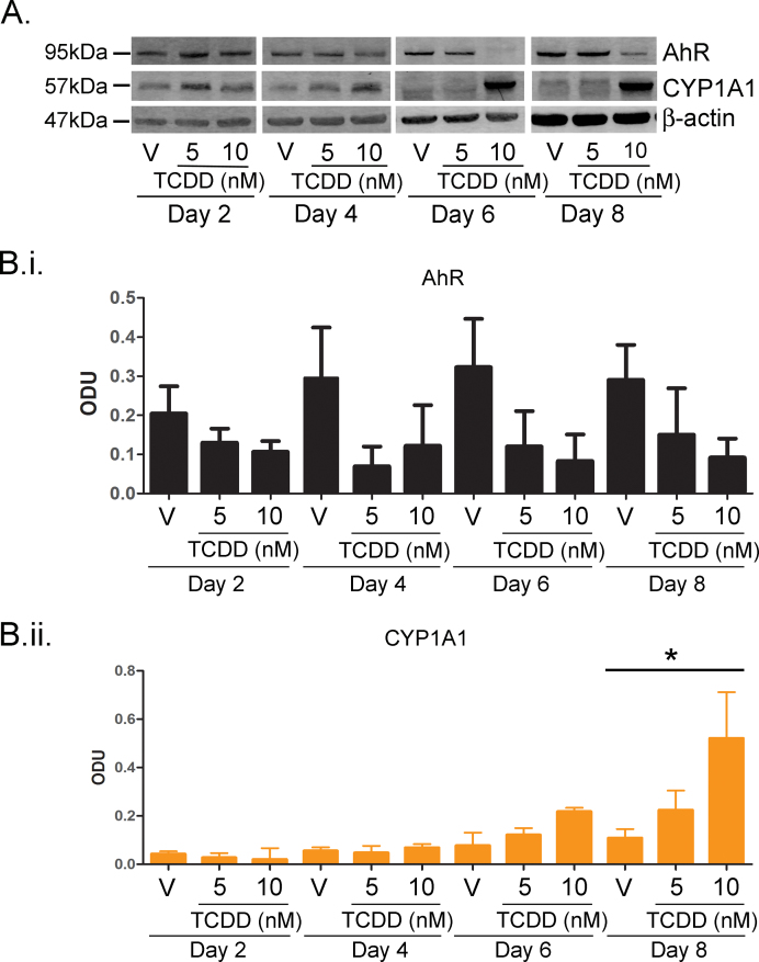
Results: In NHEKs, activation of an XRE-luciferase and CYP1A1 protein induction correlated with ligand binding affinity: TCDD>β-NF>ITE. AhR degradation was induced by all ligands. In epidermal equivalents, TCDD induced a chloracne-like phenotype, whereas β-NF or ITE did not. All three ligands induced involucrin and TGM-1 protein expression in epidermal equivalents whereas FLG protein expression decreased following treatment with TCDD and β-NF. Inhibition of AhR by α-NF blocked TCDD-induced AhR activation in NHEKs and blocked phenotypic changes in epidermal equivalents; however, AhR knock down did not reproduce the phenotype.

Conclusion: Ligand-induced CYP1A1 and AhR degradation did not correlate with their chloracnegenic potential, indicating that neither CYP1A1 nor AhR are suitable biomarkers. Mechanistic studies showed that the TCDD-induced chloracne-like phenotype depends on AhR activation whereas AhR knock down did not appear sufficient to induce the phenotype.

Keywords: 2,3,7,8-tetrachlorodibenzo-p-dioxin; 2-(1′H-indole-3′-carbonyl)-thiazole-4-carboxylic acid methyl ester; AHRR; AhR; Aryl hydrocarbon receptor; CYP1A1; Epidermal equivalent; ITE; Keratinocyte; TCDD; TGM-1; XRE; aryl hydrocarbon receptor; aryl hydrocarbon repressor protein; cytochrome P450 1A1; transglutaminase-1; xenobiotic response element; α-NF; α-naphthoflavone; β-NF; β-Naphthoflavone; β-naphthoflavone.


Figures

Fig. 1
Fig. 1
TCDD, β-NF and ITE induce AhR activation in NHEK, (A) Primary normal human epidermal keratinocytes (NHEKs) were co-transfected with XRE4-SV40-luciferase and renilla-luciferase constructs and treated with vehicle, TCDD, β-NF or ITE as indicated for 48 h. XRE-luciferase activity was measured and firefly:renilla luciferase ratio was normalised to vehicle. Data shown are mean ± sem, n = 9 (triplicate wells from 3 donors). Analysis of trend comparing vehicle to ligand, ***P < 0.0004. (B) Primary NHEKs were treated every 48 h for 8 days (on days 0, 2, 4 and 6) with vehicle or ligand as indicated. Samples were lysed and Western blotting performed. (C) Densitometry was carried out on blots probed with antibodies against AhR and CYP1A1 and normalised to β-actin. Densitometry represents mean ± sem from 3 donors; graph shows results of one-way ANOVA. Effects of TCDD on AhR: one-way ANOVA, **P = 0.007, analysis of trend P = 0.02. Effects of β-NF on AhR: one-way ANOVA, *P = 0.02, analysis of trend, NS. Effects of ITE on AhR: one-way ANOVA, ***P = 0.0002, analysis of trend P < 0.02.
Fig. 2
Fig. 2
TCDD caused decreased thickness of the viable cell layer and compaction of the stratum corneum in the epidermal equivalent model. (A) Epidermal equivalents were grown as described in materials and methods and treated with vehicle, TCDD, β-NF or ITE every 48 h for 7 days. After 7 days, equivalents were fixed, paraffin embedded and stained with H&E. (A) viable cell layer (VCL) and stratum corneum (SC) are marked by labelled black lines. Images are representative of effects in 3 donors. Scale bar = 20 μm. (B) Basket-weave formation of the stratum corneum of each section were characterised as open, mid way or compact. (C) Using Image J, 6 measurements of the viable cell layer were taken from 2 images per treatment for each donor. Individual values and mean (±sem) are shown for 3 independent donors. Dunnett's post hoc test compared vehicle to ligand, TCDD: ***P < 0.0001. (D) Compaction of the stratum corneum for each treatment per donor were characterised as demonstrated in (B). Values represent percentage of sections analysed from a minimum of 6 donors.
Fig. 3
Fig. 3
AhR activation induces dysregulated expression of involucrin, filaggrin and transglutaminase-1. Epidermal equivalents were grown and treated with vehicle, 10 nM TCDD, 15 μM β-NF or 1 μM ITE every 48 h for 7 days. (A) Western blotting was performed and blots were probed with antibodies against filaggrin (FLG), involucrin (INV) and transglutaminase-1 (TGM-1), with GAPDH as loading control. (B) Densitometry was carried out on blots probed with antibodies against FLG, INV and TGM-1 and normalised to GAPDH. Densitometry represents mean (±sem) from 2 donors. (C) Immunochemistry was performed using antibodies against FLG (left column), INV (centre column) or TGM-1 (right column) with Oregon green (488) tagged secondary antibody and To-pro-3 (blue) nuclear stain. Mid z section (TGM-1) and sum of 3 mid z (FLG, INV) images were captured by confocal microscopy and are representative of epidermal equivalents from 3 donors. Inserts show images at higher magnification (115×). Dotted white lines represent junction between basal layer and polycarbonate membrane and white arrows indicate points of interest. Scale bars = 20 μm.
Fig. 4
Fig. 4
TCDD, β-NF and ITE induce AhR activation in epidermal equivalents. Epidermal equivalents were grown and treated with vehicle, 10 nM TCDD, 15 μM β-NF or 1 μM ITE every 48 h for 7 days. (A) Immunochemistry was performed using an antibody against AhR, Oregon green (488) tagged secondary antibody and To-pro-3 (red) nuclear stain. Mid z sections were captured by confocal microscopy and some of 3 mid-sections are shown. Dotted white lines represent polycarbonate membrane and white arrows indicate points of interest. Scale bars = 20 μm. (B) Intensity of AhR fluorescence within the viable cell layer was quantified in Volocity software and normalised to number of nuclei present within the region of interest. Graph represents quantification from 2 donors. (C) mRNA was isolated from ligand treated epidermal equivalents and relative expression measured by RT-qPCR. Dunnett's post hoc test compared vehicle to ligand, ***P < 0.0001. Error bars represent mean (±sem) from 3 donors.
Fig. 5
Fig. 5
Development of the chloracne phenotype is not a direct result of AhR down-regulation. (A) Primary NHEKs were transduced with lentiviral shRNA constructs against AhR (1382, 2320), empty GFP (EGFP) or non-silencing (NS) control sequences and used to form epidermal equivalents. Samples were harvested, paraffin embedded and H&E stained. Scale bars = 20 μm. (B) 6 measurements of the viable cell layer were taken from 2 images per treatment for each donor. Individual values and mean (±sem) are shown. (C) mRNA was isolated from NHEK monolayers in parallel with epidermal equivalent cultures and knock down of AhR measured by RT-qPCR and normalised to 18S. Results presented as fold change relative to cells transfected with NS vector (NS). t-Test compared EGFP to 1382 (not significant) or 2320 (**P < 0.01) bars represent mean values from 2 donors. (D) AhR knock down epidermal equivalents were harvested in RIPA buffer and Western blotting performed. Blots were probed with antibodies against AhR and loading control β-actin. Images are representative of 3 donors. (E) Densitometry was carried out on blots probed with antibodies against AhR and normalised to β-actin. Results presented as fold change relative to cells transfected with empty GFP vector (note alternate order of bars). Densitometry represents mean (±sem) from 3 donors.
Fig. 6
Fig. 6
TCDD-induced AhR activation is inhibited by α-NF in primary NHEKs. (A) Primary NHEKs were co-transfected with XRE4-SV40-luciferase and renilla-luciferase control constructs and treated with vehicle or TCDD and/or α-NF as indicated for 48 h. Luciferase activity was measured and the firefly:renilla luciferase ratio was normalised to vehicle. Data shown are mean (±sem), n = 9 (triplicate wells from 3 donors). Two-way ANOVA: post hoc tests comparing vehicle to TCDD ± α-NF, Dunnett's: **/*, analysis of trend: ***P < 0.0001. (B) Primary NHEKs were treated with vehicle, TCDD and/or α-NF every 48 h as indicated for 8 days. Cells were lysed and proteins separated by Western blotting. Blots were probed with AhR and CYP1A1 antibodies with β-actin as loading control. Western blot is representative of duplicate blots from 3 donors. (C) Densitometry was performed on blots from 3 donors probed with antibodies against AhR (black bars) and CYP1A1 (white bars) and normalised to β-actin. Two-way ANOVA and analysis of trend: effects of TCDD on AhR, *P = 0.02. Densitometry is represents mean (±sem) from 3 donors.
Fig. 7
Fig. 7
Inhibition of AhR activation by α-NF partially blocks TCDD-induced phenotype in epidermal equivalents. (A) Epidermal equivalents were grown and treated with vehicle, 10 nM TCDD and/or 5 μM α-NF every 48 h for 7 days. After 7 days, equivalents were fixed, embedded in paraffin and stained with H&E. (B) Using Image J, 6 measurements of the viable cells layer were taken from 2 images per treatment for each donor. One-way ANOVA: Dunnett's post hoc test comparing vehicle to ligand, ***P = 0.0004. Individual values and mean (±sem) are shown for 3 donors.
Supplementary Fig. I
TCDD resulted in time and dose dependent AhR degradation and CYP1A1 induction between 2 and 8 days. (A) Primary NHEKs were treated for up to 8 days with vehicle, 5 or 10 nM TCDD as indicated. Samples were lysed and Western blotting performed. Densitometry was carried out on blots probed with antibodies against (B.i) AhR and (B.ii) CYP1A1 and normalised to β-actin. Densitometry represents mean ± sem from 3 donors. Two-way ANOVA: analysis of trend on effects of TCDD on AhR: *P = 0.02. Effects of time and TCDD on CYP1A1 induction, analysis of trend: **P < 0.002.
Supplementary Fig. II
β-NF induced CYP1A1 and AhR degradation in primary NHEKs. (A) Primary NHEKs were treated for up to 8 days with vehicle or doses of β-NF as indicated. Samples were lysed and Western blotting performed. Densitometry was carried out on blots probed with antibodies against (B.i) AhR and (B.ii) CYP1A1 normalised to β-actin. Densitometry represents mean ± sem from 3 donors. Two-way ANOVA: analysis of trend on effects of β-NF on AhR, *P < 0.02. CYP1A1 induction was not significant.
Supplementary Fig. III
ITE induced CYP1A1 and AhR degradation in primary NHEKs. (A) Primary NHEKs were treated for up to 8 days with vehicle or doses of ITE as indicated. Samples were lysed and Western blotting performed. Densitometry was carried out on blots probed with antibodies against (B.i) AhR or (B.ii) CYP1A1 normalised to β-actin. Densitometry represents mean ± sem from 3 donors. Two-way ANOVA: analysis of trend on effects of ITE on AhR, *P < 0.02. CYP1A1 induction was not significant.
Supplementary Fig. IV
AhR agonist activity of α-NF and CH-223191. (A/B) Primary NHEKs were co-transfected with XRE4-SV40-luciferase and renilla-luciferase control constructs and treated as indicated for 48h. Luciferase activity was measured and the mean firefly:renilla luciferase ratio was normalised to vehicle ± sem. n = 9, triplicate wells from 3 donors. Effects of α-NF on XRE-luciferase by one-way ANOVA: NS. Effects of CH-223191 on XRE-luciferase assay by one-way ANOVA: P < 0.0001, Dunnett's post hoc test, **/*** as indicated 5 μM ITE included as positive control, note different scale bars between (A) and (B). (C) Primary NHEKs were treated for 8 days with vehicle or α-NF as indicated. Samples were lysed and Western blotting performed. (D) Densitometry was carried out on blots probed with antibodies against AhR and CYP1A1 and normalised to β-actin. Densitometry represents mean ± sem from 3 donors. Two-way ANOVA was performed comparing vehicle to α-NF and time. α-NF significantly induced AhR degradation (P < 0.05), CYP1A1 was not significantly induced.

Similar articles

Cited by

References

    1. Abbott B.D., Schmid J.E., Brown J.G., Wood C.R., White R.D., Buckalew A.R. RT-PCR quantification of AHR, ARNT, GR, and CYP1A1 mRNA in craniofacial tissues of embryonic mice exposed to 2,3,7,8-tetrachlorodibenzo-p-dioxin and hydrocortisone. Toxicol Sci. 1999;47:76–85. - PubMed
    1. Fernandez-Salguero P.M., Ward J.M., Sundberg J.P., Gonzalez F.J. Lesions of aryl-hydrocarbon receptor-deficient mice. Vet Pathol. 1997;34:605–614. - PubMed
    1. Mimura J., Yamashita K., Nakamura K., Morita M., Takagi T.N., Nakao K. Loss of teratogenic response to 2,3,7,8-tetrachlorodibenzo-p-dioxin (TCDD) in mice lacking the Ah (dioxin) receptor. Genes Cells. 1997;2:645–654. - PubMed
    1. Caputo R., Monti M., Ermacora E., Carminati G., Gelmetti C., Gianotti R. Cutaneous manifestations of tetrachlorodibenzo-p-dioxin in children and adolescents. Follow-up 10 years after the Seveso, Italy, accident. J Am Acad Dermatol. 1988;19:812–819. - PubMed
    1. Mocarelli P., Gerthoux P.M., Patterson D.G., Jr., Milani S., Limonta G., Bertona M. Dioxin exposure, from infancy through puberty, produces endocrine disruption and affects human semen quality. Environ Health Perspect. 2008;116:70–77. - PMC - PubMed

Publication types

MeSH terms