Review
. 2014;10(2):263-79.
doi: 10.4161/hv.26902. Epub 2013 Oct 25.

Japanese encephalitis: the virus and vaccines

Affiliations
Review

Japanese encephalitis: the virus and vaccines

Sang-Im Yun et al. Hum Vaccin Immunother. 2014.

Abstract

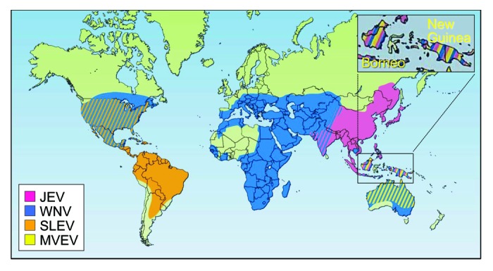
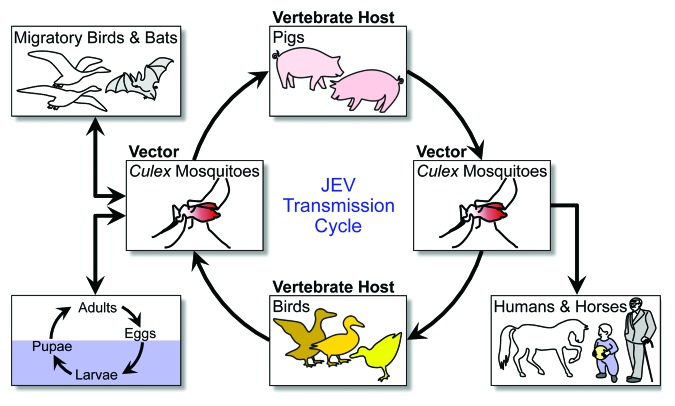
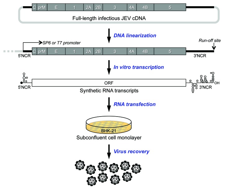
Japanese encephalitis (JE) is an infectious disease of the central nervous system caused by Japanese encephalitis virus (JEV), a zoonotic mosquito-borne flavivirus. JEV is prevalent in much of Asia and the Western Pacific, with over 4 billion people living at risk of infection. In the absence of antiviral intervention, vaccination is the only strategy to develop long-term sustainable protection against JEV infection. Over the past half-century, a mouse brain-derived inactivated vaccine has been used internationally for active immunization. To date, however, JEV is still a clinically important, emerging, and re-emerging human pathogen of global significance. In recent years, production of the mouse brain-derived vaccine has been discontinued, but 3 new cell culture-derived vaccines are available in various parts of the world. Here we review current aspects of JEV biology, summarize the 4 types of JEV vaccine, and discuss the potential of an infectious JEV cDNA technology for future vaccine development.

Keywords: Japanese encephalitis virus; biodefense; flavivirus; immunization; pathogenesis; prevention; vaccine; virulence.


Figures

None
Figure 1. Geographic distribution of four members of the JE serological group: Japanese encephalitis virus (JEV), West Nile virus (WNV), St. Louis encephalitis virus (SLEV), and Murray Valley encephalitis virus (MVEV). Adapted with permission from Macmillan Publishers Ltd: Nature Medicine, © 2004.
None
Figure 2. JEV transmission cycle. JEV is amplified in an enzootic cycle that involves mosquito vectors (mainly Culex species) and vertebrate hosts (primarily pigs and birds). Incidentally, JEV is also transmitted to dead-end hosts, such as humans and horses.
None
Figure 3. JEV genome structure and gene expression. (A) Genome structure. The plus-strand genomic RNA contains a cap structure, a 5′NCR, a long ORF, and a 3′NCR in a 5′-to-3′ direction. (B) Gene expression. The nascent polyprotein synthesized from the ORF is cleaved by host and viral proteases into at least 3 structural and 7 nonstructural proteins, as indicated. During viral maturation, prM is further processed by furin or a furin-like protease into the pr and M proteins. A derivative of NS1 (NS1') is expressed by a mechanism of frame-shifting which occurs between the codons 8 and 9 of NS2A, resulting in the addition of 52 extra amino acids.
None
Figure 4. JEV replication cycle. An infectious virion attaches to a target cell by binding to an attachment and entry receptor molecule(s) on the plasma membrane (Step 1). This interaction triggers the cell to internalize the virion by receptor-mediated endocytosis (Step 2). In the endosomes, the low pH induces significant conformational changes in the viral E glycoprotein, triggering the fusion of viral membrane with the host endosomal membrane (Step 3). Upon fusion, the viral genomic RNA is released into the cytoplasm, where it is first translated into the polyprotein precursor in association with the rough ER (Step 4). The polyprotein is processed to yield the mature viral proteins necessary for RNA replication and particle assembly. The genomic RNA is replicated in the replication complex inside virus-induced, ER-derived vesicles (Step 5). Immature progeny virions are formed by budding a complex of the newly synthesized genomic RNA and C proteins into the lumen of the ER, where they acquire the prM and E proteins on their membranes (Step 6). The immature virions are then transported to the Golgi apparatus through the secretory pathway; in the trans-Golgi network, the cleavage of prM to M leads to the maturation of the viral particles (Step 7). Finally, mature virions are released from the cell into the extracellular milieu by exocytosis (Step 8).
None
Figure 5. JEV reverse genetics. The full-length JEV cDNA is cloned in the bacterial artificial chromosome plasmid pBeloBAC11. The cloned cDNA is modified to have an SP6 or T7 promoter just upstream of the 5′-end of the viral genome and a run-off Xba I site immediately downstream of the viral 3′-end. To prepare a template for in vitro run-off transcription, the full-length cDNA is linearized by Xba I, followed by mung bean nuclease treatment to remove the 5′ overhang left by the Xba I digestion. The linearized cDNA is used as a template for run-off transcription using the SP6 or T7 RNA polymerase, as appropriate, in the presence of the m7G(5′)ppp(5′)A cap structure analog. This transcription reaction generates 5′ capped synthetic RNAs with authentic 5′ and 3′ ends of the viral genome. The synthetic RNAs are then introduced into eukaryotic cells by transfection using various methods such as DEAE-dextran, cationic liposomes, and electroporation. Typically, electroporation of the synthetic RNAs into BHK-21 cells generates a high titer of synthetic virus (~5 × 10⁶ PFU/ml) at 24 h post-transfection.

References

    1. Solomon T. Control of Japanese encephalitis--within our grasp? N Engl J Med. 2006;355:869–71. doi: 10.1056/NEJMp058263. - DOI - PubMed
    1. Endy TP, Nisalak A. Japanese encephalitis virus: ecology and epidemiology. Curr Top Microbiol Immunol. 2002;267:11–48. doi: 10.1007/978-3-642-59403-8_2. - DOI - PubMed
    1. Halstead SB, Jacobsen J. Japanese encephalitis vaccines. In: Plotkin SA, Orenstein WA, Offit PA, eds. Vaccines. Maryland Heights, MO: Saunders Elsevier, 2008:311-52.
    1. Mackenzie JS, Gubler DJ, Petersen LR. Emerging flaviviruses: the spread and resurgence of Japanese encephalitis, West Nile and dengue viruses. Nat Med. 2004;10(Suppl):S98–109. doi: 10.1038/nm1144. - DOI - PubMed
    1. Rappleye WC. Epidemiology of Japanese B encephalitis. Epidemic encephalitis: Third Report of the Matheson Commission. New York: Columbia University Press, 1939:157.

Publication types

MeSH terms

Substances