. 2013 Nov;15(11):1362-9.
doi: 10.1038/ncb2862. Epub 2013 Oct 27.

Heart field origin of great vessel precursors relies on nkx2.5-mediated vasculogenesis

Affiliations

Heart field origin of great vessel precursors relies on nkx2.5-mediated vasculogenesis

Noëlle Paffett-Lugassy et al. Nat Cell Biol. 2013 Nov.

Abstract

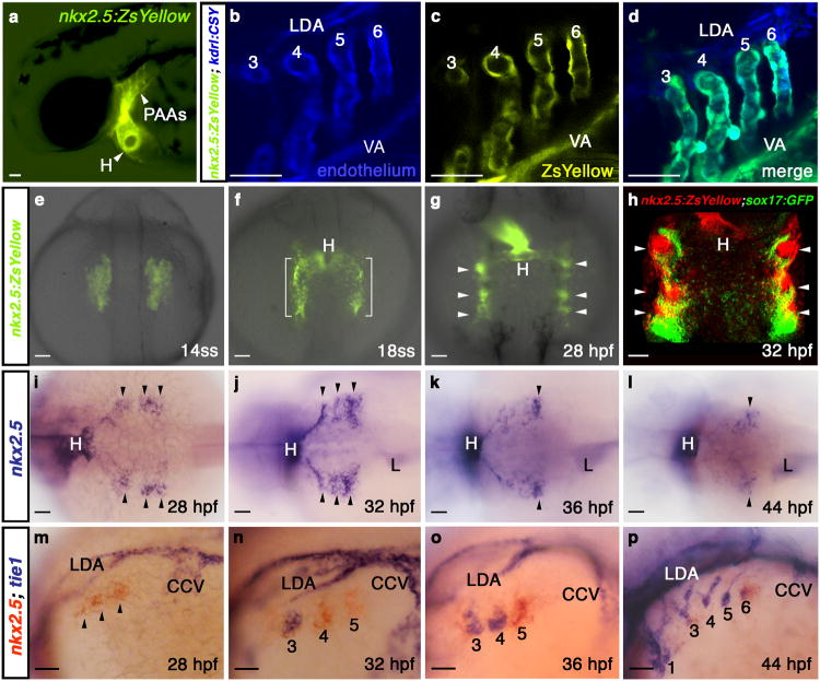
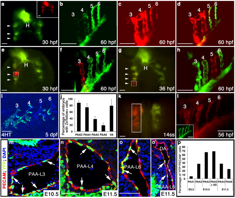
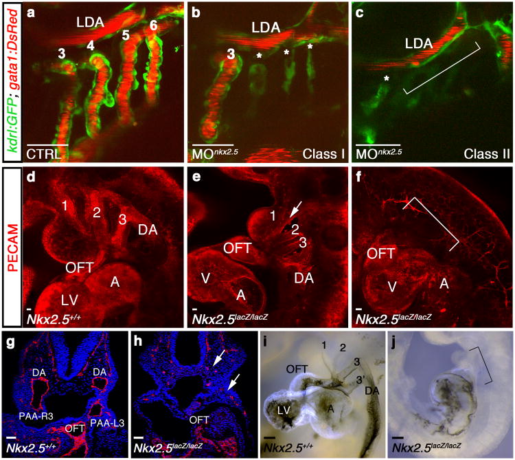
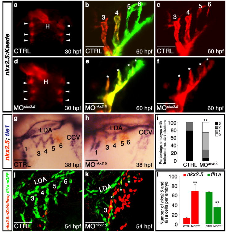
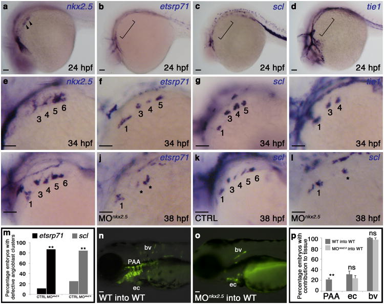
The pharyngeal arch arteries (PAAs) are transient embryonic blood vessels that make indispensable contributions to the carotid arteries and great vessels of the heart, including the aorta and pulmonary arteries. During embryogenesis, the PAAs appear in a craniocaudal sequence to connect pre-existing segments of the primitive circulation after de novo vasculogenic assembly from angioblast precursors. Despite the unique spatiotemporal characteristics of PAA development, the embryonic origins of PAA angioblasts and the genetic factors regulating their emergence remain unknown. Here, we identify the embryonic source of PAA endothelium as nkx2.5(+) progenitors in lateral plate mesoderm long considered to adopt cell fates within the heart exclusively. Further, we report that PAA endothelial differentiation relies on Nkx2.5, a canonical cardiac transcription factor not previously implicated in blood vessel formation. Together, these studies reveal the heart field origin of PAA endothelium and attribute a new vasculogenic function to the cardiac transcription factor Nkx2.5 during great vessel precursor development.


Figures

Figure 1
Figure 1. nkx2.5 is expressed in presumptive PAA endothelial progenitors
a, Tg(nkx2.5:ZsYellow) embryo at 60 hpf exhibiting fluorescence in the heart and PAAs. b-d, Tg(nkx2.5:ZsYellow); Tg(kdrl:CSY) embryo at 60 hpf with overlapping yellow and blue fluorescence in the endothelium of PAAs 3-6 and the VA. The LDA exhibits blue fluorescence exclusively. e-g, ZsYellow fluorescence in Tg(nkx2.5:ZsYellow) embryos at 14ss (e), 18ss (f), and 28 hpf (g). Brackets and arrowheads highlight non-cardiogenic nkx2.5+ cells in the pharynx. h, Tg(nkx2.5:ZsYellow); Tg(sox17:GFP) embryo at 32 hpf with nkx2.5+ pharyngeal cells (red) and sox17+ pharyngeal endoderm (green); i-l, in situ hybridization time-course of nkx2.5 expression in the pharynx. Arrowheads mark nkx2.5+ pharyngeal clusters. m-p, Double in situ hybridization time-course of nkx2.5 (red) and tie1 (blue) expression. PAA1 (labelled in p) forms earlier in development and never expresses nkx2.5. tie1+ clusters are numbered according to the mature PAA they derive. a-d, m-p, lateral views, anterior left; e-h, dorsal views, anterior up; i-l, dorsal views, anterior left; a-p, n>20 embryos per group. Abbr: VA, ventral aorta; LDA, lateral dorsal aorta; ss, somite-stage; hpf, hours post-fertilization; H, heart; L, liver; CCV, common cardinal vein.
Figure 2
Figure 2. nkx2.5+progenitors give rise to PAA endothelium in zebrafish and mouse
a-d, Tg(nkx2.5:Kaede) embryo at 30 hpf before (green) and after (red, inset) pan-photoconversion (n=3). Arrowheads highlight Kaede+ pharyngeal clusters. At 60 hpf, embryos were imaged in the green (b) and red (c) channels and these images were merged (d). e-h, Localized Kaede photoconversion of cluster 2 (e, white box, n=2) or 4 (g, white box, n=2) at the indicated developmental stages. Merged red and green images of the PAAs in the same embryos at 60 hpf are shown in (f) and (h). i, ZsYellow reporter fluorescence in PAAs 3-6 at 5 dpf in a Tg(nkx2.5:CreERT2); Tg(kdrl:CSY) embryo treated with 4HT from 10-13 hpf. j, Graph showing the average percentages of embryos with ZsYellow+ reporter fluorescence in each PAA and the VA across four experimental repliates (n=160). Error bars indicate one standard deviation. k, Left ALPM of a Tg(nkx2.5:Kaede) embryo photoconverted (white box) at 14ss and subsequently imaged in the red (l) and green channels (inset), (n=5). m-o′, Nkx2-5IRESCre; ROSAYFP embryos co-stained with PECAM1 (red) and DAPI (blue). Arrows indicate YFP+/PECAM+ lineage traced endothelial cells. o′, Left PAA 6 junction with DA. p, Graph depicting the average percentage of endothelial cells (PECAM1+) co-expressing YFP within each PAA. Cells counted across two embryos: E9.5 (PAA 1, n=244), E10.5 (PAA 2, n=216, PAA 3, n=1357, PAA3 + aortic sac, n=642), and E11.5 (PAAs 4, n=511 and 6, n=649). a,e,g,k, dorsal views, anterior up; b-d,f,h,i,l, lateral views, anterior left. Scale bar = 50μm. Abbr: hpf, hours post-fertilization; dpf, days post-fertilization; H, heart; AS, aortic sac; DA, dorsal aorta; PAA number and left (L) and right (R) designations indicated.
Figure 3
Figure 3. Nkx2.5 is required for vertebrate PAA formation
a-c, Tg(kdrl:GFP); Tg(gata1:DsRed) zebrafish embryos with green endothelial cells and red erythrocytes at 60 hpf. a, Control (CTRL) embryo with patent PAAs 3-6 and LDA (n=60/60). b, Class I nkx2.5 morphant (MOnkx2.5) in which one (shown) or two PAAs support blood flow. Asterisks label malformed PAAs (n=88/116). c, Class II nkx2.5 morphant lacking patent PAAs (bracket, n=22/116). d-f, Whole mount control (d) or Nkx2-5 null (e,f) mouse embryos stained for PECAM1 (red) at E9.5 (n=5 per group). Nkx2-5 null embryos displayed disrupted PAAs 1-3 with residual isolated endothelial cells (arrow, e) or a complete absence of PAAs altogether (bracket, f). g,h, Sections through control and Nkx2-5 null embryos stained with PECAM (red) and DAPI (blue). Arrow shows residual endothelial cells (h). i,j, Ink injections in control and Nkx2-5 null animals at E9.5 (n=5 per group). Bracket in j indicates absence of flow through the mutant OFTs. a-c, lateral views, anterior left; d-f, i,j, lateral views, anterior up; g,h, coronal sections. Scale bar = 50μm. Abbr: LDA, lateral dorsal aorta; DA, dorsal aorta; OFT, outflow tract; LV, left ventricle; A, atrium; V, ventricle; PAA number and left (L) and right (R) designations indicated.
Figure 4
Figure 4. Nkx2.5 is required for PAA progenitor cell differentiation
a-f, Control (n=3) and nkx2.5 morphant (n=5) Tg(nkx2.5:Kaede) embryos were pan-photoconverted at 30 hpf (a,d). Arrowheads show Kaede+ pharyngeal clusters. At 60 hpf, embryos were imaged in red and green channels with red (c,f) and merged (b,e) images shown. Asterisks (e,f) mark abnormal PAAs 3-6 in nkx2.5 morphant embryos. g,h, nkx2.5 (red) and tie1 (blue) transcripts evaluated by double in situ hybridization in control (g) and morphant (h) embryos. i, Quantification of tie1+ clusters in control (n=95) and nkx2.5 morphant (n=42) embryos across three experimental replicates; two-tailed t test **P=0.0001. j,k, Control and nkx2.5 morphant Tg(nkx2.5:nZsYellow); Tg(fli1a:nEGFP) embryos showing nkx2.5+ PAA progenitors (red) and fli1a+ endothelial cells (green) detected by immunohistochemistry. l, Quantification of nkx2.5+ and fli1a+ cells in control (n=4) and nkx2.5 morphant (n=4) embryos; Error bars indicate one standard deviation, two-tailed t test nkx2.5 **P=0.0073, fli1a **P=0.0008 across three independent experiments. Scale bar = 50μm. a,d, dorsal views, anterior up; b,c,e,f,g,h,j,k, lateral views, anterior left; PAA numbers indicated.
Figure 5
Figure 5. Cell-autonomous requirement for Nkx2.5 in promoting the PAA progenitor to angioblast transition
a-h, in situ hybridization analysis of nkx2.5(a,e), etsrp71(b,f), scl(c,g), and tie1(d,h) at 24 (a-d) and 34 (e-h) hpf. i-letsrp(i,j) and scl(k,l) transcripts evaluated by in situ hybridization in control (CTRL; i,k) and nkx2.5 morphant (MOnkx2.5;j,l) embryos, asterisks mark abnormal PAAs 3-6, numbers are quantified in m. (etsrp CTRL n=61, MOnkx2.5 n=53, two-tailed t test **P=0.001 across two independent experiments; scl CTRL n=106, MOnkx2.5 n=116, two-tailed t test **P=0.001 across two independent experiments). n-p, WT (n) and MOnkx2.5(o) donor-derived GFP+ PAA endothelium in unlabelled WT hosts. p, Quantification of host embryos showing GFP+ donor cells from WT (n=208) or MOnkx2.5 (n=86) embryos in the PAAs, endocardium (ec), and body vasculature (bv). Fishers exact test, PAA two tailed **P=0.0001; ec two-tailed P=0.3239 (not significant, ns); bv two-tailed P=0.2710, across four independent experiments. Scale bar = 50μm. a-l, lateral views, anterior left; PAA islands indicated; a-h n>20 embryos per group; m,p, Error bars indicate one standard deviation.

References

    1. Congdon ED. Transformation of the aortic arch system during the development of the human embryo. Contributions to Embryology. 1922;68:49–110.
    1. Moore KL, Persaud TVN. The developing human: clinically oriented embryology. 5th. W.B. Saunders; Philadelphia, PA: 1993.
    1. Anderson MJ, Pham VN, Vogel AM, Weinstein BM, Roman BL. Loss of unc45a precipitates arteriovenous shunting in the aortic arches. Developmental biology. 2008;318:258–267. - PMC - PubMed
    1. Li P, Pashmforoush M, Sucov HM. Mesodermal retinoic acid signaling regulates endothelial cell coalescence in caudal pharyngeal arch artery vasculogenesis. Developmental biology. 2012;361:116–124. - PMC - PubMed
    1. Ma Q, Zhou B, Pu WT. Reassessment of Isl1 and Nkx2-5 cardiac fate maps using a Gata4-based reporter of Cre activity. Developmental biology. 2008;323:98–104. - PMC - PubMed

Publication types

MeSH terms