Case Reports
. 2014 Mar 1;76(2):243-8.
doi: 10.1292/jvms.13-0336. Epub 2013 Oct 28.

Becker muscular dystrophy-like myopathy regarded as so-called "fatty muscular dystrophy" in a pig: a case report and its diagnostic method

Affiliations
Case Reports

Becker muscular dystrophy-like myopathy regarded as so-called "fatty muscular dystrophy" in a pig: a case report and its diagnostic method

Noriyuki Horiuchi et al. J Vet Med Sci. .

Abstract

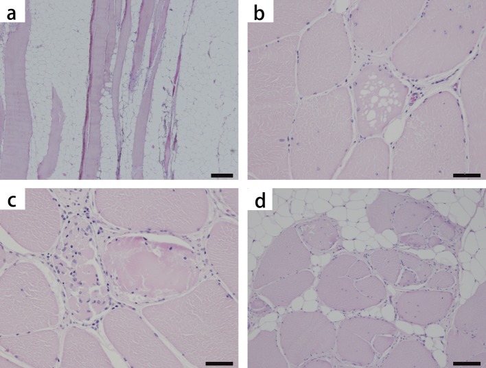
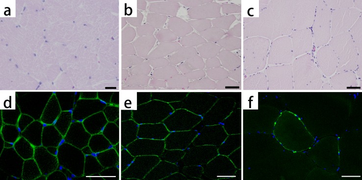
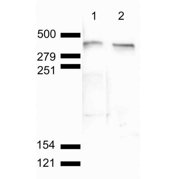
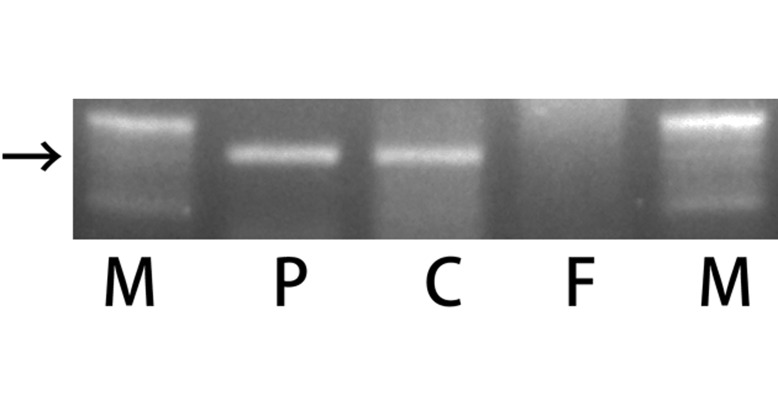
We describe a case of human Becker muscular dystrophy (BMD)-like myopathy that was characterized by the declined stainability of dystrophin at sarcolemma in a pig and the immunostaining for dystrophin on the formalin-fixed, paraffin-embedded (FFPE) tissue. The present case was found in a meat inspection center. The pig looked appeared healthy at the ante-mortem inspection. Muscular abnormalities were detected after carcass dressing as pale, discolored skeletal muscles with prominent fat infiltrations and considered so-called "fatty muscular dystrophy". Microscopic examination revealed following characteristics: diffused fat infiltration into the skeletal muscle and degeneration and regeneration of the remaining skeletal muscle fibers. Any lesions that were suspected of neurogenic atrophy, traumatic muscular degeneration, glycogen storage disease or other porcine muscular disorders were not observed. The immunostaining for dystrophin was conducted and confirmed to be applicable on FFPE porcine muscular tissues and revealed diminished stainability of dystrophin at the sarcolemma in the present case. Based on the histological observations and immunostaining results, the present case was diagnosed with BMD-like myopathy associated with dystrophin abnormality in a pig. Although the genetic properties were not clear, the present BMD-like myopathy implied the occurrence of dystrophinopathy in pigs. To the best of our knowledge, this is the first report of a natural case of myopathy associated with dystrophin abnormalities in a pig.


Figures

Fig. 1.
Fig. 1.
Pig. Skeletal muscle (rectus abdominis), hematoxylin and eosin stain (HE). Severe fat infiltration (a), vacuolar degeneration (b), hyalinized fiber and myophagy (c) and fiber splitting (d) are observed. The internal disposition of the nuclei is also seen in (b–d). Bar scales: 200 µm (a), 50 µm (b), 50 µm (c) and 100 µm (d).
Fig. 2.
Fig. 2.
Hematoxylin and eosin staining (HE) and immunofluorescence staining for dystrophin: healthy pig (a, d), necrotic muscle (b, e) and the present pig (c, f). Dystrophin is stained light green. The nuclei are stained blue. The healthy pig shows small fibers (a), because of its young age and clear stainability at the sarcolemma (d). The necrotic muscle, which consists of muscle fibers that are massively necrotized because of an infarct, shows strongly eosinophilic pale cytoplasm and declined muscle nucleus stainability in HE (b), whereas dystrophin is stained at the sarcolemma (e). In the present pig, dystrophin is faintly and partially stained at the sarcolemma (f). Bar scales: 20 µm (a) and 50 µm (b–f).
Fig. 3.
Fig. 3.
Western blot analysis of normal porcine muscle. Lane 1 is stained using pan-dystrophin antibody Dys1 (clone Dy4/6D3). Lane 2 is stained using the primary antibody (clone 1808) used in the present immunofluorescent examination. Closely spaced doublet bands of approximately 425 kDa were visualized on both lanes below the 500-kDa marker. Non-specific reactivity was not detected.
Fig. 4.
Fig. 4.
Agarose gel showing ethidium bromide-stained electrophoresis of PCR products of the SRY gene fragment, which was amplified from DNA isolated from paraffin-embedded tissue. Lane M is a ladder. Lane P is a positive control from the spleen of a male pig. Lane C is a sample from the muscular tissue of the present pig. Lane F is a sample from the spleen of a female pig. The arrow indicates 160 bp. A positive band was observed in lane P and lane C.

Similar articles

Cited by

References

    1. Arahata K., Ishiura S., Ishiguro T., Tsukahara T., Suhara Y., Eguchi C., Ishihara T., Nonaka I., Ozawa E., Sugita H.1988. Immunostaining of skeletal and cardiac muscle surface membrane with antibody against Duchenne muscular dystrophy peptide. Nature 333: 861–863. doi: 10.1038/333861a0 - DOI - PubMed
    1. Arahata K., Ishihara T., Kamakura K., Tsukahara T., Ishiura S., Baba C., Matsumoto T., Nonaka I., Sugita H.1989. Mosaic expression of dystrophin in symptomatic carriers of Duchenne’s muscular dystrophy. N. Engl. J. Med. 320: 138–142. doi: 10.1056/NEJM198901193200302 - DOI - PubMed
    1. Bordais A., Bolaños-Jimenez F., Fort P., Varela C., Sahel J. A., Picaud S., Rendon A.2005. Molecular cloning and protein expression of Duchenne muscular dystrophy gene products in porcine retina. Neuromuscul. Disord. 15: 476–487. doi: 10.1016/j.nmd.2005.03.011 - DOI - PubMed
    1. Bulfield G., Siller W. G., Wight P. A., Moore K. J.1984. X chromosome-linked muscular dystrophy (mdx) in the mouse. Proc. Natl. Acad. Sci. U.S.A. 81: 1189–1192. doi: 10.1073/pnas.81.4.1189 - DOI - PMC - PubMed
    1. Bushby K. M., Thambyayah M., Gardner-Medwin D.1991. Prevalence and incidence of Becker muscular dystrophy. Lancet 337: 1022–1024. doi: 10.1016/0140-6736(91)92671-N - DOI - PubMed

Publication types