. 2013:4:2610.
doi: 10.1038/ncomms3610.

Wnt secretion is required to maintain high levels of Wnt activity in colon cancer cells

Affiliations

Wnt secretion is required to maintain high levels of Wnt activity in colon cancer cells

Oksana Voloshanenko et al. Nat Commun. 2013.

Abstract

Aberrant regulation of the Wnt/β-catenin pathway has an important role during the onset and progression of colorectal cancer, with over 90% of cases of sporadic colon cancer featuring mutations in APC or β-catenin. However, it has remained a point of controversy whether these mutations are sufficient to activate the pathway or require additional upstream signals. Here we show that colorectal tumours express elevated levels of Wnt3 and Evi/Wls/GPR177. We found that in colon cancer cells, even in the presence of mutations in APC or β-catenin, downstream signalling remains responsive to Wnt ligands and receptor proximal signalling. Furthermore, we demonstrate that truncated APC proteins bind β-catenin and key components of the destruction complex. These results indicate that cells with mutations in APC or β-catenin depend on Wnt ligands and their secretion for a sufficient level of β-catenin signalling, which potentially opens new avenues for therapeutic interventions by targeting Wnt secretion via Evi/Wls.


Figures
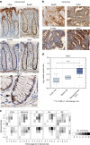
Figure 1
Figure 1. Evi/Wls is highly expressed in colon tumours.
(a,b) Immunohistochemical staining of normal or carcinoma tissue sections from colon, using the indicated antibodies. Images are representative of five cases. (a) In normal tissue, Evi/Wls expression is mostly undetectable, whereas Wnt3 is weakly expressed in most epithelial cells but more highly expressed in the Paneth-like/endocrine cells, which have a granular appearance (arrow). Keratin20 staining marks the top of the intestinal crypts and Eph receptor B2 staining the bottom. (b) In matched colon carcinomas, Evi/Wls and Wnt3 are highly expressed. (a,b) Scale bars, 60 μm, Wnt3 (enlarged) 20 μm. (c) Evi/Wls and Wnt3 are overexpressed in colon cancer. Results of immunostainings for Evi/Wls and Wnt3 on a colon cancer tissue array. Scores provided independently by three different investigators are shown as average. Staining intensity: 0=no staining, 1=weak staining, 2=moderate staining, 3=strong staining; percentage bin of cells: 0=<10%, 1=10–25%, 2=25–50%, 3=50–75%, 4=>75%). Grey scale represents the number of tumours. (d) Wnt3 is overexpressed in adenocarcinomas. Microarray-based expression data (Oncomine database, TCGA data set) were analysed for the expression of Wnt3 in normal colon(19)/rectum(3) versus adenocarcinomas (101). Preprocessed data from the array is log2-transformed and median-centred. The box plots show medians as line within the box, 25th and 75th percentiles as sides of the box, 10th and 90th percentiles as error bars, and outliers as circles. Significance of the differences in expression was calculated using the Student’s t-test. The specificity of the Wnt3 antibody was tested by knockdown experiments (Supplementary Fig. S1a). Evi/Wls antibody is described in Augustin et al.
Figure 2
Figure 2. Evi/Wls is required to maintain canonical Wnt signalling in colon cancer cell lines.
(a) Evi/Wls depletion reduces canonical Wnt signalling. DLD1 and HCT116 TCF4/Wnt reporter-colon cancer cells were transduced with lentivirus introducing a control or Evi shRNA construct. Luciferase assay was normalized to viability assessed in parallel using the CellTiterGlo assay. Results of three (for DLD1) and five (for HCT116) independent experiments are shown as mean±s.e.m. (b) Silencing of Evi/Wls regulates the Wnt target gene expression. DLD1 and HCT116 cells were transduced with a dox-inducible shmirEvi#1 construct and treated with or without dox. After 96 h, the cells were analysed for the expression of AXIN2 and EVI/WLS mRNAs by quantitative PCR. Results of four independent experiments are shown as mean±s.e.m. (c) Silencing of Evi/Wls reduces the level of active β-catenin in DLD1 and HCT116 cells, which express mutated APC or β-catenin, respectively. DLD1 cells were transfected with the indicated siRNAs for 120 h, and HCT116 cells were transfected for 72 h. Subsequently, the cells were lysed and western blotting was performed with the indicated antibodies. β-Actin served as the loading control. The reduction of total Lrp6 in β-catenin knockdown cells, which is consistently observed in DLD1 cells, may be a consequence of reduced β-catenin levels at the adherent junctions.
Figure 3
Figure 3. Wnt secretion is required to maintain canonical Wnt signalling in colon cancer cell lines.
(a) DLD1 and SW480 colon cancer cells activate the Wnt pathway in cocultured MDA-MB231 reporter cells. DLD1 or SW480 cells were cocultured with MDA-MB231 reporter cells stably transduced with a TCF/LEF reporter (1,500 cells) at the indicated ratio for 48 h. Next, cells were lysed and luciferase activity was measured. (b) Silencing of Wnt3 in DLD1 and HCT116 cells stably transfected with a TCF4/Wnt reporter reduces canonical Wnt signalling activity. Both cell lines were transfected with the indicated siRNAs. HCT116 cells were incubated for 72 h and DLD1 cells for 96 h before read-out. Luciferase levels were normalized to cell survival assessed by a CellTiterGlo assay performed in parallel. (c) Silencing of Wnt3a or Wnt3 reduces Axin2 protein expression. HCT116 cells were transfected with the indicated siRNAs for 72 h. Cell lysates were used for western blotting with indicated antibodies. β-Actin served as the loading control. (d) Evi/Wls knockdown in DLD1 cells reduces the induction of TCF/LEF reporter activity in coseeded MDA-MB231 reporter cells. Coculture of cells was conducted as described in a. Half times more DLD shEvi cells than shCtrl cells were seeded to compensate for reduced viability. (a,b,d) Data from three independent experiments are presented as mean±s.e.m.
Figure 4
Figure 4. Wnt pathway components at the receptor level are required for canonical activation in colon cancer cells.
(a) Knockdown of Lrp6, Dvl1 or Dvl3 reduces Wnt reporter activity in HCT116 and DLD1 cells. Cells stably transfected with a TCF4/Wnt reporter were reverse transfected with the indicated siRNAs and assessed for luciferase activity 72 h later. (b) Silencing of Dvl1 and Dvl3 reduces Wnt target gene expression in DLD1 cells. DLD1 cells were reverse transfected with the indicated siRNA and assessed for the expression of the Wnt target gene (AXIN2), as well as DVL1 and DVL3, by quantitative PCR 72 h later. (a,b) Data from four to five independent experiments are shown as mean±s.e.m.
Figure 5
Figure 5. Truncated APC in colon cancer cells is functional.
(a) Silencing of mutated APC in colon cancer cells increases AXIN2 expression. Schematic representation of the APC mutant proteins that are present in various colon cancer cell lines. All mutated APC proteins lack the SAMP (serine alanine methionine proline) repeats attributed to Axin binding. HT29 cells express the longest form of APC (1,555 aa); it retains all 15 AARs, as well as three of the seven 20 AARs, all of which are responsible for β-catenin binding/degradation. The indicated colon cancer cells were transfected with CTNNB1 or APC siRNAs for 72 h. Relative expression of the indicated mRNAs was determined by quantitative PCR. Data from three to four independent experiments are shown as mean±s.e.m. (b) β-Catenin binds to the main components of the destruction complex in colon cancer cells. The indicated cell lines were subjected to immunoprecipitation with anti-β-catenin antibody. Regardless of the extent of APC truncation, β-catenin co-immunoprecipitated APC, Axin1 and GSK3β. Western blottings are shown as representative of three independent experiments using the indicated antibodies.
Figure 6
Figure 6. Cells expressing truncated APC can form a functional destruction complex.
(a) β-Catenin bound to the main components of the destruction complex in DLD1 cells is phosphorylated. DLD1 cells were subjected to immunoprecipitation with anti-APC, -GSK3β or -Axin1 antibody. Western blottings are representative of three independent experiments. (b,c) A moderate decrease in APC level is not sufficient to induce Wnt signalling, but sensitizes cells to Wnt3a. RKO colon cancer cells that express wild-type APC and are stably transfected with a TCF4/Wnt reporter were transduced with the indicated shRNA constructs. (b) Luciferase activity was measured in RKO TCF4/Wnt cells treated with the indicated amounts of recombinant Wnt3a protein for 24 or 48 h. (c) Relative expression of APC and AXIN2 was measured by quantitative PCR. (b,c) Results of six (b) and three (c) independent experiments are shown as mean±s.e.m.
Figure 7
Figure 7. Evi/Wls is required for the survival of colon cancer cells in vitro and in vivo.
(a) Stable Evi/Wls knockdown reduces the clonogenic capacity of colon cancer cells. HCT116, DLD1 and SW480 cells stably transduced with a control (Ctrl) or an Evi shRNA were seeded in six-well plates at 1,000 cells per well, and were stained with crystal violet 14–20 days later. Wells are representative of three independent experiments for each cell line. (b,c) Evi/Wls is required for the formation of primary colon carcinomas in a xenograft mouse model. HCT116 or DLD1 cells were transduced as described in a, were injected subcutaneously (s.c.) into NOD/SCID mice (3 × 10⁵ per mouse) and tumour appearance was monitored over time. Kaplan–Meier plots show the probability of tumour appearance over time. (b) Results from two blinded, independent experiments are shown. CTNNB1 silencing was performed just in one of these experiments. (c) Results from one blinded experiment are shown. n, number of mice per group. *P=0.000201, **P=0.0000354 and ***P=0.0015. P-values were determined by log-rank test. (d) Evi/Wls downregulation reduces tumour volume in vivo. HCT116 cells were transduced as described in a and were injected s.c. (2 × 10⁶ per mouse) into opposite flanks of the same NOD/SCID mice. Eighteen days later, when tumour volumes had reached about 120 mm3, the mice were randomly separated into two groups: one was treated with dox (added to the drinking water) and the other was not. Tumour volume was measured on a weekly basis and the significance for each time point was calculated using the Student’s t-test. The experiment was terminated when tumours in the control group reached about 1,200 mm3. Results are presented as mean tumour volume±s.e.m.
Figure 8
Figure 8. Wnt secretion is important for the survival of colon TICs.
(a) Destruction complex is functional in primary colon cancer spheroids, independent of the mutation status of APC. Indicated cells were lysed and the main components of the destruction complex were co-immunoprecipitated with anti-β-catenin/APC antibody. P1 and P3 show APC truncation, whereas in P2 wild-type APC was detected. Representative examples of three independent experiments are shown. (b) P3 LEF/TCF cells were treated for 72+72 h with indicated amounts of IWP12. Luciferase assay was normalized to viability assessed in parallel using the CellTiterGlo assay. (c) P3 and P1 cells were treated as in b and then the CellTiterGlo assay was made. (b,c) Data from five (P3) and four (P1) independent experiments are shown as mean±s.e.m.
Figure 9
Figure 9. Schematic model on the role of Wnt secretion in APC mutant cells.
(a) In normal colon cells, Wnt signalling gradients originating at the bottom of colonic crypts determine cell identity. Low amounts of Wnt signalling lead to differentiation of epithelial cells of the colon. Under these conditions, wild-type APC serves as a scaffold, binding β-catenin and catalytic components of the destruction complex, thus sequestering it in the cytoplasm and causing its degradation. (b) In colon cancer cells, APC is truncated but retains some ability to bind β-catenin and act as a scaffold for the destruction complex. However, the efficiency of β-catenin retention is reduced in a manner related to the length of the truncated APC protein. Thus, APC truncations sensitize to canonical Wnts, allowing cancer cells to be more sensitive to activation by Wnt ligands, while retaining regulatory activity throughout the signalling cascade. Further, colon cancer cells acquire the ability to secrete Wnts in an autocrine manner, rendering the tumour increasingly independent of the original Wnt sources of the colonic crypts.

Similar articles

Cited by

References

    1. Logan C. Y. & Nusse R. The Wnt signaling pathway in development and disease. Annu. Rev. Cell Dev. Biol. 20, 781–810 (2004). - PubMed
    1. Morin P. J. et al. Activation of beta-catenin-Tcf signaling in colon cancer by mutations in beta-catenin or APC. Science 275, 1787–1790 (1997). - PubMed
    1. Cancer Genome Atlas Network. Comprehensive molecular characterization of human colon and rectal cancer. Nature 487, 330–337 (2012). - PMC - PubMed
    1. Reya T. & Clevers H. Wnt signalling in stem cells and cancer. Nature 434, 843–850 (2005). - PubMed
    1. Humphries A. & Wright N. A. Colonic crypt organization and tumorigenesis. Nat. Rev. Cancer 8, 415–424 (2008). - PubMed

Publication types

MeSH terms

LinkOut - more resources