. 2014 Feb;31(2):117-31.
doi: 10.1007/s10719-013-9505-7. Epub 2013 Oct 27.

Protein O-glucosylation in Lactobacillus buchneri

Affiliations

Protein O-glucosylation in Lactobacillus buchneri

Julia Anzengruber et al. Glycoconj J. 2014 Feb.

Abstract

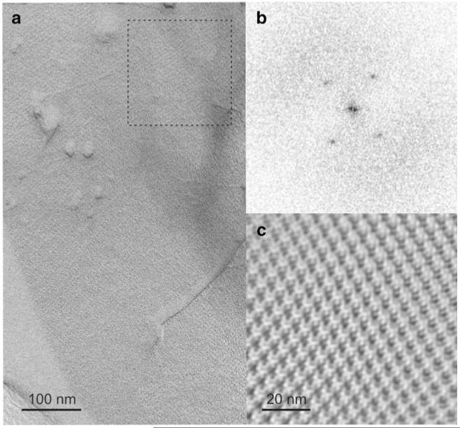
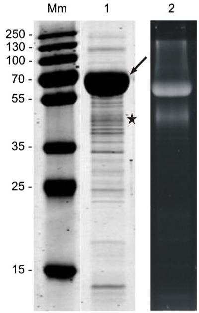
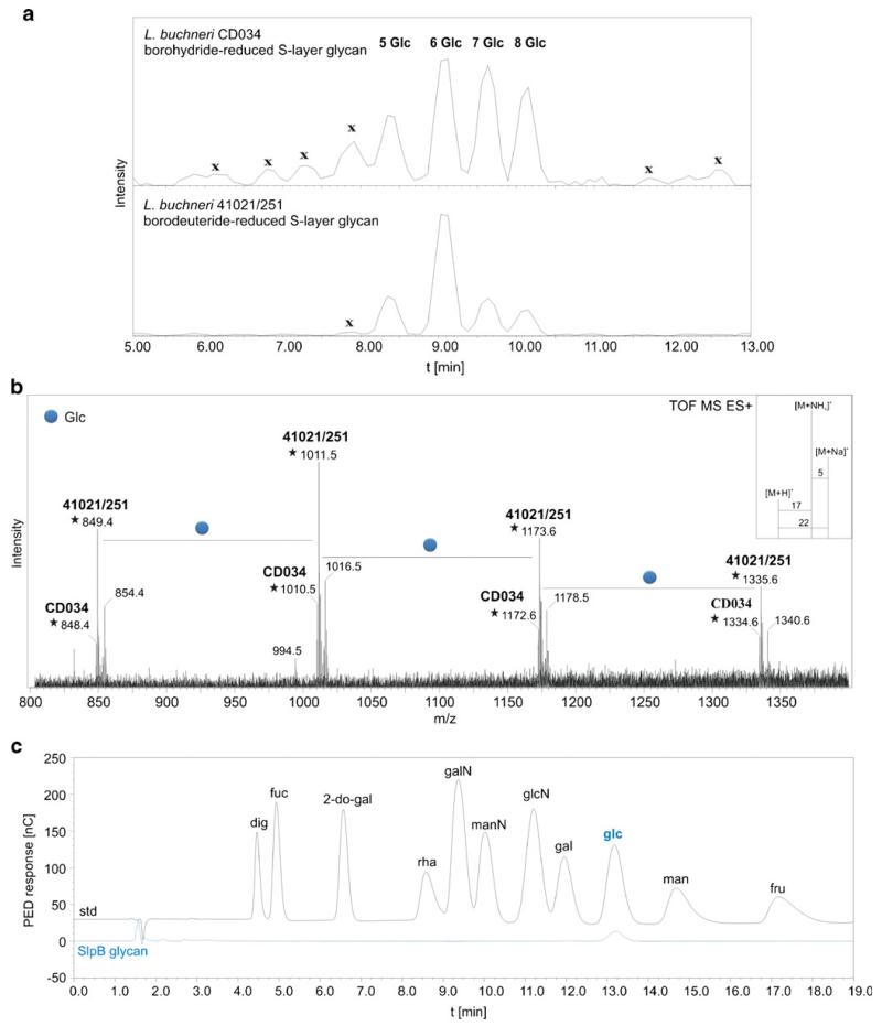
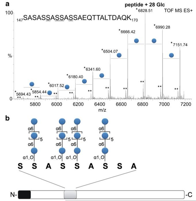
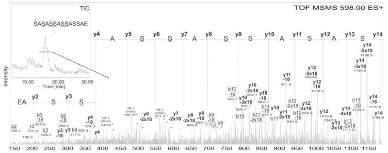
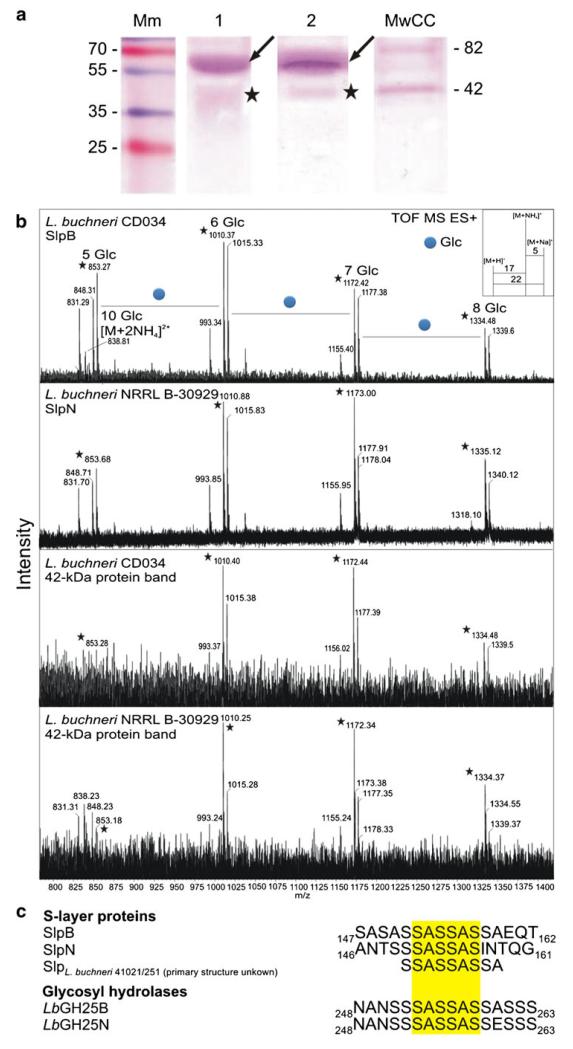
Based on the previous demonstration of surface (S-) layer protein glycosylation in Lactobacillus buchneri 41021/251 and because of general advantages of lactic acid bacteria for applied research, protein glycosylation in this bacterial species was investigated in detail. The cell surface of L. buchneri CD034 is completely covered with an oblique 2D crystalline array (lattice parameters, a = 5.9 nm; b = 6.2 nm; γ ~ 77°) formed by self-assembly of the S-layer protein SlpB. Biochemical and mass spectrometric analyses revealed that SlpB is the most abundant protein and that it is O-glycosylated at four serine residues within the sequence S(152)-A-S(154)-S(155)-A-S(157) with, on average, seven Glc(α1-6) residues, each. Subcellular fractionation of strain CD034 indicated a sequential order of SlpB export and glucosylation as evidenced by lack of glucosylation of cytosolic SlpB. Protein glycosylation analysis was extended to strain L. buchneri NRRL B-30929 where an analogous glucosylation scenario could be detected, with the S-layer glycoprotein SlpN containing an O-glycosylation motif identical to that of SlpB. This corroborates previous data on S-layer protein glucosylation of strain 41021/251 and let us propose a species-wide S-layer protein O-glucosylation in L. buchneri targeted at the sequence motif S-A-S-S-A-S. Search of the L. buchneri genomes for the said glucosylation motif revealed one further ORF, encoding the putative glycosyl-hydrolase LbGH25B and LbGH25N in L. buchneri CD034 and NRRL B-30929, respectively, for which we have indications of a glycosylation comparable to that of the S-layer proteins. These findings demonstrate the presence of a distinct protein O-glucosylation system in Gram-positive and beneficial microbes.


Figures

Fig. 1
Fig. 1
TEM micrograph of a freeze-etched and metal-carbon-shadowed preparation of L. buchneri CD034 cells. a Oblique S-layer lattice; b power spectrum of the S-layer lattice from the boxed area in (a), c lattice reconstruction
Fig. 2
Fig. 2
SDS-PAGE analysis (15 % gel) of a crude cell extract from L. buchneri CD034. Mm, PageRuler Plus pre-stained protein ladder (Thermo Scientific). Gels were stained with CBB (lane 1) and Pro-Q Emerald (lane 2). Two glyco-positive protein bands were detected. The arrow indicates the S-layer glycoprotein SlpB of L. buchneri CD034 which makes up the main portion of the whole cellular proteome. A second ~42-kDa, glyco-positive protein was detected and is indicated by an asterisk
Fig. 3
Fig. 3
Comparison of S-layer glycan profiles of L. buchneri CD034 and L. buchneri 41021/251. Glycans were released from the protein backbone via reductive β-elimination using NaBH4 and NaBD4 for L. buchneri strain CD034 and strain 41021/251, respectively. a LC profiles showing identical elution times of both glycans. Sample impurities are indicated with x. b Deconvoluted ESI-TOF-MS spectrum of released S-layer glycans from both strains premixed in one single run. Due to the use of deuterium for reduction of the L. buchneri 41021/251 S-layer glycan, a mass difference of 1 Da compared to the 1H-reduced glycan of L. buchneri CD034 was observed. As shown in (b), glycans from both strains could therefore be analyzed in one single measurement. Adducts are according to ammonium and sodium as depicted in the inset. Molecular ions indicated with an asterisk are [M+NH4]+. Peaks representing repeating glucose units were observed for both strains. c HPAEC-PED profile of SlpB glycan monosaccharides after TFA hydrolysis
Fig. 4
Fig. 4
a Mass distribution of the glycosylated peptide of the S-layer protein of L. buchneri CD034. After a tryptic digest the glycopeptide (amino acid sequence given in the figure) was applied to LC-ESI-MS. The m/z values correspond to the sum of the mass of the amino acids and the glucoses attached to the peptide. The major peak at an m/z value of 6828.51 refers to the mass of 28 glucoses distributed over the peptide, with a calculated m/z value of 6828.57. [M+H+2NH3]+ ions and [M+ NH4]+ ions are indicated by an asterisk and a double asterisk, respectively. Derivatization of glycosylated serines via ammonia based non-reductive β-elimination and ESI-TOF-MS/MS analysis (Fig. 5) revealed the presence of four glycosylation sites (underlined). b Schematic drawing of the S-layer glycoprotein of L. buchneri CD034. Based on glycan, glycopeptide as well as protein analysis, the S-layer glycoprotein possesses a signal peptide (black) consisting of 31 amino acids and an O-glycan consisting of on average seven Glc(α1–6) units attached to four serines within the acceptor sequence motif S-S-A-S-S-A-S-S-A (grey), corresponding to positions Ser152, Ser154, Ser155, and Ser157
Fig. 5
Fig. 5
Mass spectrometric determination of the O-glycosylation sites on the S-layer protein of L. buchneri CD034. The glycopeptide was subjected to RP-ESI-TOF-MS/MS after ammonia-based non-reductive β-elimination. As depicted in the inset, the deglycosylated peptide eluted at a high percentage of acetonitrile (>30 %) as a broad peak. Mainly the B- and Y- series ions were found in the MS/MS spectrum. All ions in the spectrum, if not indicated elsewise, are [M+H]+. Loss of 18 Da was deteceted for each peak originating from the Ser residues, most probably, due to loss of OH-groups. Deglycoslyted Ser was found with 86 Da (−1 Da)
Fig. 6
Fig. 6
Cellular localization of protein glycosylation and in L. buchneri CD034. L. buchneri proteins were isolated from the cell wall (lanes 1 and 3) and the cytosolic fraction (lanes 2 and 4) of L. buchneri CD034 as described in Materials and Methods. Samples were run on SDS-PAGE (15 % gel), PAS-stained for carbohydrates (lanes 1 and 2) and Western-blotted and detected with S-layer specific antiserum (lanes 3 and 4). Mm, PageRuler prestained protein ladder (Thermo Scientific)
Fig. 7
Fig. 7
Comparative analysis of protein glycosylation in L. buchneri strains. a Crude extracts of L. buchneri CD034 (lane 1) and L. buchneri NRRL B-30929 (lane 2) were run on SDS-PAGE (20 % gels) and PAS-stained for carbohydrates. The arrow and the asterisk indicate the S-layer proteins and the 42-kDa glyco-positive band, respectively. Mm, PageRuler Plus prestained protein ladder (Thermo Scientific); MmCC, Candy Cane Glycoprotein Molecular Weight Standards (Life Technologies). b Deconvoluted ESI-TOF-MS spectra of glycans released from L. buchneri CD034 and NRRL B-30929 glycoprotein bands after in-gel β-elimination in comparison with O-glycans of the S-layer protein SlpB of L. buchneri CD034. O-glycans from all proteins were found to be identical. Molecular ions indicated with an asterisk are [M+NH4]+. c Confirmed (SlpB) and predicted glycosylation sites of O-glycans in L. buchneri strains. The common sequence motif S-A-S-S-A-S (yellow background) was found in all investigated proteins
Fig. 8
Fig. 8
Apparent mass differences between native and recombinant glycosyl-hydrolases. L. buchneri CD034 and NRRL B-30929 cell extracts (lanes 1 and 3), recombinant LbGH25B and LbGH25N (lanes 2 and 4), premixed L. buchneri CD034 cell extract and recombinant LbGH25B (lane 5) and premixed L. buchneri NRRL B-30929 and recombinant LbGH25N were run on SDS-PAGE (9 % gel), Western-blotted and detected with glycosyl-hydrolase specific antiserum. Mm, PageRuler prestained protein ladder (Thermo Scientific)

References

    1. Varki A, Cummings RD, Esko JD, Freeze HH, Stanley P, Bertozzi CR, Hart GW, Etzler ME. Essentials of glycobiology. 2nd edn. Cold Spring Harbor Laboratory Press; Cold Spring Habor: 2009. p. 484. - PubMed
    1. Upreti RK, Kumar M, Shankar V. Bacterial glycoproteins: functions, biosynthesis and applications. Proteomics. 2003;3:363–379. - PubMed
    1. Schmidt MA, Riley LW, Benz I. Sweet new world: glycoproteins in bacterial pathogens. Trends Microbiol. 2003;11:554–561. - PubMed
    1. Hitchen PG, Dell A. Bacterial glycoproteomics. Microbiology. 2006;152:1575–1580. - PubMed
    1. Nothaft H, Szymanski CM. Protein glycosylation in bacteria: sweeter than ever. Nat. Rev. Microbiol. 2010;8:765–778. - PubMed

Publication types

MeSH terms

LinkOut - more resources