. 2014 Feb;21(2):310-20.
doi: 10.1038/cdd.2013.148. Epub 2013 Oct 25.

Epidermal Snail expression drives skin cancer initiation and progression through enhanced cytoprotection, epidermal stem/progenitor cell expansion and enhanced metastatic potential

Affiliations

Epidermal Snail expression drives skin cancer initiation and progression through enhanced cytoprotection, epidermal stem/progenitor cell expansion and enhanced metastatic potential

B De Craene et al. Cell Death Differ. 2014 Feb.

Abstract

Expression of the EMT-inducing transcription factor Snail is enhanced in different human cancers. To investigate the in vivo role of Snail during progression of epithelial cancer, we used a mouse model with skin-specific overexpression of Snail. Snail transgenic mice spontaneously developed distinct histological subtypes of skin cancer, such as basal cell carcinoma, squamous cell carcinoma and sebaceous gland carcinoma. Development of sebaceous gland carcinomas strongly correlated with the direct and complete repression of Blimp-1, a central regulator of sebocyte homeostasis. Snail expression in keratinocyte stem cells significantly promotes their proliferation associated with an activated FoxM1 gene expression signature, resulting in a larger pool of Mts24-marked progenitor cells. Furthermore, primary keratinocytes expressing Snail showed increased survival and strong resistance to genotoxic stress. Snail expression in a skin-specific p53-null background resulted in accelerated formation of spontaneous tumours and enhanced metastasis. Our data demonstrate that in vivo expression of Snail results in de novo epithelial carcinogenesis by allowing enhanced survival, expansion of the cancer stem cell pool with accumulated DNA damage, a block in terminal differentiation and increased proliferation rates of tumour-initiating cells.


Figures

Figure 1
Figure 1
Spontaneous tumour formation in K14-Snail mice. (a) Histological analysis of Ki67 expression in K14-Snail and WT control mice at the ages of 6 days and 4 months. Bars indicate epidermal thickness. (b) Measurement of skin thickness and Ki67-positive cell counts (right panel) in three WT and three K14-Snail mice (newborn) and five WT and four K14-Snail mice (adult). (c) Histological analysis of sections stained with H&E reveals distinct tumour types, such as sebaceous gland carcinoma (SGC) (40.%), BCC (6.67%) and SCC (13.33%). Mixed tumours were also identified (40%)
Figure 2
Figure 2
Snail expression leads to sebaceous gland hyperplasia and reduction of the transcription factor Blimp-1. (a) Confocal pictures of an immunohistological staining for adipophilin (green) and the hyperproliferation marker cytokeratin-6 (red) in WT and K14-Snail mice at the age of 6 days (left) and 3.5 weeks old (right) near the sebaceous gland (detail in insert—3 × magnified). (b) Adipohilin staining of skin sections of 4-month-old mice: transverse (upper panels) and longitudinal (lower panels) sections reveal the presence of many disorganised sebaceous gland islands in K14-Snail mice. (c) qPCR analysis reveals the strong reduction of Blimp-1 expression in K14-Snail mice compared with WT animals. Results are the average normalised relative expression values for four WT and four K14-Snail newborn mice and five WT and five K14-Snail adult mice. (d) In vitro analysis shows the significant reduction of Blimp-1 promoter activity in the presence of Snail expression in the sebocyte cell line SEB-1 (two independent experiments) and the breast carcinoma cell line MCF7 (three independent experiments). (e) Immunohistological analysis with an antibody-recognising Blimp-1 reveals the total absence of this transcription factor in the sebaceous gland of newborn and adult K14-Snail mice (presence in WT marked with arrowheads, and detail in insert—3 × magnified). However, the characteristic staining pattern in terminally differentiated keratinocytes in the suprabasal layer (marked by arrows) is retained
Figure 3
Figure 3
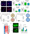
Snail expression enlarges the Mts24+ progenitor population and enhances the clonogenic capacity of CD34+ and Mts24+ cells. (a) Immunohistological staining for Mts24 in WT and K14-Snail mice (dotted lines mark the sebaceous gland boundaries). (b) qPCR analysis of Mts24 and Lgr6 expression in newborn and adult WT and K14-Snail epidermis. Results are the average normalised relative expression values for four WT and four K14-Snail newborn mice and five WT and five K14-Snail adult mice. (c) Clonogenic assay for CD34+ and Mts24+ keratinocytes from 8-week-old WT and K14-Snail mice. The relative number of colonies resulting from seeding equal numbers of CD34+ keratinocytes from WT (n=3) and K14-Snail (n=6) mice and of Mts24+ keratinocytes from WT (n=4) and K14-Snail (n=4) mice, 95% confidence intervals are indicated. Representative image showing that colonies of CD34+ and Mts24+ selected cells are larger in number and size in K14-Snail mice than in WT controls. (d) Microarray analysis for CD34+ and Mts24+ keratinocytes from 8-week-old WT and K14-Snail mice reveals an aberrant activated FoxM1 pathway in K14-Snail CD34+ stem cells. (e) A proportion of dividing stable keratinocyte cells (K38) show triple spindles when overexpressing Snail compared with luciferase control cells as shown by an alpha-tubulin staining (left). Quantification of the number of triple spindles 1 and 2 h after synchronisation of K38 cells with thymidine and nocodazole (n=5) (right)
Figure 4
Figure 4
Snail expression enhances cell viability and confers resistance to apoptosis. (a) Primary keratinocytes of pups from WT and K14-Snail animals were isolated and treated with different concentrations of DMBA (left panels) or doxorubicin (right panels). Survival was measured with an MTT assay (upper panels), and the results are presented as the average of three independent experiments of treated pooled keratinocytes from at least two different mice. Apoptosis was measured with a caspase-3/-7 Glo assay, and the results are presented as the average of two experiments of treated pooled keratinocytes from at least two different mice. (*P<0.05, **P<0.01, ***P<0.001). (b) Skin carcinogenesis in WT and K4-Snail mice after single (left) or multiple (right) DMBA treatments. The number of skin papillomas per mouse is shown. (c) Immunohistological analysis of the skin of DMBA-treated mice reveals highly invasive regions in K14-Snail animals but not in WT controls. Upper and lower panel shows the histology of two independent tumours with H&E staining
Figure 5
Figure 5
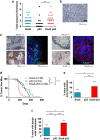
Synergistic effects of combining Snail expression with loss of the p53 tumour suppressor in vivo. (a) K14-Snail;K14Cre;Trp53f/f mice have on average more skin tumours than K14-Snail and K14Cre;Trp53f/f mice. (b) Histological analysis with a H&E staining reveals carcinosarcoma as an additional tumour type in K14-Snail;K14Cre;Trp53f/f mice. (c) (Left panel) Metastasis to lymph nodes was detected in K14-Snail mice: histological analysis with H&E staining shows cells with sebaceous characteristics (upper left); these cells stain positive for HA-tagged Snail (lower left). The infiltrating cells have retained the sebaceous cell marker adipophilin (green); no expression of the proliferation marker K6 (red) is observed. (Right panel) Metastasis to lungs was observed in K14-Snail mice. The nodules show a high degree of keratinisation (upper left) and are partially positive for HA-tagged Snail (lower left). The nodules are strongly K6-positive (red) but do not express adipohilin (green) (left). (d) Kaplan–Meier tumour-free survival curves for K14-Snail, K14Cre;Trp53f/f and K14-Snail;K14Cre;Trp53f/f mice. (e) Incidence of lung metastasis in mice with skin tumours. Bars represent 95% confidence intervals. (f) Incidence of lymph node metastasis in mice with skin tumours. Bars represent 95% confidence intervals. (Statistical significance used in this figure: *P<0.05, **P<0.01, ***P<0.001)
Figure 6
Figure 6
Summary model: Snail expression drives skin cancer progression. (Left) WT overview and detail map of the skin regions of interest studied here in more detail: bulge region containing CD34+ stem cells (red), progenitor region containing Mts24+ progenitor cells (yellow), and sebaceous gland region (blue) with terminally differentiated Blimp-1+ cells (dark blue). (Right) (a) At onset, Snail expression certainly can affect the in vivo proliferative capacity of keratinocyte stem cells, leading to the expansion of the Mts24+ progenitor region and (b) the block in their terminal differentiation. (c) Snail acts as a survival factor in non-terminally differentiated cells accumulating genetic aberrations (stress, mutations) allowing (d) progression to carcinoma. Snail acts at this stage as EMT inducer enabling the cells to invade and migrate, which eventually leads to metastasis

References

    1. Friedl P, Wolf K. Tumour-cell invasion and migration: diversity and escape mechanisms. Nat Rev. 2003;3:362–374. - PubMed
    1. De Craene B, Berx G. Regulatory networks defining EMT during cancer initiation and progression. Nat Rev. 2013;13:97–110. - PubMed
    1. Cano A, Perez-Moreno MA, Rodrigo I, Locascio A, Blanco MJ, del Barrio MG, et al. The transcription factor snail controls epithelial-mesenchymal transitions by repressing E-cadherin expression. Nat Cell Biol. 2000;2:76–83. - PubMed
    1. Batlle E, Sancho E, Franci C, Dominguez D, Monfar M, Baulida J, et al. The transcription factor snail is a repressor of E-cadherin gene expression in epithelial tumour cells. Nat Cell Biol. 2000;2:84–89. - PubMed
    1. De Craene B, Gilbert B, Stove C, Bruyneel E, van Roy F, Berx G. The transcription factor snail induces tumor cell invasion through modulation of the epithelial cell differentiation program. Cancer Res. 2005;65:6237–6244. - PubMed

Publication types

Substances