Review
. 2013 Oct;59(5):431-6.
doi: 10.1262/jrd.2013-068.

Roles of cohesin and condensin in chromosome dynamics during mammalian meiosis

Affiliations
Review

Roles of cohesin and condensin in chromosome dynamics during mammalian meiosis

Jibak Lee. J Reprod Dev. 2013 Oct.

Abstract

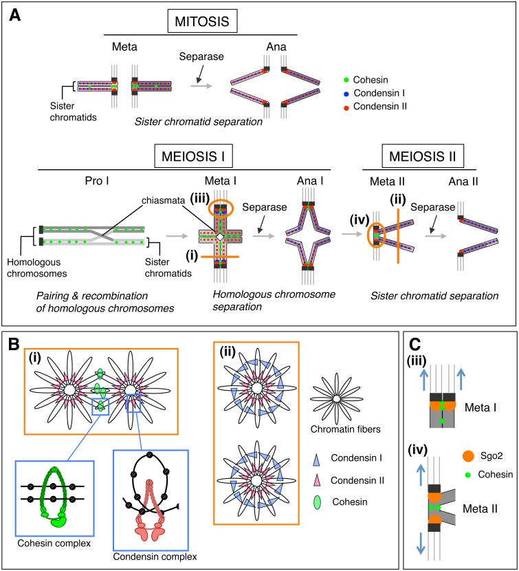
Meiosis is a key step for sexual reproduction in which chromosome number is halved by two successive meiotic divisions after a single round of DNA replication. In the first meiotic division (meiosis I), homologous chromosomes pair, synapse, and recombine with their partners in prophase I. As a result, homologous chromosomes are physically connected until metaphase I and then segregated from each other at the onset of anaphase I. In the subsequent second meiotic division (meiosis II), sister chromatids are segregated. Chromosomal abnormality arising during meiosis is one of the major causes of birth defects and congenital disorders in mammals including human and domestic animals. Hence understanding of the mechanism underlying these unique chromosome behavior in meiosis is of great importance. This review focuses on the roles of cohesin and condensin, and their regulation in chromosome dynamics during mammalian meiosis.


Figures

Fig. 1.
Fig. 1.
(A) Spatiotemporal dynamics of cohesin and condensins I and II in mitotic and meiotic divisions. In mitosis, cohesin maintains sister chromatid cohesion mainly at centromeres, while most cohesin is removed from chromosome arms until metaphase (meta). Condensins I and II are recruited to the chromatid axes and participate to construct mitotic chromosomes. Sister chromatids separate from each other by the action of Separase to cleave a cohesin subunit, RAD21, at anaphase (ana). In meiosis, homologous chromosomes pair, juxtapose, and recombine with their partners in prophase I (pro I). Theoretically, cohesin localizing arm regions distal to chiasmata contributes to the physical connection between homologous chromosomes. At metaphase I (meta I), meiotic cohesin containing REC8 maintains sister chromatid cohesion at both centromeres and chromosome arms. At this stage, condensin I localizes at the vicinity of centromeres, while condensin II localizes along chromatid axes. At anaphase I (ana I), cohesin along arms is removed by the action of Separase, while centromeric cohesin is protected. In parallel, condensin I becomes localized along arms. At the onset of anaphase II (ana II), the remaining cohesin at centromeres is now cleaved by the action of Separase. (B) Cross-sectional views of chromosomes along lines (i) and (ii) in Fig. 1A are shown. In the insets, the models of how cohesin and condensin complexes regulate the higher structure of chromosomes are proposed; cohesin complexes are thought to hold nucleosomes from sister chromatids, whereas condensin may connect the separate segments of the nucleosome of a single chromatid. (C) Localization of Sgo2 and cohesin containing REC8 around centromeres, indicated by ellipses (iii) and (iv) in Fig. 1A, is shown. When sister kinetochores are pulled towards the same spindle poles at meta I, no or little tension is exerted between the kinetochores. In such a case, Sgo2 is localized close to the centromeric cohesin. When sister kinetochores are pulled towards opposite directions at metaphase II (meta II) (or in mitotic metaphase), tension is exerted between the kinetochores. In this situation, Sgo2 relocates from the inner centromere to the outer kinetochores. This relocation of Sgo2 leads to spatial separation from centromeric cohesin, thereby stopping its protective function of centromeric cohesion.
Fig. 2.
Fig. 2.
Differentially expressed cohesin subunits during meiotic prophase I. Newly identified cohesin subunit RAD21L (green), the canonical mitotic cohesin subunit RAD21 (red), and the synaptonemal complex protein SYCP3 (blue) are immunofluorescently labeled in mouse testicular cells. DNA (silver) was counterstained with DAPI. (a) SYCP3, (b) RAD21L, (c) RAD21, (d) merged image. It is noteworthy that RAD21L, but little RAD21, is detected along the synaptonemal complex in some spermatocytes (arrows), and vice versa in other spermatocytes (arrowheads).

Similar articles

Cited by

References

    1. Peters JM, Tedeschi A, Schmitz J. The cohesin complex and its roles in chromosome biology. Genes Dev 2008; 22: 3089–3114 - PubMed
    1. Revenkova E, Eijpe M, Heyting C, Gross B, Jessberger R. Novel meiosis-specific isoform of mammalian SMC1. Mol Cell Biol 2001; 21: 6984–6998 - PMC - PubMed
    1. Prieto I, Suja JA, Pezzi N, Kremer L, Martinez AC, Rufas JS, Barbero JL. Mammalian STAG3 is a cohesin specific to sister chromatid arms in meiosis I. Nat Cell Biol 2001; 3: 761–766 - PubMed
    1. Lee J, Iwai T, Yokota T, Yamashita M. Temporally and spatially selective loss of Rec8 protein from meiotic chromosomes during mammalian meiosis. J Cell Sci 2003; 116: 2781–2790 - PubMed
    1. Lee J, Hirano T. RAD21L, a novel cohesin subunit implicated in linking homologous chromosomes in mammalian meiosis. J Cell Biol 2011; 192: 263–276 - PMC - PubMed

Publication types

MeSH terms