. 2013 Nov 7;503(7474):115-20.
doi: 10.1038/nature12600. Epub 2013 Oct 27.

Dendritic spikes enhance stimulus selectivity in cortical neurons in vivo

Affiliations

Dendritic spikes enhance stimulus selectivity in cortical neurons in vivo

Spencer L Smith et al. Nature. .

Abstract

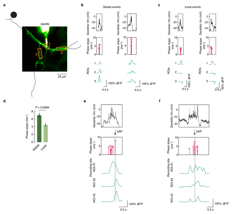
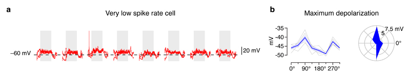
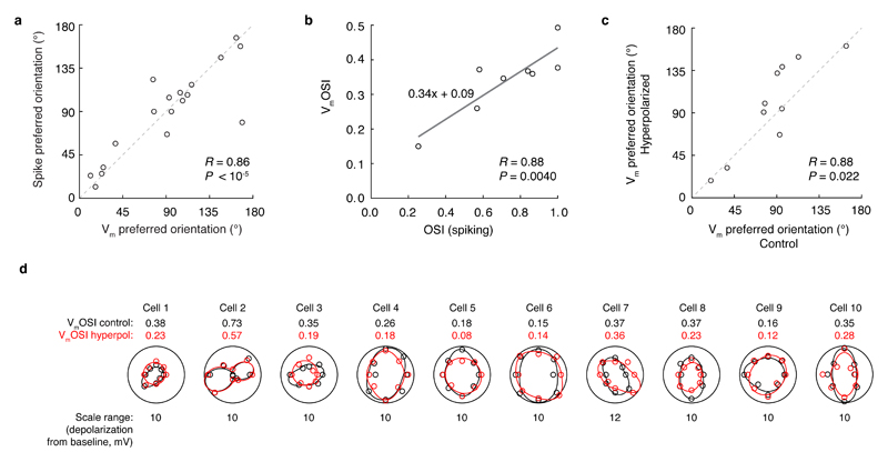
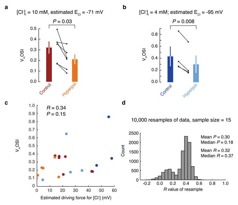
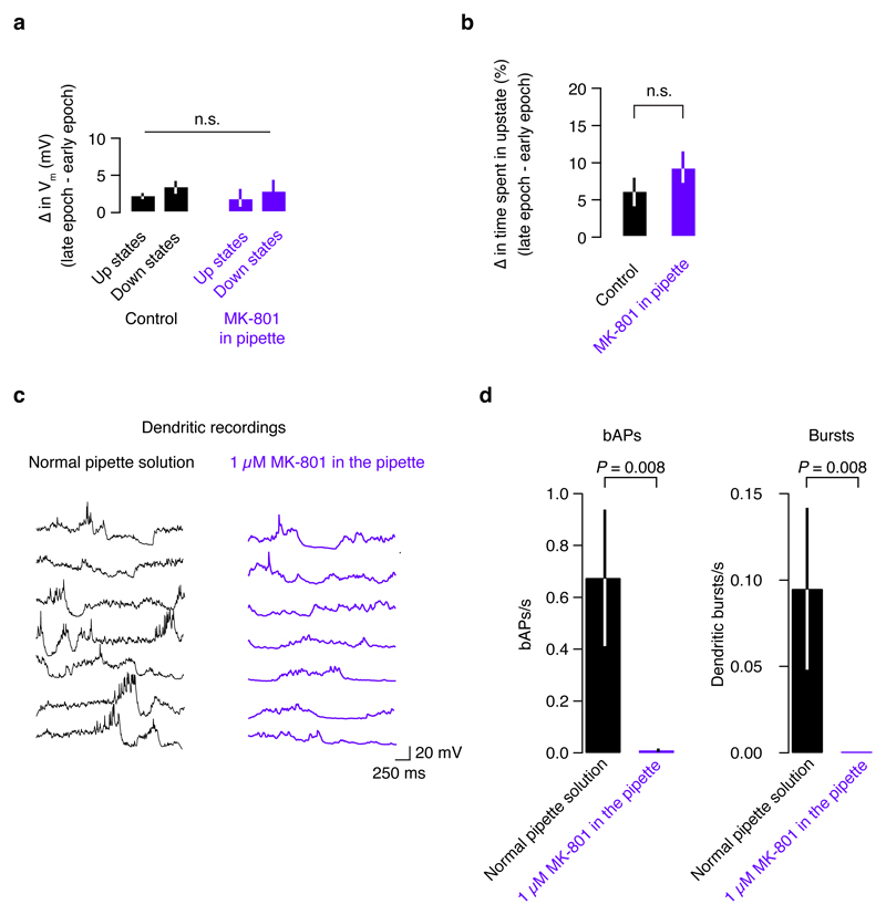
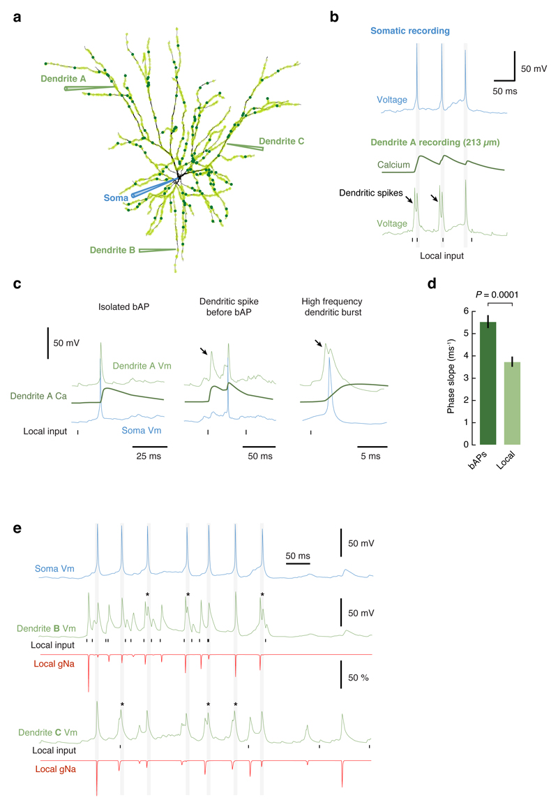
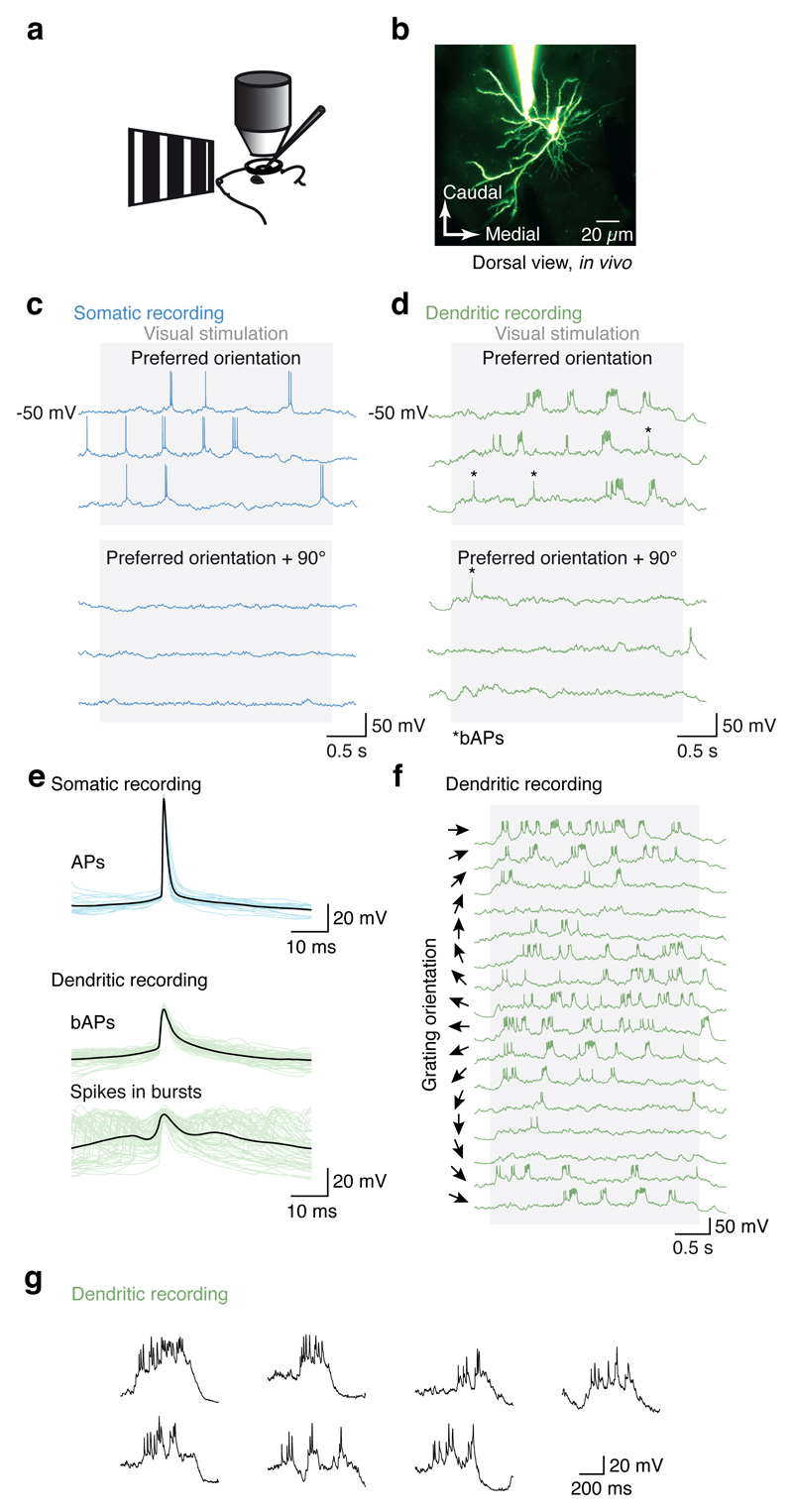
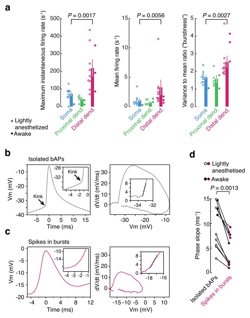
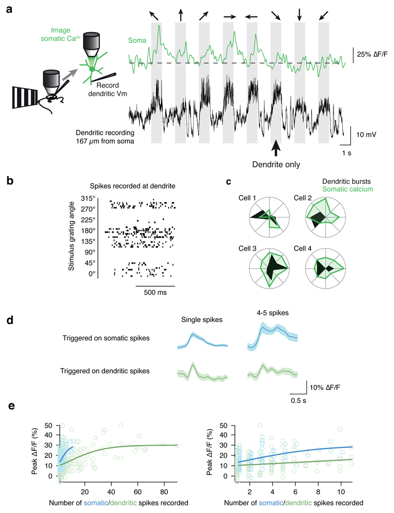
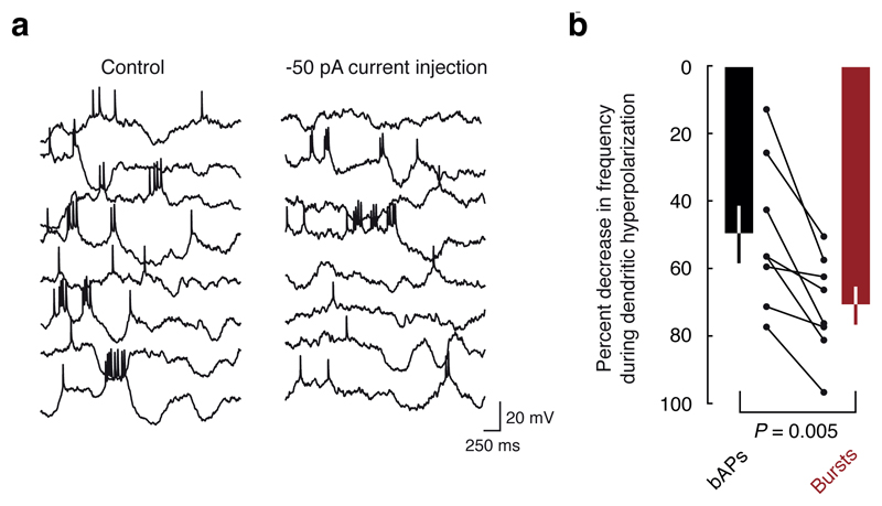
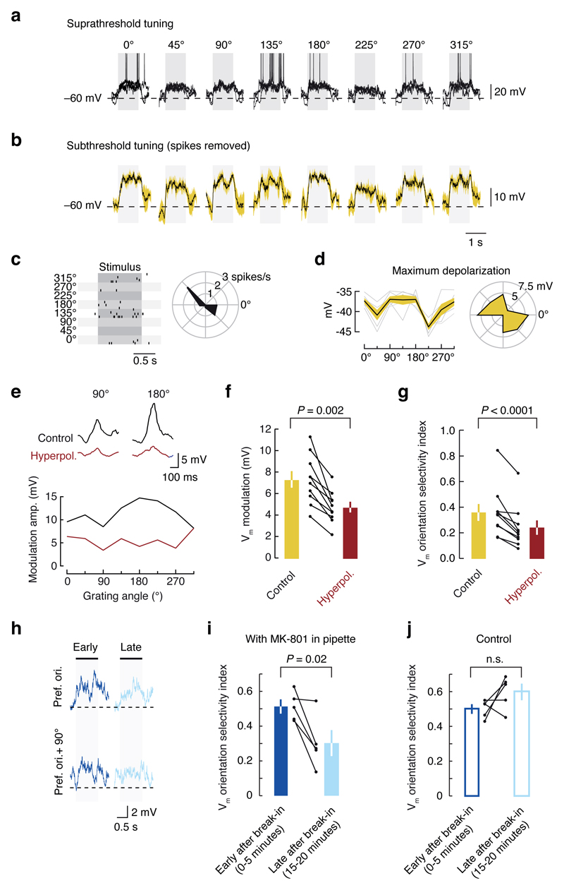
Neuronal dendrites are electrically excitable: they can generate regenerative events such as dendritic spikes in response to sufficiently strong synaptic input. Although such events have been observed in many neuronal types, it is not well understood how active dendrites contribute to the tuning of neuronal output in vivo. Here we show that dendritic spikes increase the selectivity of neuronal responses to the orientation of a visual stimulus (orientation tuning). We performed direct patch-clamp recordings from the dendrites of pyramidal neurons in the primary visual cortex of lightly anaesthetized and awake mice, during sensory processing. Visual stimulation triggered regenerative local dendritic spikes that were distinct from back-propagating action potentials. These events were orientation tuned and were suppressed by either hyperpolarization of membrane potential or intracellular blockade of NMDA (N-methyl-d-aspartate) receptors. Both of these manipulations also decreased the selectivity of subthreshold orientation tuning measured at the soma, thus linking dendritic regenerative events to somatic orientation tuning. Together, our results suggest that dendritic spikes that are triggered by visual input contribute to a fundamental cortical computation: enhancing orientation selectivity in the visual cortex. Thus, dendritic excitability is an essential component of behaviourally relevant computations in neurons.


Figures

Extended Data Figure 1
Extended Data Figure 1. Electrophysiological features of L2/3 dendrites in vivo.
a, The input resistance of distal dendrites was typically 100-300 MΩ, sometimes larger (up to 600 MΩ). Input resistance increased as function of dendritic distance from the soma, approximately doubling every 300 µm. The grey point indicates the input resistance measured in somatic patch clamp recordings (mean ± S.E.M). b, During a dendritic recording 150 μm from the soma, hyperpolarizing current steps did not reveal a voltage sag, thus there is likely little to no hyperpolarization-activated cation current, Ih, in the dendrites of layer 2/3 pyramidal neurons in vivo. c, The peak voltage response plotted against hyperpolarizing current step amplitude in an I-V plot was well fit by a linear function, confirming the lack of Ih. d, Representative dendritic bursts evoked by visual stimulation at the optimal orientation in 9 different dendritic recordings at progressively further distances from the soma. All right-hand scale bars are 20 mV. e, Compared to action potentials recorded at the soma, bAPs were lower amplitude, and f, prolonged in time, and both of these trends were more pronounced with increasing dendritic distance from the soma (error bars indicate S.D.). Both the amplitude and width were significantly different among the three groups (P < 0.01, unpaired t-tests with the Bonferroni correction for multiple comparisons).
Extended Data Figure 2
Extended Data Figure 2. Orientation tuning curves of dendritic bursts compared to bAPs.
Tuning curves for dendritic spike bursts and bAPs recorded at distal dendritic locations (> 75 μm from soma). Tuning curves for dendritic bursts match the tuning curves for isolated bAPs. The statistical significance of dendritic burst tuning curves were tested by randomly shuffling responses (details in Supplementary Methods) and found to be significant (P < 0.05) for 7 out of 9 cells (dendritic burst tuning in cells 6 and 9 were not significant). Curves are normalized to maximal values, shown at the bottom right of each polar plot. The small qualitative differences may be due to dendrites topologically distant from the dendritic recording site exhibiting slightly different tuning curves. The grating drift direction that elicited the largest response is indicated with an arrow. The difference between these directions between is indicated at the bottom of each polar plot. The cross correlation between dendritic bursts and isolated bAPs was highly significant: R = 0.54, P = 0.000013, n = 9, paired t-test. When only the spikes in bursts with rise times in the slowest quartile of the distribution were considered dendritic in origin, the preferred orientation of bAPs and the slowest quartile were still matched within individual dendritic recordings (difference in preferred orientation: 41.5 ± 58.1°, P = 0.49, n = 9, paired t-test).
Extended Data Figure 3
Extended Data Figure 3. Dendritic recordings in awake mice exhibit dendritic bursts.
a, Awake, headfixed mice viewed drifting gratings during electrophysiological recordings. b, Two photon image of the patched dendrite of a layer 2/3 pyramidal neuron in mouse visual cortex filled with Alexa 594 via the dendritic patch clamp pipette (117 µm from the soma). c, Dendritic bursts were observed when the preferred orientation was presented. d, Tuning curves for the isolated bAPs and dendritic bursts. e, Example bursts from three different distal dendritic recordings in awake mice. Calibration bars are 25 mV.
Extended Data Figure 4
Extended Data Figure 4. The diversity of onset dynamics vs. membrane potential.
a, Spikes (both isolated bAPs, in black, and spikes in dendritic burst events, in red) from each distal dendritic recording were normalized such that isolated bAPs had an mean phase slope of 1. The mean baseline membrane potential (Vm) of isolated bAPs was subtracted from the mean baseline Vm of all spikes. Although many spikes in bursts had depolarized baseline Vms relative to isolated spikes, there was overlap between the two populations around ± 3 mV. b, Expansion of panel a to show spikes at ± 3 mV relative to the mean baseline Vm of isolated bAPs. c, Histograms of the two populations reveal a tendency towards lower phase slope values for spikes in bursts (P = 0.041; KS test; n = 211 bAPs, 80 spikes in bursts). d, An example of bAPs and a spike in a burst (both from the same distal dendritic recording) show how although the bAP has a more depolarized baseline Vm, it still exhibits a steeper phase slope (a kink at the foot of the voltage waveform), indicative of a propagated action potential.
Extended Data Figure 5
Extended Data Figure 5. Calcium imaging at the site of dendritic recording reveals that global calcium signals are associated with faster onset spikes
a, During dendritic recordings, calcium signals were simultaneously imaged at the site of the recording and nearby dendrites. b, In dendritic bursts with global calcium signals that were simultaneously observed at all ROIs, the spikes recorded at the dendrite exhibited steep onsets, indicating that they were likely bAPs. c, In local calcium signals that were only observed in the ROI at the site of the recording, the dendritic spikes exhibit slower onsets, indicating that they were likely locally generated. d, The maximum phase slope of spikes occurring during global calcium events was higher than for spikes occurring during local calcium events (P = 0.0069, t-test). e,f, When global calcium signals occurred during ongoing local calcium signals, the initiation was associated with a steep-onset spike.
Extended Data Figure 6
Extended Data Figure 6. Non-firing cells exhibit subthreshold orientation tuning.
a, Raw data for an example cell in which subthreshold orientation tuning is observed although zero spikes were fired during the stimulus presentations. b, In this case, the tuning width of the subthreshold membrane potential was quite sharp and confined to two directions.
Extended Data Figure 7
Extended Data Figure 7. Tuning of APs and subthreshold membrane potential.
a, In individual cells, the orientation tuning of spikes and membrane potential were highly correlated, indicating that the tuning of the subthreshold responses was not spurious (mean difference in preferred orientation: 14.8 ± 5.3°). b, In individual cells, the orientation selectivity index based on the membrane potential response (VmOSI) was highly correlated with the conventional spiking-based OSI. c, In individual cells, the preferred orientation of the control subthreshold response was correlated with the preferred orientation of the subthreshold response during hyperpolarization. d, The black curve is the fitted subthreshold orientation tuning curve (black circles are raw data points), and the red curve is subthreshold tuning curve during hyperpolarization (red circles are raw data points). The VmOSI values for control and hyperpolarized conditions are shown next to each plot. The radial axes are linear and start at zero. The maximal radial axis range is shown below each polar plot. The differences in VmOSI are quantified in Fig. 5g.
Extended Data Figure 8
Extended Data Figure 8. Changes in the driving force for chloride do not account for the effects of hyperpolarization on VmOSI.
a, When the pipette solution contains 10 mM of chloride, the ECl is estimated to be -71 mV (based on the assumption that natural cerebrospinal fluid contains a similar amount of chloride as the artificial cerebrospinal fluid we use). In this situation, hyperpolarization decreased the orientation selectivity index. b, Even with low chloride solution (4 mM; estimated ECl = -95 mV), the result is the same. c, There was not a significant correlation between the driving force for Cl- and VmOSI. d, Resampling the data (15 of the data points in panel c were selected at random, and the R and P values for that set of data points were calculated; this process was repeated 10,000 times) confirmed that the result from the correlational analysis in panel c was not biased by a small subset of the data points (the mean R and P values from the resampling analysis match well with those for the full dataset in panel c).
Extended Data Figure 9
Extended Data Figure 9. The effect of intracellular MK-801 on Up - Down states and dendritic spikes.
a, To determine whether MK-801 that may have leaked out of the pipette during patching affects network circuitry, we examined the dynamics of Up and Down states in our recordings where MK-801 was in the pipette, and control recordings with no MK-801. b, Although generally membrane potential drifted up slightly (< 5 mV on average), and time spent in the Up state increased over long recordings (possibly due to the anesthesia wearing off), these trends were identical with or without MK-801 in the patch pipette. c, When 1 µM MK-801 was included in the recording pipette, the visually evoked responses contained fewer bAPs and bursts. This trend was clear in a, individual cells, as well as d, across the population. This reduction in spiking confirms that dendritic bursts do not occur when NMDA receptors are blocked. Since the low firing rate in MK-801 recordings prevented a reliable measurement of orientation tuning in the MK-801 dendritic patch recordings, we averaged over all stimulus presentations for both conditions, resulting in a lower average firing rate for bAPs and dendritic bursts.
Extended Data Figure 10
Extended Data Figure 10. Compartmental modeling of dendritic events.
a, A detailed reconstruction of a L2/3 pyramidal cell was used in the simulations. Light green circles over the dendritic tree represent background synapses and dark green are signal synapses (the model had 1100 synapses, not all are illustrated). Voltage was recorded at the soma and at all dendritic branches simultaneously. b, Activation of signal synapses at 5 Hz produces high-frequency dendritic bursts, composed of local dendritic spikes and backpropagating action potentials. These bursts were always accompanied by dendritic calcium transients. The timing of activation of excitatory synapses on the recorded dendritic branch is illustrated. Note how the local EPSPs are clearly smaller than the dendritic spikes. c, Examples of specific features consistently observed in the model. Isolated backpropagating action potentials were associated with global calcium transients and had “kinky” onsets. Dendritic spikes often preceded somatic action potentials, had smooth onsets and calcium transients that were localized to the branches where the spikes were recorded, and clearly started before the global transients associated with bAPs. Local dendritic spikes initiated in the dendrite could often be recorded in multiple electrotonically close dendritic branches. Pairs of local spikes and bAPs reached very high frequencies; the example shows a pair at > 400 Hz. When NMDA receptors were removed from the simulations no dendritic spikes were observed and the soma failed to reach threshold for action potential firing. The same occurred when there were no dendritic voltage-activated sodium channels, indicating that the generation of dendritic spikes is required for producing axonal output. d, Quantification of spike onset for local dendritic spikes and bAPs in the model reproduced the experimentally observed effect reported in Extended Data Fig. 5d. e, Example trial showing the somatic voltage and recordings for two dendrites indicated in panel a. For each dendrite the local voltage, sodium channel conductance (gNa, expressed as a fraction of the maximum conductance) and the timing of activation of excitatory synapses on the recorded dendrite are shown. The gNa traces show that there is significant local sodium channel inactivation after the first spike, and that subsequent spikes are associated with varying degrees of sodium channel conductance. The * symbols show extreme cases when a bAP followed a local dendritic spike at very high frequency and did not recruit any local gNa, therefore indicating that the propagation into the recorded branch was passive.
Figure 1
Figure 1. Dendritic patch-clamp recordings from visual cortex pyramidal neurons in vivo.
a, Schematic of recording and imaging setup for in vivo dendritic patch-clamp recordings under two-photon microscopy. b, Two-photon image of a layer 2/3 pyramidal neuron in mouse visual cortex in vivo filled with Alexa 594 via a dendritic patch-clamp recording 100 µm from the soma (maximum intensity projection). c, Somatic spiking and dendritic activity (d) evoked by presenting square wave grating visual stimuli both exhibited reliable, orientation-tuned burst spiking events. The asterisks mark bAPs. e, Spikes within dendritic burst events were highly variable compared to the more stereotyped bAPs and somatically recorded APs. f, Dendritic burst event frequency varied with orientation. g, individual burst events were highly variable in amplitude and kinetics (dendritic recording 150 µm from the soma).
Figure 2
Figure 2. Visually evoked dendritic burst events are local.
a, Whole cell patch clamp recordings were performed at the soma (n = 9), proximal dendritic locations (< 50 µm from soma; n = 5), or distal dendritic locations (> 75 µm from soma; n = 9). Stimulus-evoked event frequency at the soma and proximal dendritic locations were similar. However, event frequency and burstiness (variance-to-mean ratio, or Fano factor) was significantly higher at distal dendritic locations. (P-values from the Wilcoxon rank sum test). Data from awake recordings (filled circles, n = 6 total) exhibit the same trends. b, The inflection in membrane potential for backpropagated spikes recorded at distal dendritic locations (> 100 µm from the soma) exhibited a sharp kink, consistent with propagated spikes, as visible both in voltage vs. time plots (left), and dV/dt vs. voltage phase plots (right). c, By contrast, spikes in bursts exhibited a slower onset, consistent with local generation. d, Across the population of dendritic recordings, isolated spikes consistently exhibited sharper inflections at onset (measured as the initial slope in the dV/dt vs. voltage phase plots; dashed lines in b and c) than spikes in bursts. Distal dendritic recordings from awake mice (black-filled symbols) exhibited the same trend.
Figure 3
Figure 3. Simultaneous dendritic recording and calcium imaging at the soma shows that dendritic bursts are local.
a, To infer spiking activity at the soma during dendritic patch recordings, neurons were filled with a calcium indicator and somatic calcium signals were imaged. During simultaneous dendritic voltage recordings and somatic calcium imaging, dendritic bursts were sometimes not accompanied by robust somatic calcium signals (marked with an arrow). b, Dendritic bursting was well tuned, and (c) overlapped with the orientation tuning at the soma (data from Cell 1 is shown in panels a and b). d, To calibrate the calcium signals seen at the soma, we generated spike-triggered averages of the somatic calcium signal for well-isolated single spikes, and bouts of 4-5 spikes (within 640 ms). The somatic calcium signal amplitude was similar for single spikes whether the spikes were recorded at the soma or the dendrite. For bouts of 4-5 spikes recorded at the dendrite, no difference was found compared to single spikes recorded at the soma, suggesting that some dendritic spikes were local. e, Across the population of events, even though the calcium signals saturated around the same magnitude (~ 0.3 ΔF/F, not different between the two configurations, P = 0.99), the somatic calcium signal as a function of number of dendritic spikes rose much more slowly than that for somatically-recorded spikes. The right-hand plot shows an expansion of the x-axis to highlight the data from responses with < 10 spikes.
Figure 4
Figure 4. Hyperpolarization decreases the frequency of dendritic bursts.
a, An example dendritic recording shows that hyperpolarization decreased the frequency of bursts (from 0.32 Hz to 0.14 Hz; -68%) more than the frequency of bAPs (0.51 Hz to 0.36 Hz: -30%). b, Population data showing that bursts and bAPs are both suppressed (P = 3.0 x 10⁻⁶, and P = 0.0004, respectively, t-test), and bursts are suppressed to a greater degree than bAPs by hyperpolarization (P = 0.005, paired t-test).
Figure 5
Figure 5. Dendritic mechanisms contribute to the selectivity of subthreshold orientation tuning.
a, Somatic whole cell recording from a layer 2/3 pyramidal neuron, exhibiting robust visually-evoked spiking (3 sweeps overlaid; stimulus duration indicated by grey bar) and b, subthreshold responses (50 ms windows around spikes were blanked, and the same results were obtained when blanking window was decreased to 20 ms) which were orientation tuned as evidenced by c, spikes rasters, and d, polar plots of maximal depolarization. e, Hyperpolarization decreased the amplitude of the stimulus-evoked membrane potential modulation, and decreased its tuning selectivity. Hyperpolarization decreased f, Vm modulation amplitude and g, orientation selectivity index across the population. To investigate the mechanisms involved in the dendritic spike contributing to orientation tuning, whole cell somatic patch clamp recordings were performed with 1 µM MK-801 in the pipette solution. h, Orientation tuning selectivity progressively decreased during the recording as MK-801 diffused into the cell and blocked NMDA channels. In these example traces, the response to the preferred orientation decreased from early in the recording to late in the recording. i, Across the population, subthreshold orientation tuning was strongly inhibited at late time points in the recording, compared to early time points. j, In control recordings, with no blockers in the pipette solution, orientation tuning selectivity was not significantly different (P > 0.05, two sample t-test) from the early period of MK 801 recordings. Furthermore, tuning did not significantly change during long recordings.

Comment in

  • Dendrites: Dendritic processors.
    Yates D. Yates D. Nat Rev Neurosci. 2013 Dec;14(12):815. doi: 10.1038/nrn3644. Nat Rev Neurosci. 2013. PMID: 24400337 No abstract available.

References

    1. Johnston D, Narayanan R. Active dendrites: colorful wings of the mysterious butterflies. Trends Neurosci. 2008;31:309–316. - PubMed
    1. London M, Häusser M. Dendritic computation. Annu Rev Neurosci. 2005;28:503–532. - PubMed
    1. Spruston N. Pyramidal neurons: dendritic structure and synaptic integration. Nat Rev Neurosci. 2008;9:206–221. - PubMed
    1. Larkum ME, Zhu JJ, Sakmann B. A new cellular mechanism for coupling inputs arriving at different cortical layers. Nature. 1999;398:338–341. - PubMed
    1. Schiller J, Major G, Koester HJ, Schiller Y. NMDA spikes in basal dendrites of cortical pyramidal neurons. Nature. 2000;404:285–289. - PubMed

Publication types

Substances