Review
. 2014 Jan 1;10(1):86-95.
doi: 10.4161/org.26766. Epub 2013 Oct 25.

Wnt and planar cell polarity signaling in cystic renal disease

Affiliations
Review

Wnt and planar cell polarity signaling in cystic renal disease

Paraskevi Goggolidou. Organogenesis. .

Abstract

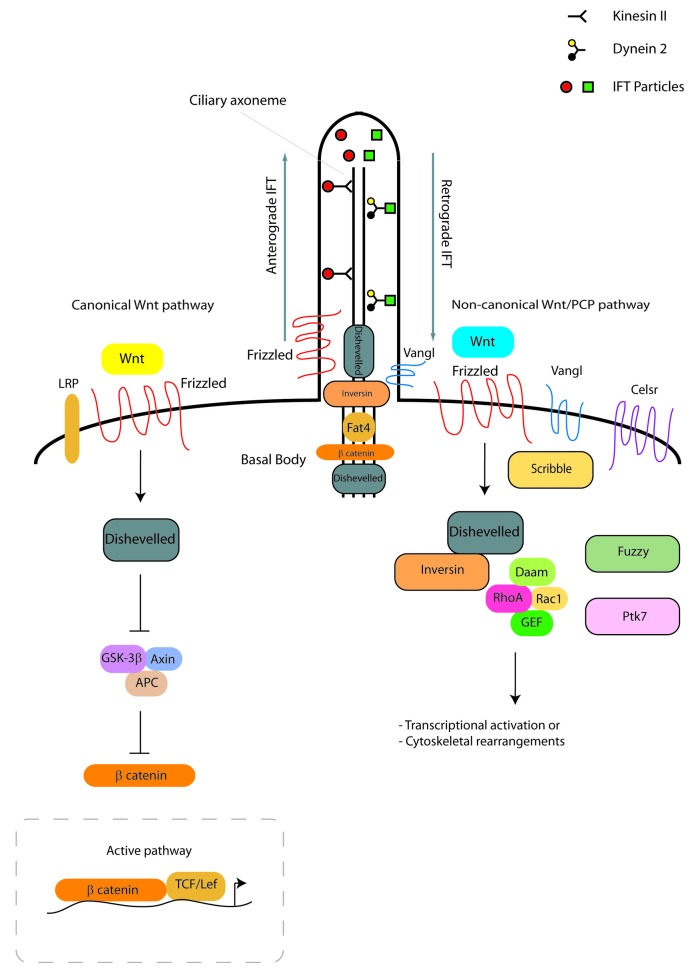
Cystic kidney diseases can cause end stage renal disease, affecting millions of individuals worldwide. They may arise early or later in life, are characterized by a spectrum of symptoms and can be caused by diverse genetic defects. The primary cilium, a microtubule-based organelle that can serve as a signaling antenna, has been demonstrated to have a significant role in ensuring correct kidney development and function. In the kidney, one of the signaling pathways that requires the cilium for normal development is Wnt signaling. In this review, the roles of primary cilia in relation to canonical and non-canonical Wnt/PCP signaling in cystic renal disease are described. The evidence of the associations between cilia, Wnt signaling and cystic renal disease is discussed and the significance of planar cell polarity-related mechanisms in cystic kidney disease is presented. Although defective Wnt signaling is not the only cause of renal disease, research is increasingly highlighting its importance, encouraging the development of Wnt-associated diagnostic and prognostic tools for cystic renal disease.

Keywords: Wnt signalling; cilia; cystic renal disease; kidney morphogenesis; planar cell polarity.


Figures

None
Figure 1. Metanephric ureteric bud branching and nephron formation. Metanephric kidney development initiates upon invasion of the ureteric bud (black lines) in the metanephric mesenchyme (dark blue cells). Signals from the ureteric bud induce differentiation of the nephron progenitors to form the renal vesicles, while further ureteric bud branching is taking place. Renal vesicles will re-arrange into the distinct comma- and S-shaped bodies (dark blue cells). Specific cell populations of these structures will give rise to the glomeruli, the proximal and distal tubules and the loops of Henle, while the ureteric bud develops into the collecting duct. Cilia are present in the renal vesicles, comma-shaped and S-shaped bodies and nephrons (blue lines) and also form in the branching ureteric bud and colleting duct cells (black lines).
None
Figure 2. Ciliogenesis and Wnt signaling in the kidney. Ciliogenesis depends on anterograde and retrograde transport involving kinesin II, dynein 2 and IFT proteins. The canonical Wnt signaling pathway (left side of figure) initiates when a Wnt ligand binds to a Frizzled receptor in the presence of LRP. This activates Dishevelled that inhibits the β-catenin destruction complex (GSK3β, APC, Axin). In the active pathway, β-catenin accumulates in the cytosol and causes the transcriptional activation of Wnt target genes. In the non-canonical Wnt/PCP signaling pathway in the kidney (right side of figure), the binding of a Wnt ligand will result in the activation of Frizzled and Dishevelled, leading to downstream cytoskeletal rearrangements or transcriptional activation through the activation of RhoA or Rac1. Core molecules (Vangl, Celsr, Scribble) are important for non-canonical Wnt/PCP signaling, while effector PCP molecules (Fuzzy, Fat4, Ptk7) are significant for kidney morphogenesis. A number of Wnt signaling proteins localize to the basal body (Dishevelled, β-catenin, Fat4) or base (Inversin, Dishevelled, Frizzled, Vangl) of kidney primary cilia.

References

    1. Carroll TJ, Das A. Planar cell polarity in kidney development and disease. Organogenesis. 2011;7:180–90. doi: 10.4161/org.7.3.18320. - DOI - PMC - PubMed
    1. Karner CM, Chirumamilla R, Aoki S, Igarashi P, Wallingford JB, Carroll TJ. Wnt9b signaling regulates planar cell polarity and kidney tubule morphogenesis. Nat Genet. 2009;41:793–9. doi: 10.1038/ng.400. - DOI - PMC - PubMed
    1. Saburi S, Hester I, Fischer E, Pontoglio M, Eremina V, Gessler M, Quaggin SE, Harrison R, Mount R, McNeill H. Loss of Fat4 disrupts PCP signaling and oriented cell division and leads to cystic kidney disease. Nat Genet. 2008;40:1010–5. doi: 10.1038/ng.179. - DOI - PubMed
    1. Fischer E, Legue E, Doyen A, Nato F, Nicolas JF, Torres V, Yaniv M, Pontoglio M. Defective planar cell polarity in polycystic kidney disease. Nat Genet. 2006;38:21–3. doi: 10.1038/ng1701. - DOI - PubMed
    1. Luyten A, Su X, Gondela S, Chen Y, Rompani S, Takakura A, Zhou J. Aberrant regulation of planar cell polarity in polycystic kidney disease. J Am Soc Nephrol. 2010;21:1521–32. doi: 10.1681/ASN.2010010127. - DOI - PMC - PubMed

LinkOut - more resources