. 2014 Jan 21;129(3):359-72.
doi: 10.1161/CIRCULATIONAHA.113.003000. Epub 2013 Oct 25.

Vascular progenitors from cord blood-derived induced pluripotent stem cells possess augmented capacity for regenerating ischemic retinal vasculature

Affiliations

Vascular progenitors from cord blood-derived induced pluripotent stem cells possess augmented capacity for regenerating ischemic retinal vasculature

Tea Soon Park et al. Circulation. .

Abstract

Background: The generation of vascular progenitors (VPs) from human induced pluripotent stem cells (hiPSCs) has great potential for treating vascular disorders such as ischemic retinopathies. However, long-term in vivo engraftment of hiPSC-derived VPs into the retina has not yet been reported. This goal may be limited by the low differentiation yield, greater senescence, and poor proliferation of hiPSC-derived vascular cells. To evaluate the potential of hiPSCs for treating ischemic retinopathies, we generated VPs from a repertoire of viral-integrated and nonintegrated fibroblast and cord blood (CB)-derived hiPSC lines and tested their capacity for homing and engrafting into murine retina in an ischemia-reperfusion model.

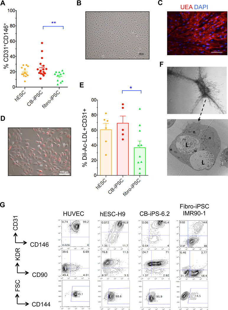
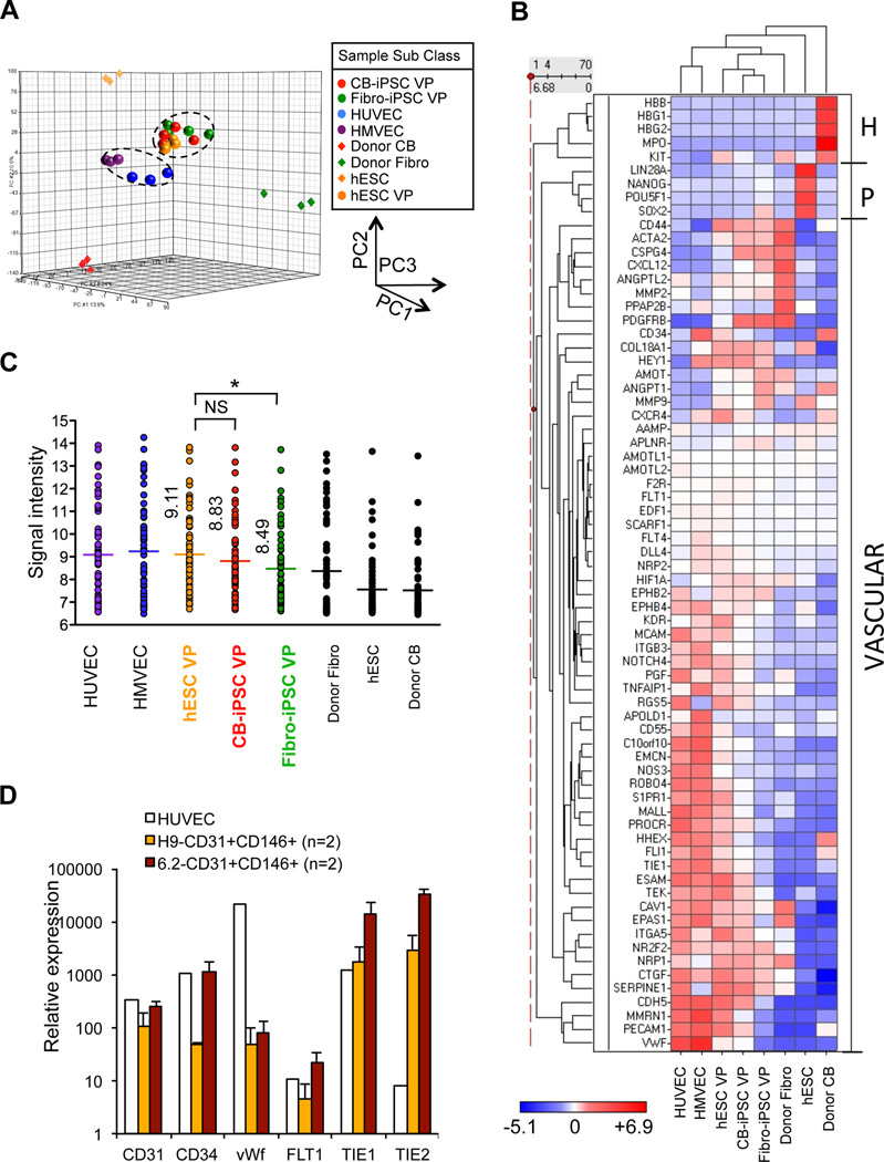
Methods and results: VPs from human embryonic stem cells and hiPSCs were generated with an optimized vascular differentiation system. Fluorescence-activated cell sorting purification of human embryoid body cells differentially expressing endothelial/pericytic markers identified a CD31(+)CD146(+) VP population with high vascular potency. Episomal CB-induced pluripotent stem cells (iPSCs) generated these VPs with higher efficiencies than fibroblast-iPSC. Moreover, in contrast to fibroblast-iPSC-VPs, CB-iPSC-VPs maintained expression signatures more comparable to human embryonic stem cell VPs, expressed higher levels of immature vascular markers, demonstrated less culture senescence and sensitivity to DNA damage, and possessed fewer transmitted reprogramming errors. Luciferase transgene-marked VPs from human embryonic stem cells, CB-iPSCs, and fibroblast-iPSCs were injected systemically or directly into the vitreous of retinal ischemia-reperfusion-injured adult nonobese diabetic-severe combined immunodeficient mice. Only human embryonic stem cell- and CB-iPSC-derived VPs reliably homed and engrafted into injured retinal capillaries, with incorporation into damaged vessels for up to 45 days.

Conclusions: VPs generated from CB-iPSCs possessed augmented capacity to home, integrate into, and repair damaged retinal vasculature.

Keywords: blood supply; diabetic retinopathy; embryonic stem cells; induced pluripotent stem cells; reperfusion injury; stem cells; transplantation.


Conflict of interest statement

Conflict of Interest Disclosures: Under a licensing agreement between Life Technologies and the JHU, ETZ is entitled to a share of royalty received by the University for licensing of stem cells. The terms of this arrangement are managed by JHU in accordance with its Conflict of Interest policies. This does not alter the authors’ adherence to journal policies on sharing data and materials.

Figures

Figure 1
Figure 1
Efficient generation of embryonic VP populations from hPSC. A, Schema for vascular differentiation and expansion of VP. B, Flow cytometry plot of day 8 hEB cells from H9-hESC following expansion in EGM2 for 4 days. The average percentage of four indicated quadrants ± SEM is shown (n=13 experiments). C, Percentage Dil-Ac-LDL uptake (Mean ± SEM) of FACS-purified and EGM2-expanded populations differentiated from two hESC lines (gold), three CB-iPSC lines (red) and six Fibro-iPSC lines (green). Each data point represents an independent, replicate experiment. D, In vitro Matrigel assays of purified and expanded populations. Scale = 500 µm. E, Representative Matrigel plugs consisted of vascular structures formed by indicated CB-iPSC-6.2 hEB-derived populations, and immunostained with anti-CD31 (brown). Scale bars =100 µm. F, Measurements in Matrigel plug sections: (left panel) percentages of blood vessels >30 μm diameter per microscopic field (mean ±SEM); (right panel) total number of blood vessels per microscopic field (mean ±SEM) (Two-tailed t-tests, *: p <0.05, **: p <0.01).
Figure 2
Figure 2
Characterization of VP generated from hPSC. A, Efficiency of hEB differentiation (%mean ± SEM) of hPSC to CD31+CD146+ VP. Data are from four hESC lines (H1, H7, H9, ES03; n=13 experiments (gold)), eight fibroblast-iPSC lines (IMR90-1, IMR90-4, HUF3, HUF5, 7ta, WT2, WT4, fF6.1); n=11 experiments (green)), and six CB-iPSC lines (6.2, 6.13, 19.11, E5C3, E12C5, E17C6; n=17 experiments (red)). Two-tailed t-tests: **: p <0.01. B, Phase contrast image of FACS-purified, expanded CD31+CD146+ VP cells differentiated from CB-iPSC-6.2; C, with Ulex europaeus agglutinin (UEA) and DAPI co-staining D, and with Dil-Ac-LDL uptake staining E, Percentage Dil-Ac-LDL uptake (mean ± SEM) of expanded CD31+CD146+ VP from individual differentiations of hESC (gold), CB-iPSC (red), and fibroblast-iPSC (green). Mann-Whitney tests: *: p<0.05. F, TEM image CB-iPSC-derived VP forming vascular tubes in collagen gel via cooperating endothelial and pericytic-like cells; all border on lumens and are potential EC in apparent bifurcation. L: lumen, n: nuclei. G, Representative surface marker analyses of FACS-purified/expanded hPSC-derived CD31+CD146+ VP and HUVEC.
Figure 3
Figure 3
Expression signatures of hPSC-derived VP. A, PCA of genome-wide expression signatures for indicated samples of embryonic CD31+CD146+ VP generated from hESC, CB-iPSC, and fibroblast-iPSC; adult endothelial cells (HUVEC, HMEC); hiPSC donor cells; hESC lines. Data were generated from samples in Figure S10. Pearson coefficient R2 for hESC-VP vs. CB-iPSC-VP = 0.974; hESC-VP vs. fibroblast-iPSC-VP = 0.958. B, Heatmap-dendrogram of Ilumina expression array data of the vascular lineage-specific genes indicated. Individual RNA samples from independent differentiations were obtained from HUVEC (n=3), HMVEC (n=3), hESC-VP (n=4), CB-iPSC-VP (n=4), fibroblast-iPSC-VP (n=4), donor fibroblast (Fibro, n=3), hESC (n=3), and donor CB (n=3). H: hematopoietic-specific genes, P: pluripotency-specific genes, VASCULAR: vascular lineage-specific genes. C, Quantitation of expression of vascular lineage-specific genes in B. Mean value of the gene signal intensity is shown (*: p<0.05; mean expression among hESC-VP, CB-iPSC-VP and Fib-iPSC-VP differed significantly (one-way ANOVA p = 0.001). Fib-iPSC-VP also significantly differed by individual comparison than hESC-VP (p =0.0002, Bonferroni correction threshold p < 0.016), while CB-iPSC-VP vs. hESC-VP did not (p = 0.09). List of genes analyzed: Table S8. D, Q-RT-PCR analysis of CD31+CD146+ VP from hESC-H9, CB-iPSC-6.2, and HUVEC. Relative expression of CD31, CD34, vWF, FLT1, TIE1, and TIE2 of replicate samples was normalized to expression in undifferentiated hESC-H9 (n=2 experiments).
Figure 4
Figure 4
Senescence and DNA damage sensitivity of expanded VP. A, Representative β-galactosidase senescence staining in hPSC classes; legend symbols are as before. Scale bars = 100 µm. B, % senescent cells (mean ±SEM of n=12 microscopic fields per each line of each hPSC class; n=3 per class). C. Western blots of p53 expression before, and 24 hours following 2Gy irradiation of VP from 1: H9 (p46), 2: ES03 (p88), 3: 6.2 (P20), 4: 6.2 (P23), 5: 19.11 (P19), 6: E17C6 (P29), 7: HUF3 (P44), 8: IMR90-1 (P71), 9: IMR90-1 (p72). D, Fold percent change in p53 protein expression levels by Western blot densitometry; >1.0 (increase) or <1.0 (decrease) above baseline). Two-tailed t-tests *: p<0.05.
Figure 5
Figure 5
In vivo migration, homing, and engraftment of luciferase-transgenic VP cells into I/R-damaged mouse retina. A, Experimental design for quantifying human VP engraftment into NOD-SCID mouse retinas. (Left panel) Anatomy of mouse eye indicating I/R location at anterior chamber and site of human cell injections into vitreous body. (Right panel) Timeline of in vivo engraftment analysis. B, Representative immuno-fluorescent retinal sections of I/R-damaged eyes injected with hESC-luciferase-transgenic (green) CD31+CD146+ VP (left) and CD31+CD146 (right) cells at 3 days post-injection. CD31+CD146+ cells readily migrated into deep retinal layers whereas CD31+CD146 cells remained primarily in vitreous. (VB: vitreous body, ILM: internal limiting membrane, PR: photoreceptor) C, Quantification of (a) cell migration into retina and (b) number of engrafted human cells visualized per retinal cross sections following injection of hESC derived CD31+CD146+ VP and CD31+CD146 cells (n=15 and 9 sections evaluated, respectively; two-tailed t-tests: * p<0.005; ** p<0.001). D, CD31+CD146+ hESC-VP injected to I/R-damaged (injury, left) and normal eye (no injury, right) demonstrating that cells do not migrate into retinal layers without injury signals. (ONH: optic nerve head) E, CD31+CD146+ hESC-VP (left) and CD31+CD146+ CB-iPSC-VP (right) engrafted into both venules and micro-capillaries (arrows) in this flat mount retina at post-injection day 3. Scale bars 50 µm.
Figure 6
Figure 6
In vivo engraftment of CB-iPSC-VP into retinal vasculature. Luciferase-transgenic human VP were injected directly into the eye (intra-vitreal) (A–H), or systemically IV (orbital sinus) (I). G, Transgenic CB-iPSC-VP (green) homed to both damaged capillaries (arrows), and larger blood vessels (retinal flat mounts). C, Damaged host vessels lacked murine ECs (lack of blue signal from anti-mouse CD31), but their Coll IV+ basement membranes remained intact (red). D–F, Human cells (green) were often observed in retinal cross-sections at pericytic positions surrounding host murine ECs (blue) in capillaries. Engrafted human CB-iPSC-VP detected in murine retina at 14 days (G), 21 days (H), and 45 days (I) post injection. The degree of damage was more severe and the density of functional blood vessels was reduced with time following I/R. Scale bars are 20 µm (A, G–I) and 10 µm (D). Shown are representative experiments from Table 1.
Figure 7
Figure 7
Quantification of human VP engraftment into murine retinal vasculature. A, Representative experiments (from Table 1) demonstrating CB-iPSC-VP (green) engrafting into murine vessels following orbital sinus (OS) or tail-vein (TV) injections. Scale bars = 20 µm. B, Representative experiments showing detection of CB-iPSC-VP injected via orbital sinus (left) or tail vein (right). Retinas were harvested at post-injection day 7, and whole flat mount retinas were scanned and human cells quantified in superficial (near vitreous body, red) and deep retinal vasculatures (blue) layers. C, Representative quantification of a retinal engraftment experiment demonstrated that systemic injections attracted higher numbers of homing CD31+CD146+ VP into damaged deep retinal blood vessels (OS>TV).

Similar articles

Cited by

References

    1. Paxhia MJ, Ting TD, Fekrat S. Ischemic central retinal vein occlusion and retinitis pigmentosa: Lower risk of neovascularization? Retina. 2001;21:179–180. - PubMed
    1. Awdeh RM, Elsing SH, Deramo VA, Stinnett S, Lee PP, Fekrat S. Vision-related quality of life in persons with unilateral branch retinal vein occlusion using the 25-item national eye institute visual function questionnaire. Br J Ophthalmol. 2010;94:319–323. - PubMed
    1. Cao J, McLeod S, Merges CA, Lutty GA. Choriocapillaris degeneration and related pathologic changes in human diabetic eyes. Arch Ophthalmol. 1998;116:589–597. - PubMed
    1. Zheng L, Gong B, Hatala DA, Kern TS. Retinal ischemia and reperfusion causes capillary degeneration: Similarities to diabetes. Invest Ophthalmol Vis Sci. 2007;48:361–367. - PubMed
    1. Kim SY, Johnson MA, McLeod DS, Alexander T, Otsuji T, Steidl SM, Hansen BC, Lutty GA. Retinopathy in monkeys with spontaneous type 2 diabetes. Invest Ophthalmol Vis Sci. 2004;45:4543–4553. - PubMed

Publication types

MeSH terms