. 2014 Jan 1;306(1):H109-20.
doi: 10.1152/ajpheart.00137.2013. Epub 2013 Oct 25.

Postprandial VLDL lipolysis products increase monocyte adhesion and lipid droplet formation via activation of ERK2 and NFκB

Affiliations

Postprandial VLDL lipolysis products increase monocyte adhesion and lipid droplet formation via activation of ERK2 and NFκB

Laura J den Hartigh et al. Am J Physiol Heart Circ Physiol. .

Abstract

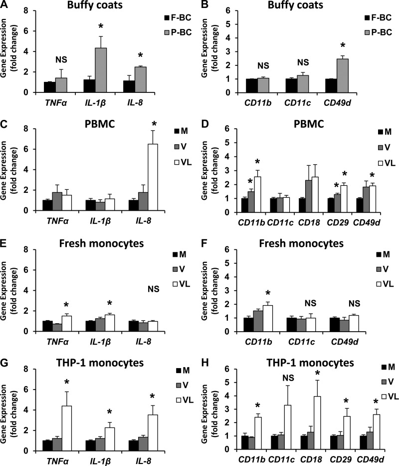
Postprandial lipemia is characterized by a transient increase in circulating triglyceride-rich lipoproteins such as very low-density lipoprotein (VLDL) and has been shown to activate monocytes in vivo. Lipolysis of VLDL releases remnant particles, phospholipids, monoglycerides, diglycerides, and fatty acids in close proximity to endothelial cells and monocytes. We hypothesized that postprandial VLDL lipolysis products could activate and recruit monocytes by increasing monocyte expression of proinflammatory cytokines and adhesion molecules, and that such activation is related to the development of lipid droplets. Freshly isolated human monocytes were treated with VLDL lipolysis products (2.28 mmol/l triglycerides + 2 U/ml lipoprotein lipase), and monocyte adhesion to a primed endothelial monolayer was observed using a parallel plate flow chamber coupled with a CCD camera. Treated monocytes showed more rolling and adhesion than controls, and an increase in transmigration between endothelial cells. The increased adhesive events were related to elevated expression of key integrin complexes including Mac-1 [α(m)-integrin (CD11b)/β2-integrin (CD18)], CR4 [α(x)-integrin (CD11c)/CD18] and VLA-4 [α4-integrin (CD49d)/β1-integrin (CD29)] on treated monocytes. Treatment of peripheral blood mononuclear cells (PBMCs) and THP-1 monocytes with VLDL lipolysis products increased expression of TNFα, IL-1β, and IL-8 over controls, with concurrent activation of NFkB and AP-1. NFκB and AP-1-induced cytokine and integrin expression was dependent on ERK and Akt phosphorylation. Additionally, fatty acids from VLDL lipolysis products induced ERK2-dependent lipid droplet formation in monocytes, suggesting a link to inflammatory signaling pathways. These results provide novel mechanisms for postprandial monocyte activation by VLDL lipolysis products, suggesting new pathways and biomarkers for chronic, intermittent vascular injury.

Keywords: adhesion molecules; fatty acids; inflammation; lipoprotein lipase.


Figures

Fig. 1.
Fig. 1.
Monocyte adhesive interactions with human aortic endothelial cells are increased by very low-density lipoproteins (VLDL) lipolysis products. Human aortic endothelial cells (HAECs) were treated with TNFα while freshly isolated monocytes (A–C) or THP-1 monocytes (D) were treated with media, lipoprotein lipase (LpL), VLDL, or VLDL + LpL. Monocytes were either injected into a flow chamber for image capture (A and B) and adhesive event quantification (C), or incubated with HAECs under static conditions and adherence quantified (D). A: image of the flow chamber 10 s into the image acquisition sequence with VLDL + LpL-treated monocytes. B: the same frame 45 s after the beginning of image acquisition. Examples of rolling (r1–r2), adherent (a1–a4), and transmigrating (t1–t3) monocytes are labeled. C: counted monocytes that are rolling, adherent, or transmigrating, expressed as the average number of cells per min + SE, from 5 different experiments. D: static adhesion events counted using THP-1 monocytes with indicated treatments. *P < 0.05 from the media control.
Fig. 2.
Fig. 2.
Several monocytic populations show increased gene expression of cytokines and integrins in response to VLDL lipolysis products. A and B: buffy coats were isolated from normal human subjects after an overnight fast (“F”) and after consumption of a moderately high-fat meal (“P”). Peripheral blood mononuclear cells (PBMCs) (C and D), freshly isolated monocytes (E and F), or THP-1 monocytes (G and H) were treated for 3 h with media (M), VLDL (V), or VLDL + LpL (VL) for 3 h. Gene expression levels of inflammatory cytokines (A, C, E, G) and integrins (B, D, F, H) were quantified and normalized to beta-actin and expressed as a fold change from fasting levels (A and B) or media control (C–H) + SE (n = 3–5). *P < 0.05 from the fasting or media control. NS, not significant.
Fig. 3.
Fig. 3.
Integrin and cytokine protein is also increased by VLDL lipolysis products. Surface CD11b levels were observed from THP-1 monocytes treated with media (M), LpL (L), VLDL (V), or VLDL + LpL (VL) using immunofluorescence and flow cytometry (A and B). A: representative immunofluorescent images of treated THP-1 monocytes stained for CD11b (green) and nuclear DAPI (blue), presented in triplicate (40X). B: relative quantification of CD11b-positive monocytes using flow cytometry (n = 3). Gates were set using unstained cells assuming a background autofluorescence of 4–5%. C: secreted TNFα and IL-1β from treated monocytes was measured by ELISA, and expressed as pg protein/ml + SE (n = 3). *P < 0.05 from the media control.
Fig. 4.
Fig. 4.
VLDL lipolysis products stimulate NFκB and AP-1 nuclear translocation and activation. THP-1 monocytes were treated with media (M), LpL (L), VLDL (V), or VLDL + LpL (VL) for 2 h, then nuclear protein was isolated. A: 10 μg total nuclear protein was used for Western blots. Band densities relative to the media control (arbitrary units) are shown below each lane (n = 3). B and C: electrophoretic mobility shift assay (EMSAs) for NFκB and AP-1. Arrows illustrate probe shifts (n = 3).
Fig. 5.
Fig. 5.
Akt, IκBα, ERK1/2, p38, and JNK1/2 become phosphorylated by VLDL lipolysis products. THP-1 monocytes were treated with LpL, VLDL, or VLDL + LpL for 0, 15, 30, 60, or 120 min. A: 10 µg total protein was used for Western blots for each treatment at each time point. BF: desitometry is represented as mean band intensity of the indicated phosphorylated protein (p-) normalized first to the unmodified protein ± SE, then normalized to the media control (time 0), n = 3. *P < 0.05 from the media control.
Fig. 6.
Fig. 6.
VLDL lipolysis products increase Akt-, ERK1/2-, and NFkB-mediated expression of CD11b, CD11c, CD18, CD29, and CD49d integrins from THP-1 monocytes. THP-1 monocytes were pretreated with inhibitors to NFκB (MG), Akt (LY), ERK1/2 (MEK), p38 (SB), or JNK (SP) for 1 h, then treated for 3 h with VLDL lipolysis products (VL). Gene expression levels of integrins Cd11b (A), Cd11c (B), Cd29 (C), and Cd49d (D) were normalized to beta-actin and expressed as a percentage of the media control + SEM (n = 5). *P < 0.05 from the media control, #P < 0.05 from the VL treatment. NS, not significant.
Fig. 7.
Fig. 7.
Akt and ERK1/2 activation are required for VLDL lipolysis product-induced cytokine expression. THP-1 monocytes were pretreated with inhibitors to NFκB (MG), Akt (LY), ERK1/2 (MEK), p38 (SB), or JNK (SP) for 1 h, then treated for 3 h with VLDL lipolysis products (VL). Gene expression levels of cytokines Tnfα (A), Ill1β (B), and Il8 (C) were normalized to beta-actin and expressed as a percentage of the media control + SE (n = 5). *P < 0.05 from the media control, #P < 0.05 from the VL treatment. NS, not significant.
Fig. 8.
Fig. 8.
VLDL lipolysis product-induced lipid droplet accumulation within THP-1 monocytes is ERK2-dependent. THP-1 monocytes were treated for 3 h with media, LpL, VLDL (V), VLDL + LpL (VL), or isolated free fatty acids (FFA). For some treatments, THP-1 monocytes were pretreated with an ERK2 inhibitor (Ste-MEK113, 25 or 50 μM as indicated), a neutralizing CD11b antibody, and NFkB inhibitor (MG), or an ERK1/2 inhibitor (MEK) for 1 h. A–F: images of Oil Red O-stained cells were captured using an Olympus BX41 phase-contrast microscope (60X, NA 0.80) with an Olympus Qcolor3 digital CCD camera. Images shown are representative of 3 independent experiments. G–I: relative gene expression analysis was performed to quantify Tnfα, Il1β, and Il8, normalized to beta-actin and presented as a fold change from the media control. J: static adhesion of treated THP-1 monocytes after pretreatment with inhibitors was quantified and presented as adherent cells per frame + SEM (n = 3). K: CD11b surface expression was quantified using flow cytometry and presented as a percentage of cells positive for CD11b (n = 3). *P < 0.05 from the media control, #P < 0.05 from the VL treatment.

Similar articles

Cited by

References

    1. Ali S, Mann DA. Signal transduction via the NF-kappaB pathway: a targeted treatment modality for infection, inflammation and repair. Cell Biochem Funct 22: 67–79, 2004 - PubMed
    1. Alipour A, van Oostrom AJ, Izraeljan A, Verseyden C, Collins JM, Frayn KN, Plokker TW, Elte JW, Castro Cabezas M. Leukocyte activation by triglyceride-rich lipoproteins. Arterioscler Thromb Vasc Biol 28: 792–797, 2008 - PubMed
    1. Aung HH, Lame MW, Gohil K, An CI, Wilson DW, Rutledge JC. Induction of ATF3 gene network by triglyceride-rich lipoprotein lipolysis products increases vascular apoptosis and inflammation. Arterioscler Thromb Vasc Biol 33: 2088–2096, 2013 - PMC - PubMed
    1. Bermudez B, Lopez S, Varela LM, Ortega A, Pacheco YM, Moreda W, Moreno-Luna R, Abia R, Muriana FJ. Triglyceride-rich lipoprotein regulates APOB48 receptor gene expression in human THP-1 monocytes and macrophages. J Nutr 142: 227–232, 2012 - PubMed
    1. Bouwens M, Grootte Bromhaar M, Jansen J, Müller M, Afman LA. Postprandial dietary lipid-specific effects on human peripheral blood mononuclear cell gene expression profiles. Am J Clin Nutr 91: 208–217, 2010 - PubMed

Publication types

MeSH terms

LinkOut - more resources