

Structural determinants of human APOBEC3A enzymatic and nucleic acid binding properties
- PMID: 24163103
- PMCID: PMC3902935
- DOI: 10.1093/nar/gkt945
Structural determinants of human APOBEC3A enzymatic and nucleic acid binding properties
Abstract
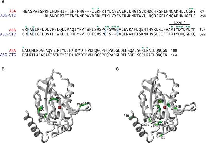
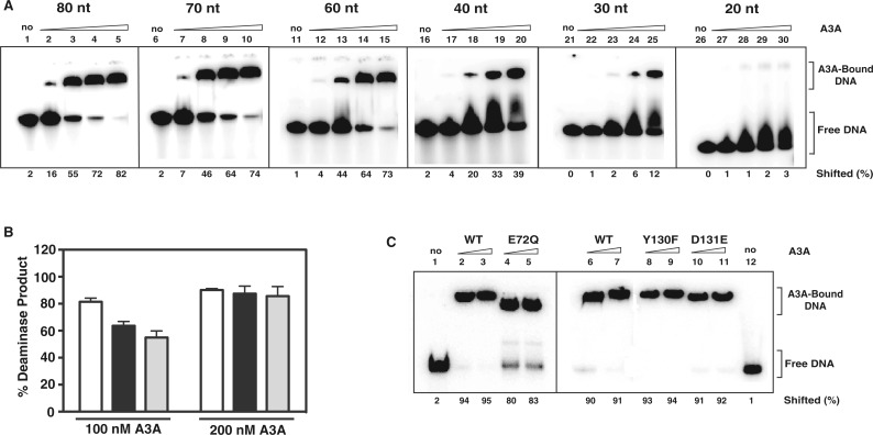
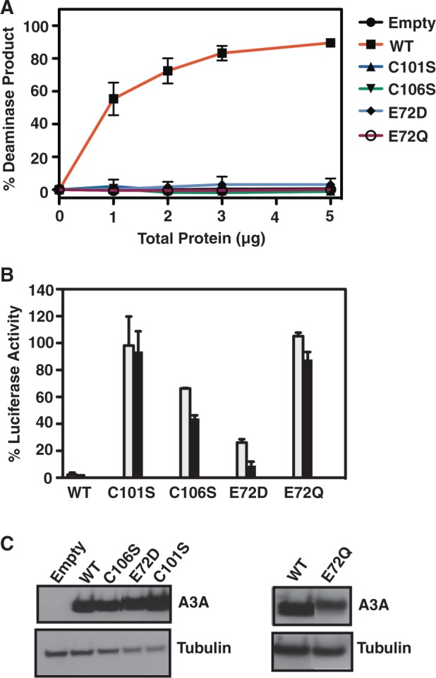
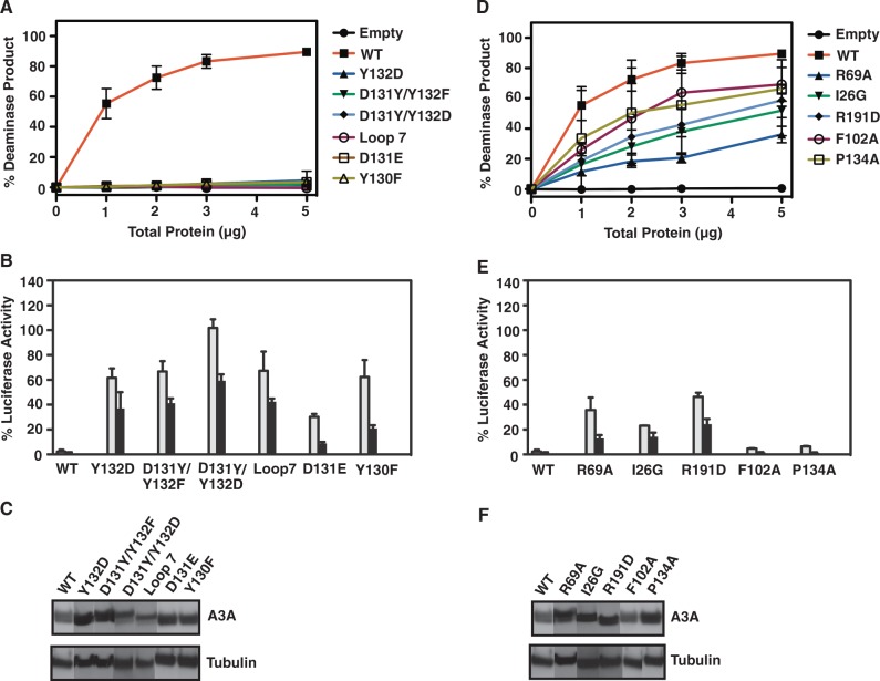
Human APOBEC3A (A3A) is a single-domain cytidine deaminase that converts deoxycytidine residues to deoxyuridine in single-stranded DNA (ssDNA). It inhibits a wide range of viruses and endogenous retroelements such as LINE-1, but it can also edit genomic DNA, which may play a role in carcinogenesis. Here, we extend our recent findings on the NMR structure of A3A and report structural, biochemical and cell-based mutagenesis studies to further characterize A3A's deaminase and nucleic acid binding activities. We find that A3A binds ssRNA, but the RNA and DNA binding interfaces differ and no deamination of ssRNA is detected. Surprisingly, with only one exception (G105A), alanine substitution mutants with changes in residues affected by specific ssDNA binding retain deaminase activity. Furthermore, A3A binds and deaminates ssDNA in a length-dependent manner. Using catalytically active and inactive A3A mutants, we show that the determinants of A3A deaminase activity and anti-LINE-1 activity are not the same. Finally, we demonstrate A3A's potential to mutate genomic DNA during transient strand separation and show that this process could be counteracted by ssDNA binding proteins. Taken together, our studies provide new insights into the molecular properties of A3A and its role in multiple cellular and antiviral functions.
Figures


Similar articles
-
Creating RNA Specific C-to-U Editase from APOBEC3A by Separation of Its Activities on DNA and RNA Substrates.ACS Synth Biol. 2021 May 21;10(5):1106-1115. doi: 10.1021/acssynbio.0c00627. Epub 2021 May 2. ACS Synth Biol. 2021. PMID: 33938211
-
Substrate sequence selectivity of APOBEC3A implicates intra-DNA interactions.Sci Rep. 2018 May 14;8(1):7511. doi: 10.1038/s41598-018-25881-z. Sci Rep. 2018. PMID: 29760455 Free PMC article.
-
Single-stranded DNA binding proteins influence APOBEC3A substrate preference.Sci Rep. 2021 Oct 25;11(1):21008. doi: 10.1038/s41598-021-00435-y. Sci Rep. 2021. PMID: 34697369 Free PMC article.
-
Functions and regulation of the APOBEC family of proteins.Semin Cell Dev Biol. 2012 May;23(3):258-68. doi: 10.1016/j.semcdb.2011.10.004. Epub 2011 Oct 6. Semin Cell Dev Biol. 2012. PMID: 22001110 Free PMC article. Review.
-
Site directed mutagenesis as a tool to understand the catalytic mechanism of human cytidine deaminase.Protein Pept Lett. 2013 May;20(5):538-49. doi: 10.2174/0929866511320050007. Protein Pept Lett. 2013. PMID: 23033855 Review.
Cited by
-
Control of Human Anelloviruses by Cytosine to Uracil Genome Editing.mSphere. 2022 Dec 21;7(6):e0050622. doi: 10.1128/msphere.00506-22. Epub 2022 Nov 14. mSphere. 2022. PMID: 36374042 Free PMC article.
-
1.92 Angstrom Zinc-Free APOBEC3F Catalytic Domain Crystal Structure.J Mol Biol. 2016 Jun 5;428(11):2307-2316. doi: 10.1016/j.jmb.2016.04.026. Epub 2016 Apr 30. J Mol Biol. 2016. PMID: 27139641 Free PMC article.
-
Modeling the Embrace of a Mutator: APOBEC Selection of Nucleic Acid Ligands.Trends Biochem Sci. 2018 Aug;43(8):606-622. doi: 10.1016/j.tibs.2018.04.013. Epub 2018 May 23. Trends Biochem Sci. 2018. PMID: 29803538 Free PMC article. Review.
-
The ssDNA Mutator APOBEC3A Is Regulated by Cooperative Dimerization.Structure. 2015 May 5;23(5):903-911. doi: 10.1016/j.str.2015.03.016. Epub 2015 Apr 23. Structure. 2015. PMID: 25914058 Free PMC article.
-
The roles of APOBEC-mediated RNA editing in SARS-CoV-2 mutations, replication and fitness.Sci Rep. 2022 Sep 13;12(1):14972. doi: 10.1038/s41598-022-19067-x. Sci Rep. 2022. PMID: 36100631 Free PMC article.
References
-
- Harris RS, Liddament MT. Retroviral restriction by APOBEC proteins. Nat. Rev. Immunol. 2004;4:868–877. - PubMed
-
- Chiu YL, Greene WC. The APOBEC3 cytidine deaminases: an innate defensive network opposing exogenous retroviruses and endogenous retroelements. Annu. Rev. Immunol. 2008;26:317–353. - PubMed
-
- Holmes RK, Malim MH, Bishop KN. APOBEC-mediated viral restriction: not simply editing? Trends Biochem. Sci. 2007;32:118–128. - PubMed
Publication types
MeSH terms
Substances
Grants and funding
LinkOut - more resources
Full Text Sources
Other Literature Sources
