. 2014 Mar 15;23(6):1479-91.
doi: 10.1093/hmg/ddt535. Epub 2013 Oct 24.

Absence of cell surface expression of human ACE leads to perinatal death

Affiliations

Absence of cell surface expression of human ACE leads to perinatal death

Annie Michaud et al. Hum Mol Genet. .

Abstract

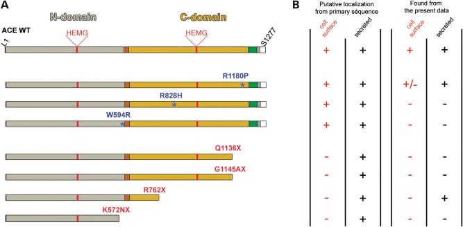
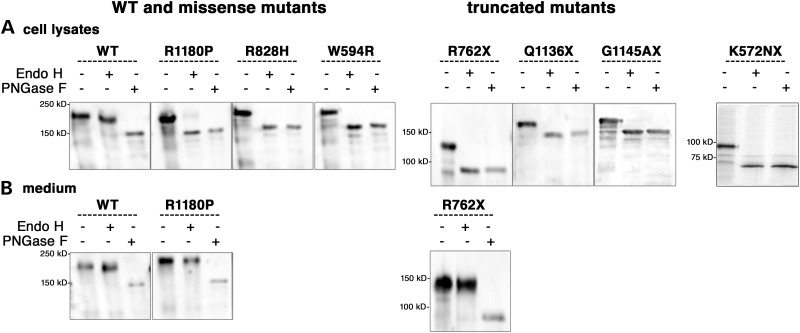
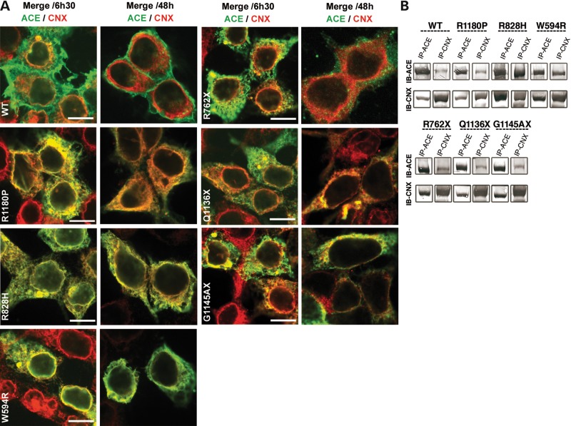
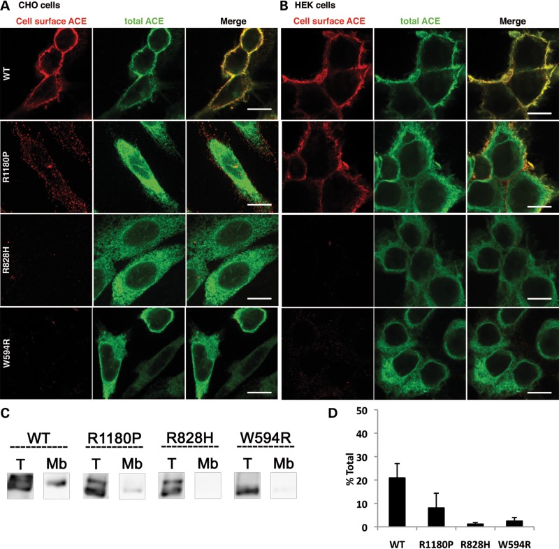
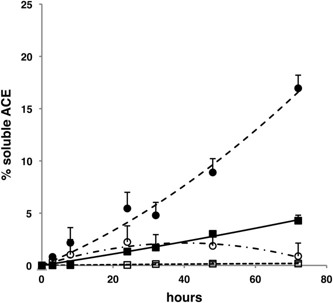
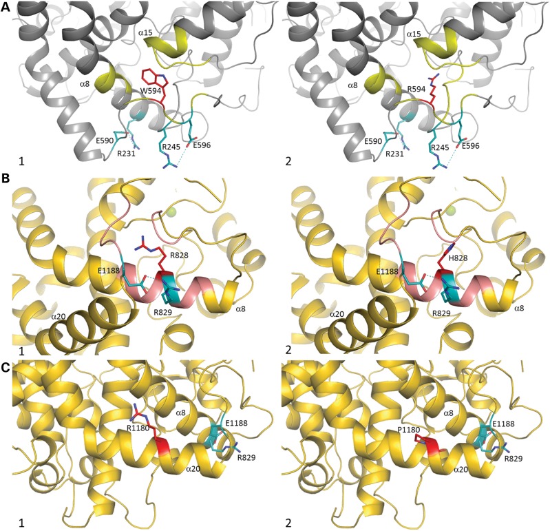
Renal tubular dysgenesis (RTD) is a recessive autosomal disease characterized most often by perinatal death. It is due to the inactivation of any of the major genes of the renin-angiotensin system (RAS), one of which is the angiotensin I-converting enzyme (ACE). ACE is present as a tissue-bound enzyme and circulates in plasma after its solubilization. In this report, we present the effect of different ACE mutations associated with RTD on ACE intracellular trafficking, secretion and enzymatic activity. One truncated mutant, R762X, responsible for neonatal death was found to be an enzymatically active, secreted form, not inserted in the plasma membrane. In contrast, another mutant, R1180P, was compatible with life after transient neonatal renal insufficiency. This mutant was located at the plasma membrane and rapidly secreted. These results highlight the importance of tissue-bound ACE versus circulating ACE and show that the total absence of cell surface expression of ACE is incompatible with life. In addition, two missense mutants (W594R and R828H) and two truncated mutants (Q1136X and G1145AX) were also studied. These mutants were neither inserted in the plasma membrane nor secreted. Finally, the structural implications of these ACE mutations were examined by molecular modelling, which suggested some important structural alterations such as disruption of intra-molecular non-covalent interactions (e.g. salt bridges).


Figures

Figure 1.
Figure 1.
(A) Schematic diagram of ACE WT and RTD mutants. The ACE N-domain (L1-P602) is shown in grey, the linker region (P602-D612) in brown, the C-domain (L613-P1193) in yellow, the stalk region (Q1194-R1227) in green, the transmembrane segment (V1228-S1248) in grey, and the ic domain (Q1249-S1277) in white. Amino acids corresponding to the catalytic sites of N- and C-domains are H361–H365 and H959–H963, respectively. The positions of disease-causing missense mutations are shown by blue asterisks. The truncated mutations are indicated in red. (B) On the left, the table summarizes the putative cellular localization of ACE mutants according to their primary sequence and on the right, the actual localization of mutants found in cell culture in this study.
Figure 2.
Figure 2.
Expression of WT ACE and RTD mutants in stably transfected Chinese Hamster Ovary (CHO) cells. (A) Immunoblot analysis: cell lysates (C) and culture medium (M) collected 24 h after a switch to serum-free medium were analysed by immuno-blot with the ACE Ab Y1. (B) Pulse-chase analysis: CHO cells were labelled for 30 min and subjected to a 2–28 h chase period for WT and all mutants, except the R762X mutant which was chased from 15 to 120 min. Proteins were immunoprecipitated from cell lysates and culture medium with the ACE Ab HKCE.
Figure 3.
Figure 3.
Glycosylation of WT ACE and RTD mutants in stably transfected CHO cells. Proteins were immunoprecipitated with the anti-ACE Ab HKCE from cell lysates (A) and culture medium (B). The samples were treated with buffer alone, PNGase F or Endo H and analysed by immunoblotting with the anti-ACE Ab Y1.
Figure 4.
Figure 4.
Interaction of WT ACE and RTD mutants with CNX in human embryonic kidney (HEK) and CHO cells. (A) Confocal analysis in HEK cells: HEK cells, transiently expressing WT and the six different mutants, were fixed 6 h 30 min and 48 h after transfection, permeabilized and stained with the HKCE Ab (green) and a CNX Ab (red). Only merge images are shown. Scale bar 10 µm. (B) Co-immunoprecipitations (IP) of WT and RTD mutants with CNX in CHO cells. IP of cell lysates was performed by incubation with anti-ACE Ab HKCE (IP-ACE) or anti-CNX Ab (IP-CNX). Immune complexes were precipitated with Protein G-Sepharose and analysed by immuno-blot (IB) with anti-ACE Ab Y1 (IB ACE) or anti-CNX Ab (IB-CNX).
Figure 5.
Figure 5.
Cell surface expression of WT ACE and RTD missense mutants. WT ACE, R1180P, R828H and W594R mutants were stably and transiently expressed in CHO (A, C and D) and HEK cells (B), respectively. (A and B) IF analysis. CHO cells (A) and HEK cells (B) were grown on glass coverslips, fixed and stained with the rabbit anti-ACE Ab Y1 shown in red (cell-surface ACE). Cells were then permeabilized and the total ACE pool was stained with the sheep anti-ACE Ab HKCE shown in green. Scale bar 10 µm. (C and D) Detection of ACE surface expression. (C) Stably transfected CHO cells grown in 6-well cell culture plates, were fixed with 0.4% PFA and then incubated for 1 h with the anti-ACE Ab HKCE, solubilized and immunoprecipitated to obtain expression at the cell membrane (Mb). Total amounts (T) were obtained with cells which were fixed, solubilized and then immunoprecipitated with the HKCE Ab. Proteins were analysed by IB with the anti-ACE Ab Y1. (D) ACE protein bands were quantified by densitometry and expressed as percentages of the total ACE amount. Results shown are mean ± SD of four experiments.
Figure 6.
Figure 6.
Shedding of WT ACE and of R1180P mutant in CHO cells. WT ACE (black square) and R1180P mutant (black circle) secretion in CHO cells was quantified with a human ACE ELISA in cell lysates and culture media. ACE is expressed as a percentage of the total amount of ACE in cell lysates and in culture medium. The effect of the sheddase inhibitor TAPI-1 on the solubilization rate of WT ACE (open squares) and the R1180P mutant (open circles) is indicated. Results shown are means of four points corresponding to two separate experiments.
Figure 7.
Figure 7.
(A) Crystal structure of N-domain ACE (PDB code 3NXQ) (32) in grey. Residue W594 is shown in red, with salt bridges R245–E596 and R231–E590 in cyan (bond in dash lines). Interactions with W594 are highlighted in yellow. (1) Close-up view of the W594 area. (2) Modelled W594R mutation (red) with its potential interaction (yellow). (B and C) Crystal structure of C-domain ACE (PDB code 1O8A) (12) in gold. (B) Residue R828 is in red, with salt bridge R829–E1188 in cyan (bond in dash lines). Interactions with R828 highlighted in pink. (1) Close-up view of the R828 area. (2) Modelled R828H mutation (red) with its potential interaction (pink). (C) Residue R1180 in red, with salt bridge R829–E1188 in cyan (bond in dash lines). Interactions with R1180 are highlighted in pink. (1) Close-up view of the R1180 area. (2) Modelled R828H mutation (red). Secondary structure numbering is based on C-domain alone (12).

References

    1. Bernstein K.E., Ong F.S., Blackwell W.L., Shah K.H., Giani J.F., Gonzalez-Villalobos R.A., Shen X.Z., Fuchs S., Touyz R.M. A modern understanding of the traditional and nontraditional biological functions of angiotensin-converting enzyme. Pharmacol. Rev. 2013;65:1–46. doi:10.1124/pr.112.006809. - DOI - PMC - PubMed
    1. Wei L., Alhenc-Gelas F., Corvol P., Clauser E. The two homologous domains of human angiotensin I-converting enzyme are both catalytically active. J. Biol. Chem. 1991;266:9002–9008. - PubMed
    1. Jaspard E., Wei L., Alhenc-Gelas F. Differences in the properties and enzymatic specificities of the two active sites of angiotensin I-converting enzyme (kininase II). Studies with bradykinin and other natural peptides. J. Biol. Chem. 1993;268:9496–9503. - PubMed
    1. Rousseau A., Michaud A., Chauvet M.T., Lenfant M., Corvol P. The hemoregulatory peptide N-acetyl-Ser-Asp-Lys-Pro is a natural and specific substrate of the N-terminal active site of human angiotensin-converting enzyme. J. Biol. Chem. 1995;270:3656–3661. doi:10.1074/jbc.270.8.3656. - DOI - PubMed
    1. Ramchandran R., Sen I. Cleavage processing of angiotensin-converting enzyme by a membrane-associated metalloprotease. Biochemistry. 1995;34:12645–12652. doi:10.1021/bi00039a021. - DOI - PubMed

Publication types

MeSH terms

Supplementary concepts