

Cognitive control and its impact on recovery from aphasic stroke
- PMID: 24163248
- PMCID: PMC3891442
- DOI: 10.1093/brain/awt289
Cognitive control and its impact on recovery from aphasic stroke
Abstract
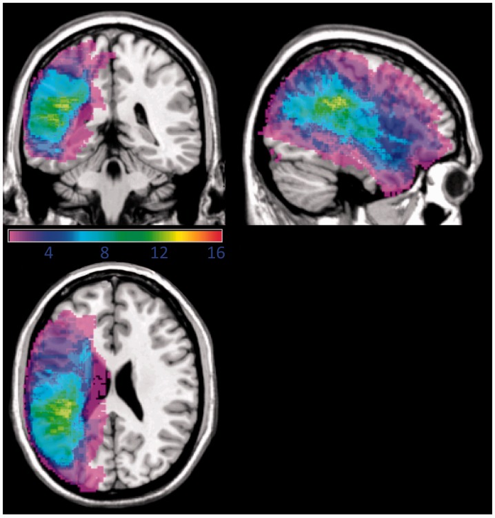
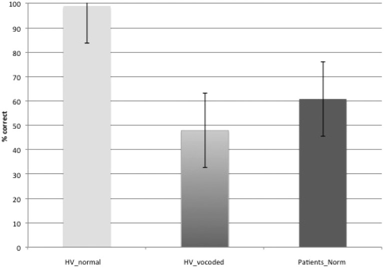
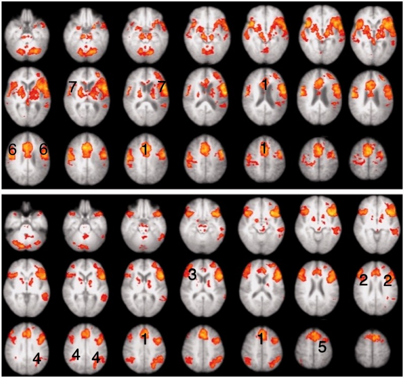
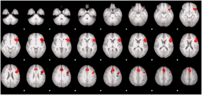
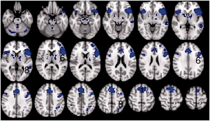
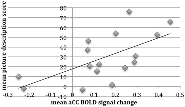
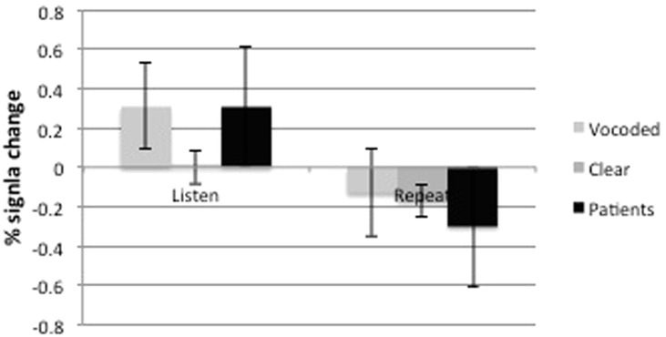
Aphasic deficits are usually only interpreted in terms of domain-specific language processes. However, effective human communication and tests that probe this complex cognitive skill are also dependent on domain-general processes. In the clinical context, it is a pragmatic observation that impaired attention and executive functions interfere with the rehabilitation of aphasia. One system that is important in cognitive control is the salience network, which includes dorsal anterior cingulate cortex and adjacent cortex in the superior frontal gyrus (midline frontal cortex). This functional imaging study assessed domain-general activity in the midline frontal cortex, which was remote from the infarct, in relation to performance on a standard test of spoken language in 16 chronic aphasic patients both before and after a rehabilitation programme. During scanning, participants heard simple sentences, with each listening trial followed immediately by a trial in which they repeated back the previous sentence. Listening to sentences in the context of a listen-repeat task was expected to activate regions involved in both language-specific processes (speech perception and comprehension, verbal working memory and pre-articulatory rehearsal) and a number of task-specific processes (including attention to utterances and attempts to overcome pre-response conflict and decision uncertainty during impaired speech perception). To visualize the same system in healthy participants, sentences were presented to them as three-channel noise-vocoded speech, thereby impairing speech perception and assessing whether this evokes domain general cognitive systems. As expected, contrasting the more difficult task of perceiving and preparing to repeat noise-vocoded speech with the same task on clear speech demonstrated increased activity in the midline frontal cortex in the healthy participants. The same region was activated in the aphasic patients as they listened to standard (undistorted) sentences. Using a region of interest defined from the data on the healthy participants, data from the midline frontal cortex was obtained from the patients. Across the group and across different scanning sessions, activity correlated significantly with the patients' communicative abilities. This correlation was not influenced by the sizes of the lesion or the patients' chronological ages. This is the first study that has directly correlated activity in a domain general system, specifically the salience network, with residual language performance in post-stroke aphasia. It provides direct evidence in support of the clinical intuition that domain-general cognitive control is an essential factor contributing to the potential for recovery from aphasic stroke.
Keywords: aphasia; cingulate; executive; functional MRI; salience.
Figures


References
-
- Abo M, Senoo A, Watanabe S, Miyano S, Doseki K, Sasaki N, et al. Language-related brain function during word repetition in post-stroke aphasics. Neuroreport. 2004;15:1891–4. - PubMed
-
- Bakheit AM, Shaw S, Carrington S, Griffiths S. The rate and extent of improvement with therapy from the different types of aphasia in the first year after stroke. Clin Rehabil. 2007;21:941–9. - PubMed
-
- Bench J, Kowal A, Bamford J. The BKB (Bamford-Kowal-Bench) sentence lists for partially- hearing children. Br J Audiol. 1979;13:108–12. - PubMed
-
- Blank SC, Bird H, Turkheimer F, Wise RJ. Speech production after stroke: the role of the right pars opercularis. Ann Neurol. 2003;54:310–20. - PubMed
Publication types
MeSH terms
Grants and funding
LinkOut - more resources
Full Text Sources
Other Literature Sources
Medical
Research Materials
