. 2014 Jan;196(1):170-9.
doi: 10.1128/JB.01148-13. Epub 2013 Oct 25.

Inefficient translation renders the Enterococcus faecalis fabK enoyl-acyl carrier protein reductase phenotypically cryptic

Affiliations

Inefficient translation renders the Enterococcus faecalis fabK enoyl-acyl carrier protein reductase phenotypically cryptic

Hongkai Bi et al. J Bacteriol. 2014 Jan.

Abstract

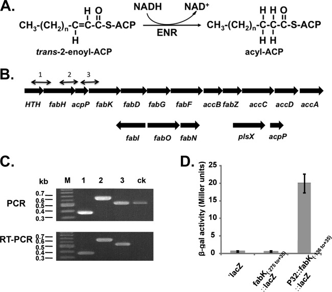
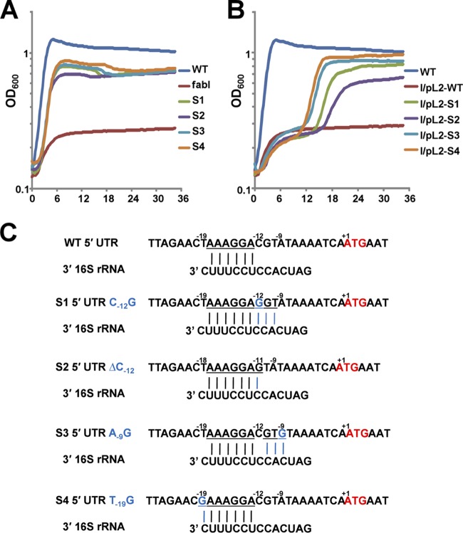
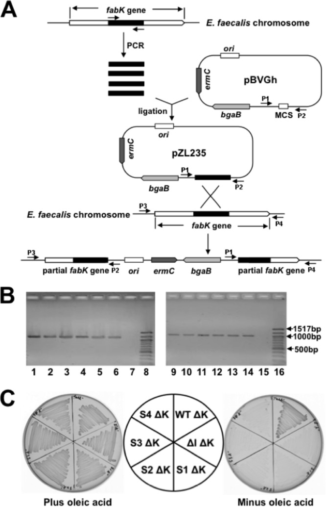
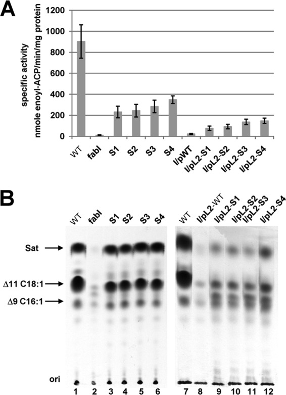
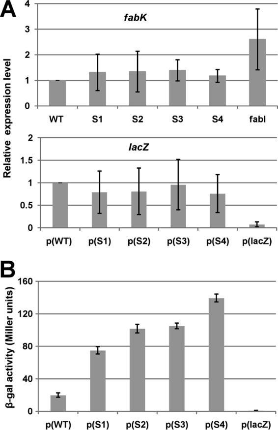
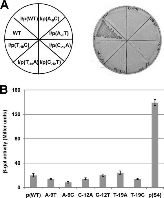
Enoyl-acyl carrier protein (ACP) reductase catalyzes the last step of the bacterial fatty acid elongation cycle. Enterococcus faecalis is unusual in that it encodes two unrelated enoyl-ACP reductases, FabI and FabK. We recently reported that deletion of the gene encoding FabI results in an unsaturated fatty acid (UFA) auxotroph despite the presence of fabK, a gene encoding a second fully functional enoyl-ACP reductase. By process of elimination, our prior report argued that poor expression was the reason that fabK failed to functionally replace FabI. We now report that FabK is indeed poorly expressed and that the expression defect is at the level of translation rather than transcription. We isolated four spontaneous mutants that allowed growth of the E. faecalis ΔfabI strain on fatty acid-free medium. Each mutational lesion (single base substitution or deletion) extended the fabK ribosome binding site. Inactivation of fabK blocked growth, indicating that the mutations acted only on fabK rather than a downstream gene. The mutations activated fabK translation to levels that supported fatty acid synthesis and hence cell growth. Furthermore, site-directed and random mutagenesis experiments showed that point mutations that resulted in increased complementarity to the 3' end of the 16S rRNA increased FabK translation to levels sufficient to support growth, whereas mutations that decreased complementarity blocked fabK translation.


Figures

FIG 1
FIG 1
The ENR reaction, the fabK and fabI genome neighborhoods, and transcriptional analyses of the operon containing fabK. (A) The enoyl-acyl-ACP reductase reaction. (B) Genetic organization of the E. faecalis fatty acid biosynthetic genes (fab). The numbered short lines (1, 2, and 3) represent the specific PCR amplicons observed in the PCR and RT-PCR assays shown in panel C. The second acpP gene (EF_3111 in strain V583) located downstream of plsX is conserved in all extant E. faecalis genome sequences. (C) PCR and RT-PCR analyses of the fab operon. The results for the four genes at the 5′ end of the operon are shown. The helix-turn-helix-encoding gene probably encodes a homologue of the FabT transcription factor found in other Firmicutes bacteria (43, 44). The primer numbering system is the same as that for panel B. Both PCR products obtained from genomic DNA and those obtained by RT-PCR were separated by electrophoresis on a 1.5% agarose gel. The ck designation denotes two neighboring genes transcribed from the opposite strand that were included as controls. (D) β-Galactosidase activities of fabK::lacZ translational fusions. The wild-type strain FA2-2 carrying each of the fusion plasmids was grown in GM17 medium to mid-log phase, and β-galactosidase activities were measured in more than three independent experiments. The error bars indicate standard deviations. The plasmids carried a promoterless lacZ (′lacZ), the −275 to +35 fragment, which includes the first 35 bp of the fabK coding sequence fused to lacZ (fabK-lacZ), or the −136 to +35 fragment driven by the P32 promoter. The fusion plasmids were pBHK322, pBHK394, and pBHK323, respectively.
FIG 2
FIG 2
Growth and sequences of the spontaneously arising mutant strains and complementation of the ΔfabI strain by plasmid-borne fragments that included the fabK mutant or wild-type alleles. (A) Growth of the four spontaneously arising mutant ΔfabI strains (S1 to S4) on GM17 medium in the absence of oleate supplementation. (B) Complementation of the ΔfabI strain FAZL1 by plasmids (Table 1) carrying a mutant fabK allele plus about 300 bp of upstream and downstream sequence. Each growth assay was carried out in triplicate in GM17 medium at 37°C, and the averages are shown. Strain FAZL1 in panel A and the strain carrying wild-type fabK in panel B were used as negative controls. Panels A and B have the same color coding for the mutant fabK alleles. (C) Alignments of 5′ untranslated regions of the wild-type gene fabK or of the four spontaneously arising mutant strains with the 3′ end of the E. faecalis 16S rRNA. The fabK genes of wild-type strain FAZL1 and the four spontaneous mutants plus about 300 bp upstream and downstream were sequenced. Each point mutation expanded the fabK ribosome binding site. The underlined letters indicate bases capable of Watson-Crick base pairing with the 16S rRNA. +1 indicates the A of the initiation ATG codon.
FIG 3
FIG 3
Construction, characterization, and growth of fabK disruption mutant strains. (A) Strategy for isolation of erythromycin-marked chromosomal E. faecalis fabK disruption mutant strains via plasmid insertion. P1, P2, P3, and P4 were the PCR primers used for characterization of the fabK disruption mutant strains. (B) Characterization of the fabK disruption strains by PCR analyses. Lanes 1 to 6, PCR products amplified from lane 1, the fabK disruption strain (FAZL6); lane 2, the fabK disruption ΔfabI double mutant strain (FAZL7); lanes 3 to 6, the S1 to S4 fabK strains in which fabK was disrupted (strains FAZL8 to -11, respectively). Genomic DNA preparations were used as templates with primer sets pBVGh For (P1) and fabK check Down (P4). Lanes 9 to 14 are the PCR products amplified using the same template DNAs as for lanes 1 to 6 but with primers pBVGh Rev (P2) and fabK check Up (P3). Lanes 7 and 15 are the PCR products formed from the genomic DNA of the wild-type strain FA2-2. Lanes 8 and 16 are molecular size standards. (C) Growth of E. faecalis mutant strains on GM17 medium plus 5 μg/ml of erythromycin at 42°C with (left) or without (right) oleic acid. Cell growth proceeded at 42°C for 36 h. ΔK, the disrupted fabK; WT, E. faecalis wild-type strain; ΔI, the E. faecalis ΔfabI gene deletion strain; S1, S2, S3, and S4, the spontaneously arising suppressors the of E. faecalis fabI deletion. The normal growth in the absence of fatty acid supplementation of the disrupted fabK derivative of the wild-type strain indicated that the disruption cassette is not polar on the downstream fatty acid synthesis genes.
FIG 4
FIG 4
The spontaneous mutations increased expression of the FabK ENR. (A) ENR activities of the wild-type FA2-2, spontaneous mutants, and the complementing plasmid-bearing strains. The 100-μl ENR reaction mixtures contained 0.1 M sodium phosphate buffer (pH 7.0), 0.15 mM NADH, and 2 μg of cell extract protein, and NADH-dependent ENR activity was assayed. The reactions were initiated by addition of 100 μM trans-2-decenoyl-ACP. The data are from three independent experiments and are expressed as means ± standard deviations. (B) Incorporation of [1-14C]acetate into the membrane phospholipids of the wild-type strain FA2-2, the spontaneous mutant strains, and the ΔfabI strain FAZL1 transformed with the wild-type fabK gene or the mutant alleles. The strains used here are the same with those in Fig. 2. The fatty acid methyl esters were prepared and analyzed by argentation thin-layer chromatography as described in Materials and Methods. Designations: Sat, saturated fatty acids; Δ9C16:1, palmitoleic (cis-9-hexadecenoic) acid; Δ11C18:1, cis-vaccenic (cis-11-octadecenoic) acid.
FIG 5
FIG 5
Effects of the spontaneous mutations on fabK transcription and translation. (A) qRT-PCR analyses of the effects on fabK and lacZ transcription. Transcription of fabK was analyzed in the wild-type strain and the spontaneous mutant strains, whereas lacZ transcription was analyzed for the wild-type strains carrying the translational fusion plasmid. Cells were grown in GM17 medium to mid-log phase, and RNA was isolated as described in Materials and Methods. The qRT-PCR data were from no less than four independent tests and are expressed as means ± standard deviations. The p(lacZ) designation denotes the promoterless ′lacZ vector. (B) Effects of the spontaneous mutations on β-galactosidase expression from a plasmid carrying a P32::fabK::lacZ translational fusion. The wild-type strain FA2-2 carrying the fusion plasmids was grown in GM17 medium to mid-log phase, and β-galactosidase activities were measured from more than three independent experiments. The error bars indicate standard deviations. p(WT), p(S1), p(S2), p(S3), p(S4), and p(lacZ) indicate fusion plasmids pBHK323, pBHK324, pBHK325, pBHK326, pBHK327, and pBHK322 carrying the −136 to +35 fragments of wild-type fabK, one of the four spontaneous fabK mutants, or the empty ′lacZ vector, respectively.
FIG 6
FIG 6
16S rRNA-mRNA complementarity is required for growth of the spontaneous mutants. (A) Growth was tested on GM17 plates incubated for 36 h at 37°C. In three of the spontaneous mutants, the unique nucleotide of the mutation was changed to two other nucleotides. Each site-directed fabK gene was cloned into the vector pTRKL2 and then transformed to the ΔfabI strain, FAZL1. (B) Effects of the nucleotide changes on β-galactosidase expression from a plasmid carrying a P32::fabK::lacZ translational fusion. The wild-type strain FA2-2 carrying the fusion plasmids was grown in GM17 medium to mid-log phase, and β-galactosidase activities were measured from more than three independent experiments. The error bars indicate standard deviations. The designations p(WT), A-9T, A-9C, C-12A, C-12T, T-19A, T-19C, and p(S4) indicate the fusion plasmids pBHK323, pBHK334, pBHK335, pBHK336, pBHK337, pBHK338, pBHK339, and pBHK327, respectively. The p(WT) and p(S4) plasmids were used as negative and positive controls, respectively.

References

    1. Lu YJ, Zhang YM, Rock CO. 2004. Product diversity and regulation of type II fatty acid synthases. Biochem. Cell Biol. 82:145–155. 10.1139/o03-076 - DOI - PubMed
    1. Cronan JE, Thomas J. 2009. Bacterial fatty acid synthesis and its relationships with polyketide synthetic pathways. Methods Enzymol. 459:395–433. 10.1016/S0076-6879(09)04617-5 - DOI - PMC - PubMed
    1. Cronan JE., Jr 2004. The structure of mammalian fatty acid synthase turned back to front. Chem. Biol. 11:1601–1602. 10.1016/j.chembiol.2004.11.011 - DOI - PubMed
    1. Cronan JE. 2006. Remarkable structural variation within fatty acid megasynthases. Nat. Chem. Biol. 2:232–234. 10.1038/nchembio0506-232 - DOI - PubMed
    1. Schweizer E, Hofmann J. 2004. Microbial type I fatty acid synthases (FAS): major players in a network of cellular FAS systems. Microbiol. Mol. Biol. Rev. 68:501–517. 10.1128/MMBR.68.3.501-517.2004 - DOI - PMC - PubMed

Publication types

MeSH terms

LinkOut - more resources