. 2014 Jan;196(1):189-201.
doi: 10.1128/JB.00918-13. Epub 2013 Oct 25.

GTP dysregulation in Bacillus subtilis cells lacking (p)ppGpp results in phenotypic amino acid auxotrophy and failure to adapt to nutrient downshift and regulate biosynthesis genes

Affiliations

GTP dysregulation in Bacillus subtilis cells lacking (p)ppGpp results in phenotypic amino acid auxotrophy and failure to adapt to nutrient downshift and regulate biosynthesis genes

Allison Kriel et al. J Bacteriol. 2014 Jan.

Abstract

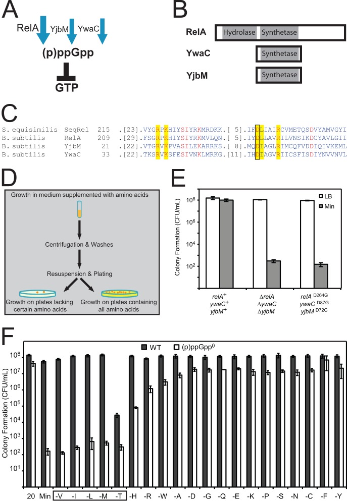
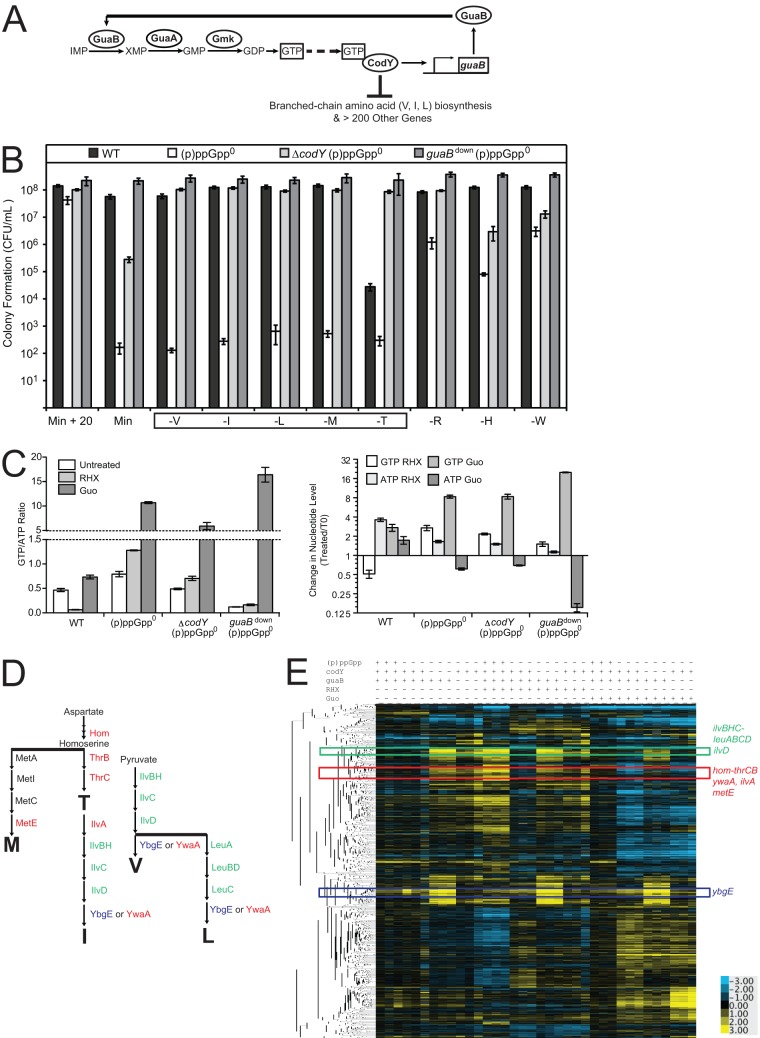
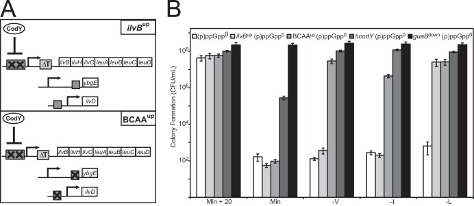
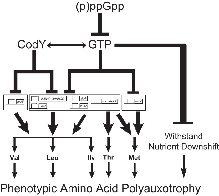
The nucleotide (p)ppGpp inhibits GTP biosynthesis in the Gram-positive bacterium Bacillus subtilis. Here we examined how this regulation allows cells to grow in the absence of amino acids. We showed that B. subtilis cells lacking (p)ppGpp, due to either deletions or point mutations in all three (p)ppGpp synthetase genes, yjbM, ywaC, and relA, strongly require supplementation of leucine, isoleucine, valine, methionine, and threonine and modestly require three additional amino acids. This polyauxotrophy is rescued by reducing GTP levels. Reduction of GTP levels activates transcription of genes responsible for the biosynthesis of the five strongly required amino acids by inactivating the transcription factor CodY, which represses the ybgE, ilvD, ilvBHC-leuABCD, ilvA, ywaA, and hom-thrCB operons, and by a CodY-independent activation of transcription of the ilvA, ywaA, hom-thrCB, and metE operons. Interestingly, providing the eight required amino acids does not allow for colony formation of (p)ppGpp(0) cells when transitioning from amino acid-replete medium to amino acid-limiting medium, and we found that this is due to an additional role that (p)ppGpp plays in protecting cells during nutrient downshifts. We conclude that (p)ppGpp allows adaptation to amino acid limitation by a combined effect of preventing death during metabolic transitions and sustaining growth by activating amino acid biosynthesis. This ability of (p)ppGpp to integrate a general stress response with a targeted reprogramming of gene regulation allows appropriate adaptation and is likely conserved among diverse bacteria.


Figures

FIG 1
FIG 1
(p)ppGpp synthesis is required for amino acid prototrophy. (A) In B. subtilis, the enzymes RelA, YwaC, and YjbM synthesize (p)ppGpp. The bifunctional enzyme RelA also hydrolyzes (p)ppGpp. (B) Conserved N-terminal (p)ppGpp synthetase and hydrolase domains of YwaC, YjbM, and RelA. (C) Sequence alignment showing the conserved residues (shown in red), active site residues (highlighted in yellow), and aspartic acid active site residue (boxed) that when mutated in the RelA protein of S. equisimilis completely abolishes (p)ppGpp synthetase activity without affecting hydrolase activity. The respective conserved aspartic acid residues (RelAD264, YwaCD87, and YjbMD72), aligned using the Conserved Domain Database (44), were mutated in each (p)ppGpp synthetase of B. subtilis in order to specifically abolish (p)ppGpp synthetase activity without affecting other potential functions of these proteins. (D) Schematic of the plating efficiency assay for determining amino acid auxotrophy. (E) Wild-type (relA+ ywaC+ yjbM+), synthetase deletion (ΔrelA ΔywaC ΔyjbM), and synthetase point mutant (relAD264G ywaCD87G yjbMD72G) cells were plated on LB and minimal medium (Min) plates, and colonies were counted the next day. “CFU/ml” indicates CFU per ml of culture, normalized to an OD600 of 1. (F) (p)ppGpp0 cells display strong requirements for valine, leucine, isoleucine, threonine, and methionine (boxed); (p)ppGpp0 cells also display weaker requirements for arginine, histidine, and tryptophan. Cells were grown in medium supplemented with Casamino Acids, washed, and then plated on defined medium plates supplemented with all 20 amino acids (20) or supplemented with only 19 amino acids (denoted as “-X” to indicate the amino acid that has not been supplemented in the indicated plates). Colonies were counted the next day. “CFU/ml” indicates CFU per ml of culture, normalized to an OD600 of 1.
FIG 2
FIG 2
Effects of (p)ppGpp, guaB, and codY on global transcription profiles of B. subtilis cells. (A) Schematic of the pathways affected by the (p)ppGpp0 suppressor mutations. GuaB, GuaA, and Gmk are enzymes in the GTP biosynthesis pathway. GTP activates CodY. CodY activates transcription of guaB and represses numerous other genes, including those involved in biosynthesis of BCAA: valine, isoleucine, and leucine. (B) Colony formation of WT, (p)ppGpp0, ΔcodY (p)ppGpp0, and guaBdown (p)ppGpp0 cells on plates supplemented with all 20 amino acids or plates lacking valine (V), isoleucine (I), leucine (L), methionine (M), threonine (T), arginine (R), histidine (H), or tryptophan (W). Cells were plated as for Fig. 1F. The box indicates strong amino acid requirements. (C) GTP/ATP ratios or GTP and ATP levels relative to those for untreated cells at t = 0 of wild-type, (p)ppGpp0, ΔcodY (p)ppGpp0, and guaBdown (p)ppGpp0 cells. 32P-labeled cells were treated with 0.5 mg/ml RHX or 1 mM Guo for 20 min. Error bars represent standard errors of the means for three independent biological replicates. (D) Schematic of the amino acid biosynthesis pathways that lead to the production of the amino acids that are strongly required by (p)ppGpp0 cells: threonine, methionine, isoleucine, valine, and leucine. (E) Hierarchical clustering analysis of transcript levels obtained from microarrays. Rows correspond to the genes; columns correspond to samples with strains and treatments labeled above the heat map for WT [(p)ppGpp +], (p)ppGpp0 [(p)ppGpp −], ΔcodY (p)ppGpp0 (codY −), and guaBdown (p)ppGpp0 (guaB −) cells that are either untreated, treated with RHX or treated with Guo. Three independent experimental replicates are shown. Transcript levels (log2) are indicated by color such that high levels are yellow and low levels are blue. Genes were hierarchically clustered using the software program Cluster and plotted with TreeView software. Colored boxes correspond to distinct clusters containing the genes involved in synthesis of the amino acids that are strongly required by (p)ppGpp0 cells, which are indicated with the corresponding colors in panel C. For an expanded view, see Fig. S1B in the supplemental material.
FIG 3
FIG 3
Transcript levels of ilvBHC-leuABCD, ilvD, and ybgE in (p)ppGpp0 cells and the effects of CodY and GTP. (A) Schematic of the BCAA (isoleucine, valine, and leucine) biosynthesis pathways. Genes in two major clusters in the hierarchical tree of the microarray data in Fig. 2D are colored accordingly: the ilvBHC-leuABCD and ilvD operons are in the green cluster; the ybgE operon clusters separately in blue. (B) A cluster in which genes in the ilvB and ilvD operons (green) are located. Transcript levels (log2) are indicated by color such that high levels are yellow and low levels are blue. (C to F) Transcript levels of ilvB (C), leuC (D), ilvD (E), or ybgE (F) in wild-type, (p)ppGpp0, ΔcodY (p)ppGpp0, and guaBdown (p)ppGpp0 cells both before and after RHX or Guo treatment. Transcript levels were normalized to a common reference, and log2 ratios are plotted. Similar trends were obtained for other genes in the ilvB operon: ilvH, ilvC, leuA, leuB, and leuD. For this and all subsequent figures, the averages of three biological replicates and the standard errors of the means are presented for each strain and treatment.
FIG 4
FIG 4
Hyperrepression by CodY causes the branched-chain amino acid requirements of (p)ppGpp0 cells. (A) Alteration of CodY-binding sites in the ilvBup and BCAAup mutants. In the ilvBup mutant, two point mutations (X) in the high-affinity CodY-binding site (rectangle) at the promoter of the ilvBHC-leuABCD operon prevent CodY from binding to the promoter. An additional deletion of the leucine-dependent terminator (T box) increases the basal expression of the ilvB operon (rectangle labeled ΔT). In the BCAAup mutant, in addition to the ilvB promoter mutations, CodY binding sites (rectangles) in the promoters of ybgE and ilvD are deleted (X) to prevent CodY from repressing transcription of the ilvBHC-leuABCD, ilvD, and ybgE operons. (B) Colony formation of (p)ppGpp0 (white), ilvBup (p)ppGpp0 (light gray), BCAAup (p)ppGpp0 (medium gray), ΔcodY (p)ppGpp0 (dark gray), and guaBdown (p)ppGpp0 (black) cells on dropout plates. Cells were plated as for Fig. 1F.
FIG 5
FIG 5
Transcript levels of ilvA, ywaA, hom-thrCB, and metE in (p)ppGpp0 cells and the effects of CodY and GTP. (A) Schematic of the amino acid biosynthesis pathways that lead to the production of those amino acids that are strongly required by (p)ppGpp0 cells: threonine, methionine, isoleucine, valine, and leucine. Proteins involved in the biosynthesis of these amino acids whose genes localized together in clustering analysis of transcriptional profiles, separately from the ilvB, ilvD, and ybgE operons, are highlighted in red. (B) A cluster of genes obtained from Fig. 2D (red), where the ilvA, ywaA, hom-thrCB, and metE operons are located. Transcript levels (log2) are indicated by color such that high levels are yellow and low levels are blue. (C to F) Transcript levels of ilvA (C), ywaA (D), hom (E), or metE (F) both before and after RHX or Guo treatment in wild-type, (p)ppGpp0, ΔcodY (p)ppGpp0, and guaBdown (p)ppGpp0 cells. Similar results were obtained for the other genes in the hom operon: thrC and thrB.
FIG 6
FIG 6
Supplementing required amino acids allows for colony formation of (p)ppGpp0 cells only in the absence of nutrient downshift. (A) Standard plating efficiency assay for determining amino acid auxotrophy involves nutrient downshift (Step 1). Cells were grown in liquid medium supplemented with Casamino Acids and then plated on defined medium supplemented with the auxotrophic requirements of (p)ppGpp0 cells: valine, isoleucine, leucine, methionine, threonine, histidine, arginine, and tryptophan (8 aa). Cells were also plated on medium supplemented with all 20 amino acids (20 aa) and on medium without amino acid supplementation (Min) for comparison. (B) Colony formation (CFU/ml) results from Step 1. (C) To determine whether supplementing the required amino acids allows colony formation of (p)ppGpp0 cells in the absence of nutrient downshift (Step 2), cells that formed colonies on medium supplemented with 8 aa were suspended in buffer and plated directly on medium supplemented with the auxotrophic requirements (8 aa). Cells were also plated on medium supplemented with all 20 amino acids (20 aa) for comparison and on minimal medium without amino acid supplementation (Min) to test for suppressor mutants. (D) Colony formation (CFU/ml) results from Step 2.
FIG 7
FIG 7
(p)ppGpp decreases GTP levels to upregulate amino acid biosynthesis and protect cellular viability during nutritional downshifts. (p)ppGpp allows growth on minimal medium by decreasing GTP levels. Decreased GTP levels allow increased transcription of various amino acid biosynthesis genes, through both CodY-dependent and CodY-independent mechanisms. Specifically, transcription of ybgE is controlled by CodY, transcription of the ilvB and ilvD operons is controlled primarily by CodY but is also affected by GTP levels, and transcription of ywaA and ilvA is likely directly regulated by CodY but also controlled by GTP levels. In the absence of (p)ppGpp-mediated regulation of GTP levels, hyperactivity of CodY is primarily responsible for causing the auxotrophies for valine, leucine, and isoleucine. Transcription of the hom-thrCB operon is likely directly regulated by CodY but is also strongly affected by GTP levels. Transcription of metE is also strongly affected by GTP levels. Thus, the inability of (p)ppGpp0 cells to properly regulate GTP levels and thus transcription of these operons likely leads to the auxotrophies for threonine and methionine. Additionally, even when all of the amino acid requirements of (p)ppGpp0 cells are met, these cells still cannot form colonies following a downshift in amino acid availability. Hence, (p)ppGpp-mediated regulation of GTP levels also allows cells to survive nutrient downshifts. This complex regulatory cascade, initiated by the inhibition of GTP biosynthesis by (p)ppGpp, allows cells to rapidly respond to changing nutrient conditions and sustain growth afterwards.

References

    1. Potrykus K, Cashel M. 2008. (p) ppGpp: still magical? Annu. Rev. Microbiol. 62:35–51. 10.1146/annurev.micro.62.081307.162903 - DOI - PubMed
    1. Xiao H, Kalman M, Ikehara K, Zemel S, Glaser G, Cashel M. 1991. Residual guanosine 3′,5′-bispyrophosphate synthetic activity of relA null mutants can be eliminated by spoT null mutations. J. Biol. Chem. 266:5980–5990 - PubMed
    1. Murphy H, Cashel M. 2003. Isolation of RNA polymerase suppressors of a (p)ppGpp deficiency. Methods Enzymol. 371:596–601. 10.1016/S0076-6879(03)71044-1 - DOI - PubMed
    1. Paul BJ, Barker MM, Ross W, Schneider DA, Webb C, Foster JW, Gourse RL. 2004. DksA: a critical component of the transcription initiation machinery that potentiates the regulation of rRNA promoters by ppGpp and the initiating NTP. Cell 118:311–322. 10.1016/j.cell.2004.07.009 - DOI - PubMed
    1. Paul BJ, Berkmen MB, Gourse RL. 2005. DksA potentiates direct activation of amino acid promoters by ppGpp. Proc. Natl. Acad. Sci. U. S. A. 102:7823–7828. 10.1073/pnas.0501170102 - DOI - PMC - PubMed

Publication types

MeSH terms