

Hemimegalencephaly: A rare cause of hemihypoperfusion on 99m technetium-ethyl cysteinate dimer brain perfusion single-photon emission computed tomography
- PMID: 24163513
- PMCID: PMC3800318
- DOI: 10.4103/0972-3919.118257
Hemimegalencephaly: A rare cause of hemihypoperfusion on 99m technetium-ethyl cysteinate dimer brain perfusion single-photon emission computed tomography
Abstract
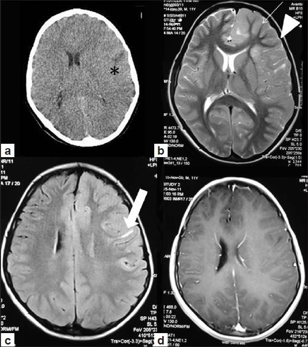
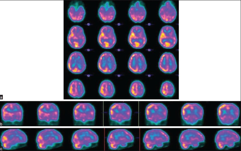
Hemimegalencephaly is a rare congenital neuronal migration disorder that can presents with the equally rare finding of hemihypoperfusion on brain perfusion single-photon emission computed tomography (SPECT). It is an extremely rare cause of intractable epilepsy. Technetium-99m ethyl cysteinate dimer (ECD) brain perfusion SPECT is useful in excluding other foci of hypoperfusion in the contralateral since hemispherectomy has been suggested to be the treatment of choice. Furthermore, hemimegalencephaly may present with hyper as well as hypoperfusion on ECD SPECT. We present the case of an 11-year-old male child with intractable seizures who showed hemihypoperfusion in the hemimegalecephalic hemisphere.
Keywords: 99m technetium-ethyl cysteinate dimer brain single-photon emission computed tomography; hemihypoperfusion; hemimegalencephaly.
Conflict of interest statement
Figures
Similar articles
-
Localization of cerebral hypoperfusion in dogs with refractory and non-refractory epilepsy using [99mTc] ethyl cysteinate dimer and single photon emission computed tomography.J Vet Med Sci. 2020 May 15;82(5):553-558. doi: 10.1292/jvms.19-0372. Epub 2020 Mar 19. J Vet Med Sci. 2020. PMID: 32188799 Free PMC article.
-
Regional brain perfusion in epileptic dogs evaluated by technetium-99m-ethyl cysteinate dimer SPECT.Vet Radiol Ultrasound. 2009 Nov-Dec;50(6):655-9. doi: 10.1111/j.1740-8261.2009.01600.x. Vet Radiol Ultrasound. 2009. PMID: 19999353
-
Abnormal regional cerebral blood flow found by technetium-99m ethyl cysteinate dimer brain single photon emission computed tomography in systemic lupus erythematosus patients with normal brain MRI findings.Clin Rheumatol. 2002 Nov;21(6):516-9. doi: 10.1007/s100670200126. Clin Rheumatol. 2002. PMID: 12447638
-
Neuronuclear assessment of patients with epilepsy.Semin Nucl Med. 2008 Jul;38(4):227-39. doi: 10.1053/j.semnuclmed.2008.02.004. Semin Nucl Med. 2008. PMID: 18514079 Review.
-
[A case of 55-year-old man with first-ever generalized seizure diagnosed with Sturge-Weber syndrome type III by characteristic MRI findings].Rinsho Shinkeigaku. 2017 May 27;57(5):214-219. doi: 10.5692/clinicalneurol.cn-001006. Epub 2017 Apr 27. Rinsho Shinkeigaku. 2017. PMID: 28450688 Review. Japanese.
References
-
- Bar-Sever Z, Connolly LP, Barnes PD, Treves ST. Brain SPECT evaluation of cerebral perfusion in hemimegalencephaly. Clin Nucl Med. 1997;22:250–2. - PubMed
-
- Tanaka Y, Hamano S, Nara T, Nakanishi Y. A case of hemimegalencephaly: Ictal EEG and SPECT. No To Hattatsu. 1994;26:528. - PubMed
-
- Uematsu M, Haginoya K, Togashi N, Hino-Fukuyo N, Nakayama T, Kikuchi A, et al. Unique discrepancy between cerebral blood flow and glucose metabolism in hemimegalencephaly. Epilepsy Res. 2010;92:201–8. - PubMed
-
- Tutuncuoglu S, Tekgul H, Duman Y, Kayalioglu M. 99m Tc HMPAO-SPECT in patients with neuronal migration anomalies. Turkish Journal of Nuclear Medicine. 1996;5:165–9.
-
- Podreka I, Suess E, Goldenberg G, Steiner M, Brücke T, Müller C, et al. Initial experience with technetium-99m HM-PAO brain SPECT. J Nucl Med. 1987;28:1657–66. - PubMed
Publication types
LinkOut - more resources
Full Text Sources
Other Literature Sources
