Cone beam computed tomography in oral implants
- PMID: 24163545
- PMCID: PMC3800380
- DOI: 10.4103/0975-5950.117811
Cone beam computed tomography in oral implants
Abstract
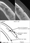
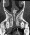
Cone beam computed tomography (CBCT) scanners for the oral and maxillofacial region were pioneered in the late 1990s independently by Arai et al. in Japan and Mozzo et al. CBCT has a lower dose of radiation, minimal metal artifacts, reduced costs, easier accessibility, and easier handling than multislice computed tomography (MSCT); however, the latter is still considered a better choice for the analysis of bone density using a Hounsfield unit (HU) scale. Oral implants require localized area of oral and maxillofacial area for radiation exposure; so, CBCT is an ideal choice. CBCT scans help in the planning of oral implants; they enable measurement of the distance between the alveolar crest and mandibular canal to avoid impingement of inferior alveolar nerve, avoid perforation of the mandibular posterior lingual undercut, and assess the density and quality of bone, and help in planning of the oral implant in the maxilla with special attention to the nasopalatine canal and maxillary sinus. Hence, CBCT reduces the overall exposure to radiation.
Keywords: Cone beam computed tomography; Hounsfield units; multislice computed tomography.
Conflict of interest statement
Figures


References
-
- De Vos W, Casselman J, Swennen GR. Cone-beam computerized tomography (CBCT) imaging of the oral and maxillofacial region: A systematic review of the literature. Int J Oral Maxillofac Surg. 2009;38:609–25. - PubMed
-
- Arai Y, Tammisalo E, Iwai K, Hashimoto K, Shinoda K. Development of a compact computed tomographic apparatus for dental use. Dentomaxillofac Radiol. 1999;28:245–8. - PubMed
-
- Suomalainen A, Vehmas T, Kortesniemi M, Robinson S, Peltola J. Accuracy of linear measurements using dental cone beam and conventional multislice computed tomography. Dentomaxillofac Radiol. 2008;37:10–7. - PubMed
-
- Tyndall DA, Brooks SL. Selection criteria for dental implant site imaging: A position paper of the American academy of oral and maxillofacial radiology. Oral Surg Oral Med Oral Pathol Oral Radiol Endod. 2000;9:630–7. - PubMed
-
- United Kingdom: ICRP Publication 60. Ann ICRP; 1991. International Commission on Radiological Protection. Recommendations of the International Commission on Radiological Protection; p. 21.
Publication types
LinkOut - more resources
Full Text Sources
Other Literature Sources

