

Incidental finding of isolated colonic neurofibroma
- PMID: 24163647
- PMCID: PMC3806709
- DOI: 10.1159/000355163
Incidental finding of isolated colonic neurofibroma
Abstract
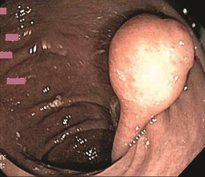
Neurofibromatosis is a genetic disorder manifested by characteristic cutaneous lesions called neurofibromas. There are two distinct neurocutaneous syndromes named neurofibromatosis type 1 (also called von Recklinghausen disease or NF1) and neurofibromatosis type 2 (NF2). NF1 is by far the most common presentation and is caused by an autosomal dominant mutation in the NF1 gene mapped to chromosome 17q11.2. The literature shows that gastrointestinal involvement is noted in systemic neurofibromatosis in up to 25% of patients, but isolated intestinal neurofibromatosis is a very rare manifestation. We herein present the case of a 70-year-old woman who was diagnosed with an isolated colonic neurofibroma without any systemic signs of neurofibromatosis; only a few case reports of this condition have been published to date.
Keywords: Colonic polyps; Intestinal; Neurofibroma; Neurofibromatosis.
Figures


References
-
- Korf BR. Neurofibromatosis. Handb Clin Neurol. 2013;111:333–340. - PubMed
-
- Gibson JA, Hornick JL. Mucosal Schwann cell ‘hamartoma’: clinicopathologic study of 26 neural colorectal polyps distinct from neurofibromas and mucosal neuromas. Am J Surg Pathol. 2009;33:781–787. - PubMed
-
- Hochberg FH, Dasilva AB, Galdabini J, Richardson EP., Jr Gastrointestinal involvement in von Recklinghausen's neurofibromatosis. Neurology. 1974;24:1144–1151. - PubMed
-
- Kim HR, Kim YJ. Neurofibromatosis of the colon and rectum combined with other manifestations of von Recklinghausen's disease: case report. Dis Colon Rectum. 1998;41:1187–1192. - PubMed
-
- Fuller CE, Williams GT. Gastrointestinal manifestations of type 1 neurofibromatosis. Histopathology. 1991;19:1–11. - PubMed
Publication types
LinkOut - more resources
Full Text Sources
Other Literature Sources
Research Materials
Miscellaneous
