

Age-related changes in trabecular meshwork imaging
- PMID: 24163814
- PMCID: PMC3791583
- DOI: 10.1155/2013/295204
Age-related changes in trabecular meshwork imaging
Abstract
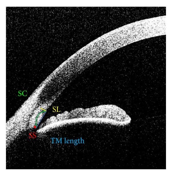
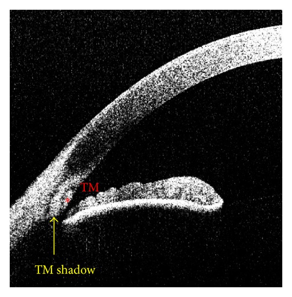
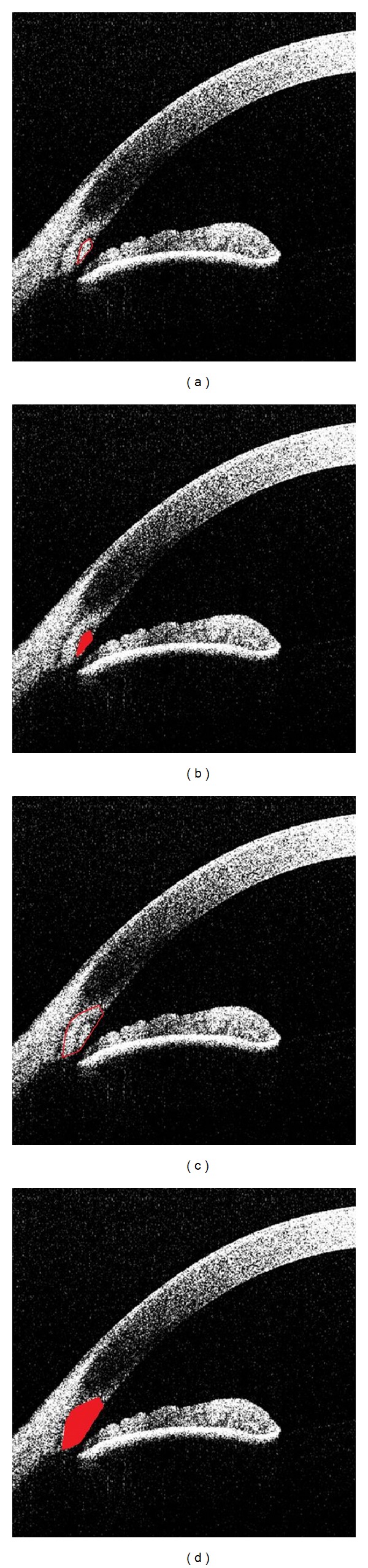
Purpose: To evaluate the normal aging effects on trabecular meshwork (TM) parameters using Fourier domain anterior segment optical coherence tomography (ASOCT) images.
Patients and methods: One eye from 45 participants with open angles was imaged. Two independent readers measured TM area, TM length, and area and length of the TM interface shadow from 3 age groups (18-40, 41-60, and 61-80). Measurements were compared using stepwise regression analysis.
Results: The average TM parameters were 0.0487 (± 0.0092) mm(2) for TM area, 0.5502 (± 0.1033) mm for TM length, 0.1623 (± 0.341) mm(2) for TM interface shadow area, and 0.7755 (± 0.1574) mm for TM interface shadow length. Interobserver reproducibility coefficients ranged from 0.45 (TM length) to 0.82 (TM area). TM area and length were not correlated with age. While the TM interface shadow length did not correlate with age, the TM interface shadow area increased with age. Race, sex, intraocular pressure, and gonioscopy score were not correlated with any TM parameters.
Conclusion: Although the TM measurements were not correlated with age, the TM interface shadow area increased with age. Further study is required to determine whether there is any relationship between the age-related ASOCT findings of the TM interface shadow area and physiologic function.
Figures
References
-
- McMenamin PG, Lee WR, Aitken DAN. Age-related changes in the human outflow apparatus. Ophthalmology. 1986;93(2):194–209. - PubMed
-
- Alvarado J, Murphy C, Polansky J, Juster R. Age-related changes in trabecular meshwork cellularity. Investigative Ophthalmology and Visual Science. 1981;21(5):714–727. - PubMed
-
- Garcia JPS, Jr., Rosen RB. Anterior segment imaging: optical coherence tomography versus ultrasound biomicroscopy. Ophthalmic Surgery Lasers and Imaging. 2008;39(6):476–484. - PubMed
-
- Nolan W. Anterior segment imaging: ultrasound biomicroscopy and anterior segment optical coherence tomography. Current Opinion in Ophthalmology. 2008;19(2):115–121. - PubMed
Publication types
MeSH terms
Grants and funding
LinkOut - more resources
Full Text Sources
Other Literature Sources
Medical
