. 2012 Nov 1;1(2):10.4172/2324-9110.1000102.
doi: 10.4172/2324-9110.1000102.

Expression of Voltage-Gated Sodium Channel Nav1.8 in Human Prostate Cancer is Associated with High Histological Grade

Affiliations

Expression of Voltage-Gated Sodium Channel Nav1.8 in Human Prostate Cancer is Associated with High Histological Grade

Simeng Suy et al. J Clin Exp Oncol. .

Abstract

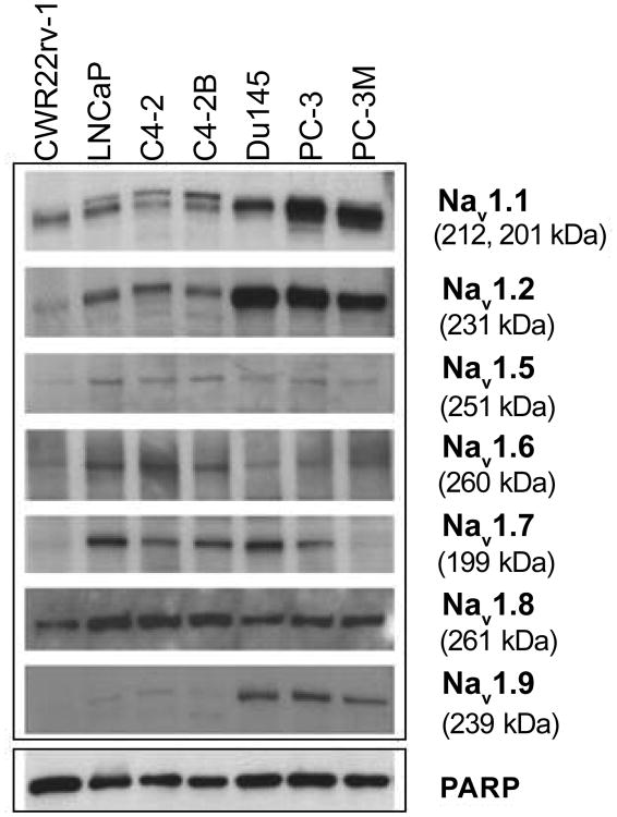
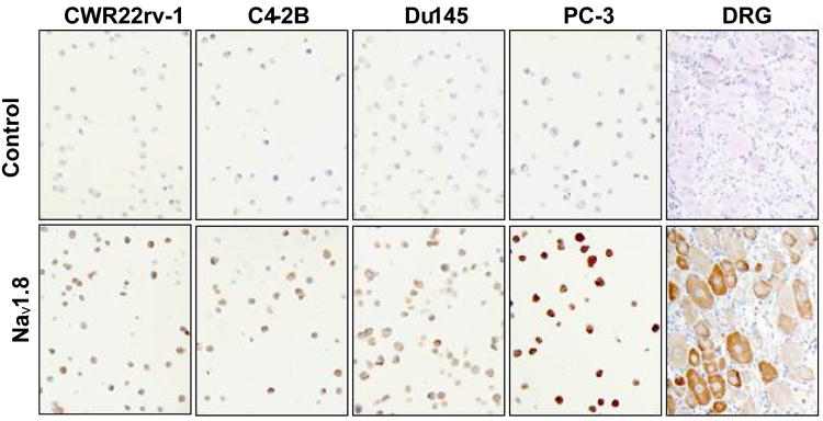
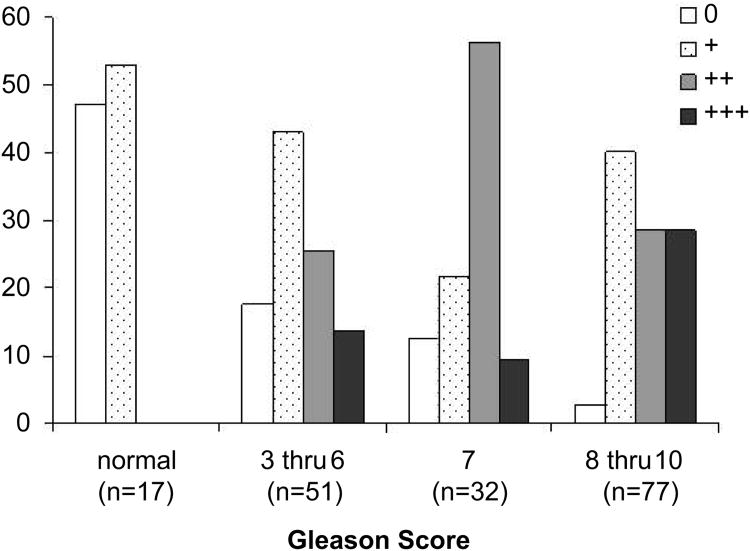
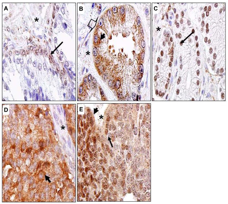
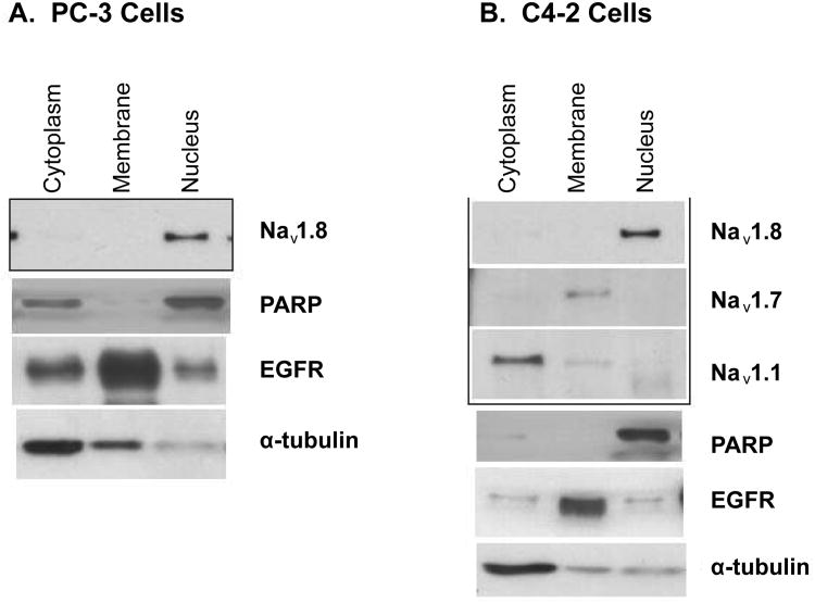
Voltage-gated sodium (Nav) channels are required for impulse conductance in excitable tissues. Navs have been linked to human cancers, including prostate. The expression and distribution of Nav isoforms (Nav1.1-Nav1.9) in human prostate cancer are not well established. Here, we evaluated the expression of these isoforms and investigated the expression of Nav1.8 in human prostate cancer tissues. Nav1.8 was highly expressed in all examined cells. Expression of Nav1.1, Nav1.2, and Nav1.9 were high in DU-145, PC-3 and PC-3M cells compared to LNCaP (hormone-dependent), C4-2, C4-2B, and CWR22Rv-1 cells. Nav1.5 and Nav1.6 were expressed in all cells examined. Nav1.7 expression was absent in PC-3M and CWR22Rv-1, but expressed in the other cells examined. Immunohistochemistry revealed intensive Nav1.8 staining correlated with more advanced pathologic stage of disease. Increased intensity of nuclear Nav1.8 correlated with increased Gleason grade. Our results revealed that Nav1.8 is universally expressed in human prostate cancer cells. Nav1.8 expression statistically correlated with pathologic stage (P=0.04) and Gleason score (P=0.01) of human prostate tissue specimens. The aberrant nuclear localization of Nav1.8 with advanced prostate cancer tissues warrant further investigation into use of Nav1.8 as a potential biomarker to differentiate between early and advanced disease.

Keywords: Gleason score; Prostate biomarker; Prostate cancer; Voltage-gated sodium channel.


Conflict of interest statement

Conflict of Interest: The authors do not have conflict of interest for this study.

Figures

Figure 1
Figure 1. Expression of Nav isoforms in human prostate cancer cells
Western blot analyses of extracts from human prostate cancer cell lines. Cell lysates were analyzed by immunoblotting with anti-Nav1.1, Nav1.2, Nav1.5, Nav1.6, Nav1.7, Nav1.8, and Nav1.9. Blots were probed with anti-PARP-1 antibody to normalize for protein loading.
Figure 2
Figure 2. Nav1.8 immunohistochemical analysis of human prostate cancer cell lines
DRG from rat tissue was used as a positive control for Nav1.8. Negative control (top panels) represents neutralization of Nav1.8 antibody with Nav1.8 peptide.
Figure 3
Figure 3. Expression of Nav 1.8 in human prostate cancer tissues
Human prostate tissue specimens consist of normal (n=17) and malignant (n=160) were analyzed by immunohistochemical staining using a polyclonal anti- Nav 1.8 antibody. Representative samples are shown. A. Normal human prostate specimen with very low Nav1.8 staining. B. Prostatic adenocarcinoma specimen GS 4 with very low Nav1.8 staining. Prostatic adenocarcinoma specimens with moderate Nav1.8 staining, GS 6 (C) and GS 7(D). Prostatic adenocarcinoma specimens with strong Nav1.8 staining, GS 8 (E) and GS10 (F). Images were taken at 40× (Olympus BX61 Camera/DP70 inverted microscope) using DP Controller Software. Arrows indicate prostate epithelium. Asterisks indicate stroma. GS, Gleason score.
Figure 4
Figure 4. Relationship between Nav 1.8 staining intensity and Gleason score
Human prostate tissue specimens consist of normal (n=17) and malignant (n=160) were analyzed by immunohistochemical staining using a polyclonal anti- Nav 1.8 antibody. Prostate tissue specimens were divided into four groups (normal, GS 3-6, GS 7, and GS 8-10) and individually scored for Nav 1.8 staining: 0 = no detectable, 1+ = low Nav 1.8 staining, 2+ = moderate Nav 1.8 staining, 3+ = strong Nav 1.8 staining.
Figure 5
Figure 5. Localization of Nav 1.8 in human prostate cancer tissues
Human prostate tissue specimens consist of normal (n=17) and malignant (n=160) were analyzed by immunohistochemical staining using a polyclonal anti- Nav 1.8 antibody. Representative samples are shown. A. Normal human prostate specimen, nuclei of prostatic acinar basal cells with very low Nav1.8 staining. B. Prostatic adenocarcinoma specimen GS 6 with moderate Nav1.8 staining in the plasma membrane (arrow head) and cytosol (bold thick arrow). C. Nuclear staining of a case of GS 7. D. Cytosolic staining of a case of GS10. E. A case of GS10 with mixed cytosolic and nuclear staining. Images were taken at 100× (Olympus BX61 Camera/DP70 inverted microscope) using DP Controller Software. Asterick represents stroma. Long thin arrows denote nuclei.
Figure 6
Figure 6. Nav 1.8 is localized to the nucleus
Human prostate cancer cell lines were fractionated into plasma membrane, cytosolic and nuclear enriched fractions. Proteins were separated on a 4-12% SDS-PAGE gradient gel. A. Subcellular fractions of PC-3 cells immunoblotted with Nav 1.8. B. Subcellular fractions of C4-2 cells immunoblotting with Nav 1.1 and 1.7 and 1.8. Purity of the enriched fractions were shown using anti-α-tubulin for the cytoplasm, EGFR for the plasma membrane, and PARP for the nucleus.

References

    1. Jemal A, Siegel R, Ward E, Hao Y, Xu J, et al. Cancer statistics, 2008. CA Cancer J Clin. 2008;58:71–96. - PubMed
    1. Anderson JD, Hansen TP, Lenkowski PW, Walls AM, Choudhury IM, et al. Voltage-gated sodium channel blockers as cytostatic inhibitors of the androgen-independent prostate cancer cell line PC-3. Mol Cancer Ther. 2003;2:1149–1154. - PubMed
    1. Bennett ES, Smith BA, Harper JM. Voltage-gated Na+ channels confer invasive properties on human prostate cancer cells. Pflugers Arch. 2004;447:908–914. - PubMed
    1. Smith P, Rhodes NP, Shortland AP, Fraser SP, Djamgoz MB, et al. Sodium channel protein expression enhances the invasiveness of rat and human prostate cancer cells. FEBS Lett. 1998;423:19–24. - PubMed
    1. Yu FH, Yarov-Yarovoy V, Gutman GA, Catterall WA. Overview of molecular relationships in the voltage-gated ion channel superfamily. Pharmacol Rev. 2005;57:387–395. - PubMed

LinkOut - more resources