. 2014 Feb;48(2):200-5.
doi: 10.3109/10715762.2013.859386. Epub 2013 Nov 19.

Hemin uptake and release by neurons and glia

Affiliations

Hemin uptake and release by neurons and glia

J Chen-Roetling et al. Free Radic Res. 2014 Feb.

Abstract

Hemin accumulates in intracerebral hematomas and may contribute to cell injury in adjacent tissue. Despite its relevance to hemorrhagic CNS insults, very little is known about hemin trafficking by neural cells. In the present study, hemin uptake and release were quantified in primary murine cortical cultures, and the effect of the hemin-binding compound deferoxamine (DFO) was assessed. Net uptake of (55)Fe-hemin was similar in mixed neuron-glia, neuron, and glia cultures, but was 2.6-3.6-fold greater in microglia cultures. After washout, 40-60% of the isotope signal was released by mixed neuron-glia cultures into albumin-containing medium within 24 h. Inhibiting hemin breakdown with tin protoporphyrin IX (SnPPIX) had minimal effect, while release of the fluorescent hemin analog zinc mesoporphyrin was quantitatively similar to that of (55)Fe-hemin. Isotope was released most rapidly by neurons (52.2 ± 7.2% at 2 h), compared with glia (15.6 ± 1.3%) and microglia (17.6 ± 0.54%). DFO did not alter (55)Fe-hemin uptake, but significantly increased its release. Mixed cultures treated with 10 μM hemin for 24 h sustained widespread neuronal loss that was attenuated by DFO. Concomitant treatment with SnPPIX had no effect on either enhancement of isotope release by DFO or neuroprotection. These results suggest that in the presence of a physiologic albumin concentration, hemin uptake by neural cells is followed by considerable extracellular release. Enhancement of this release by DFO may contribute to its protective effect against hemin toxicity.


Conflict of interest statement

Declaration of Interest

This study was supported by a grant from the National Institutes of Health (NS079500) to R.F.R. The authors have no financial or consulting interests which could influence this work.

Figures

Figure 1
Figure 1
Time course of hemin release from neuron-glia cultures, and effect of HO inhibition. A) Percentage 55Fe signal in cell lysate and culture medium at indicated time points after 55Fe-hemin loading; B) Effect of indicated concentrations (μM) of the HO inhibitor tin protoporphyrin IX (Sn) on percentage isotope release into the culture medium at 4 and 24 hours after 55Fe-hemin loading; C) Percentage of zinc mesoporphyrin fluorescence signal in cell lysate and culture medium at indicated time points after loading. All values represent mean ± S.E.M. (*P < 0.05 vs. mean value in corresponding control cultures (CTRL) lacking tin protoporphyrin IX, Bonferroni multiple comparisons test, n = 6 per condition).
Figure 2
Figure 2
Comparison of 55Fe-hemin uptake and release in neurons, glia, and microglia. A) 55Fe-hemin signal (± S.E.M.) in cell lysates of indicated culture types after 2 hour treatment with 5 μM 55Fe-hemin. B–D) % 55Fe signal in cell lysate and culture medium at indicated time points after 55Fe-hemin loading. ***P < 0.001 vs. mean value in microglia cultures, n = 8–10/condition).
Figure 3
Figure 3
Deferoxamine (DFO) increases release of cell hemin. A) Uptake: bars represent mean cell 55Fe signal in mixed cortical cultures after two hour incubation with 5 μM 55Fe-hemin alone (CTRL) or with 100 μM DFO or 2,2′-dipyridyl (DP); Release: mean medium 55Fe signal at 24 hours after 55Fe-hemin washout followed by incubation in medium containing 0.67 mg/ml bovine serum albumin alone (CTRL) or with 100 μM DFO or DP. B) Medium 55Fe signal at 24 hours after 55Fe-hemin washout followed by incubation in BSA-free medium alone (CTRL) or with indicated concentrations of DFO. C) As in A, testing the effect of concomitant treatment with the HO inhibitor tin protoporphyrin IX (Sn, 60 μM) on enhancement of 55Fe-hemin release by DFO, in medium containing BSA. **P < 0.01, ***P < 0.001 vs. mean value in corresponding CTRL condition.
Figure 4
Figure 4
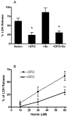
Deferoxamine attenuates hemin neurotoxicity. A) Bars represent mean LDH release (± S.E.M., n = 10–12/condition) in cultures treated for 24 hours with 10 μM hemin or with 10 μM hemin plus 100 μM DFO, 60 μM tin protoporphyrin IX (Sn) or both DFO and tin protoporphyrin IX. B) Cultures (n= 6/condition) were treated with indicated concentrations of hemin for 2 hours without DFO; after hemin washout, cultures were placed into medium containing or lacking 100 μM DFO. *P < 0.05, **P < 0.01 vs. corresponding condition treated with hemin without DFO. LDH values are normalized to those in sister cultures treated with NMDA for 24 hours (=100), which kills all neurons in these cultures. The LDH signal in controls exposed to washes only was subtracted from all values to calculate the signal due to hemin toxicity.

References

    1. Letarte PB, Lieberman K, Nagatani K, Haworth RA, Odell GB, Duff TA. Hemin: levels in experimental subarachnoid hematoma and effects on dissociated vascular smooth muscle cells. J Neurosurg. 1993;79(2):252–255. - PubMed
    1. Khan AA, Quigley JG. Heme and FLVCR-related transporter families SLC48 and SLC49. Mol Aspects Med. 2013;34(2–3):669–82. - PMC - PubMed
    1. Yuan X, Fleming MD, Hamza I. Heme transport and erythropoiesis. Curr Opin Chem Biol. 2013;17(2):204–11. - PMC - PubMed
    1. Robinson SR, Dang TN, Dringen R, Bishop GM. Hemin toxicity: a preventable source of brain damage following hemorrhagic stroke. Redox Rep. 2009;14(6):228–35. - PubMed
    1. Gutteridge JMC, Smith A. Antioxidant protection by haemopexin of haem-stimulated lipid peroxidation. Biochem J. 1988;256:861–865. - PMC - PubMed

Publication types

MeSH terms

LinkOut - more resources