Review
. 2014 Apr;129(2):221-34.
doi: 10.1111/jnc.12506. Epub 2013 Nov 17.

Actin dynamics in growth cone motility and navigation

Affiliations
Review

Actin dynamics in growth cone motility and navigation

Timothy M Gomez et al. J Neurochem. 2014 Apr.

Abstract

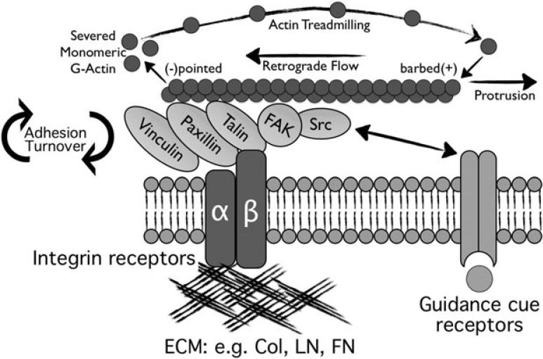
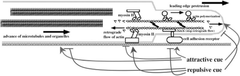
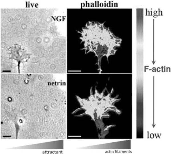
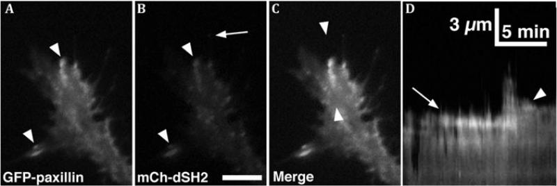
Motile growth cones lead growing axons through developing tissues to synaptic targets. These behaviors depend on the organization and dynamics of actin filaments that fill the growth cone leading margin [peripheral (P-) domain]. Actin filament organization in growth cones is regulated by actin-binding proteins that control all aspects of filament assembly, turnover, interactions with other filaments and cytoplasmic components, and participation in producing mechanical forces. Actin filament polymerization drives protrusion of sensory filopodia and lamellipodia, and actin filament connections to the plasma membrane link the filament network to adhesive contacts of filopodia and lamellipodia with other surfaces. These contacts stabilize protrusions and transduce mechanical forces generated by actomyosin activity into traction that pulls an elongating axon along the path toward its target. Adhesive ligands and extrinsic guidance cues bind growth cone receptors and trigger signaling activities involving Rho GTPases, kinases, phosphatases, cyclic nucleotides, and [Ca++] fluxes. These signals regulate actin-binding proteins to locally modulate actin polymerization, interactions, and force transduction to steer the growth cone leading margin toward the sources of attractive cues and away from repellent guidance cues.

Keywords: actin; actin-binding proteins; axon guidance; growth cone.


Figures

Figure 1
Figure 1
Left panel is a confocal microscope image of a growth cone of a chick dorsal root ganglion neuron showing F-actin labeled with fluorescent phalloidin. Actin filaments fill the filopodia and small veil-like lamellipodia of the growth cone leading margin. The right panel is an electron micrograph of the branched network and bundles of actin filaments in the growth cone periphery in an area similar to that marked with an asterisk in the left panel. Right image is courtesy of Dr. Lorene Lanier, University of Minnesota.
Figure 2
Figure 2
A,B. GFP-Utrophin (GFP-Utr; F-actin probe) and TMR-Kabiramide C (TMR-KabC; F-actin barbed end binding) fluorescent images of a Xenopus growth cone on PDL-laminin collected with a TIRF microscope. Note bright TMR-KabC fluorescence at the periphery (arrow in B), indicating a high concentration of F-actin barbed ends. C. Merged image of the growth cone in (A,B) shows strong co-localization of GFP-Utr and TMR-KabC in central domain, but primarily TMR-KabC in the peripheral domain. Weak labeling of peripheral actin with GFP-Utr is consistent with the slow association rate of GFP-Utr onto recently polymerized F-actin. D. Single line kymograph constructed from region indicated by the white line in (C). The slope of the diagonal bands of GFP-Utr and TMR-KabC fluorescence (arrows) indicates a retrograde flow rate of ~ 4 μm/min. Scale bars, 5 μm or as indicated. With permission from Santiago-Medina et al. (2012) Dev. Neurobiol. 72, 585-599.
Figure 3
Figure 3
Immunofluorescent images of two dorsal root ganglion growth cones multiple-labeled to show (upper panels) F-actin barbed ends (Rh-actin), ADF/cofilin, and F-actin (phalloidin) or (lower panels) barbed ends (Rh-actin, phosphorylated ERM proteins (pERM) and F-actin (phalloidin) in the growth cone leading margin after global exposure to an attractive guidance cue. With permission from Marsick et al., (2012b) J. Neurosci. 32, 282-196.
Figure 4
Figure 4
Immunofluorescence images of chick dorsal root ganglion growth cones multiple-labeled to show (left panel) microtubules, F-actin and myosin II and (right panels) microtubules, F-actin and paxillin. Myosin II is the motor molecule that exerts tension on adhesive sites and growth cone structures, and paxillin marks adhesive contacts of filopodia and lamellipodia.
Figure 5
Figure 5
Key components of growth cone point contact adhesions. Integrin αβ heterodimeric receptors (dark blue lines) bind to extracellular matrix proteins, such as collagen, laminin and fibronectin. Integrin activation leads to the assembly of scaffolding proteins, such as talin, paxillin and vinculin with the cytoplasmic tail of integrins. In addition, kinases FAK and Src are activated, and they modulate the adhesions through phosphorylation of key residues that allow for assembly of additional proteins (not shown). Several proteins bind directly to actin filaments (red), which is believed to restrain retrograde flow and allow the force of actin polymerization to generate membrane protrusion. Guidance cue receptors (orange) can regulate adhesion-associated proteins through binding and activation of FAK and Src. Cross-talk through FAK/Src signaling modulates adhesion dynamics, as well as actin dynamics. With permission from Myers et al., (2011) Dev. Neurobiol. 71, 901-923.
Figure 6
Figure 6
Left panel is a whole-mount electron micrographs from the leading margin of a dorsal root ganglion growth cone. Right panel is a thin section from a similar region. These images show the relationship between microtubules and associated organelles that have advanced into the P-domain (arrows) and bundles of actin filaments (arrowheads), which might determine where the microtubules advance. Left panel with permission from Letourneau, P.C. (1979) Exp. Cell Res. 124, 127-138, right panel from Letourneau, P.C. (1983) J. Cell Biol. 97, 963-973.
Figure 7
Figure 7
A model of growth cone navigation. Actin polymerization pushes the leading margin of the growth cone forward. Forces generated by myosin II pull actin filaments backwards, where filaments are disassembled. When growth cone receptors make adhesive contacts, a ‘clutch’ links the adhesive contact to actin filaments, and the retrograde flow of actin filaments slows. This permits the advance of microtubules and organelles and promotes axonal elongation. Intracellular signals generated by attractive and repulsive axonal guidance cues interact with the mechanisms of actin polymerization, myosin II force generation, adhesive contacts, and microtubule advance to regulate the paths of growth cone migration.
Figure 8
Figure 8
Phase-contrast and psuedocolor images depicting the higher density of F-actin in the P-domain regions of growth cones that are closer to a micropipette releasing an attractive guidance cue. Upper panel shows a chick dorsal root ganglion growth cone exposed to nerve growth factor, and lower panel shows a chick retinal growth cone exposed to netrin. With permission from Marsick et al., (2010) Dev. Neurobiol. 70, 565-588.
Figure 9
Figure 9
A, B. Total Internal Reflection Fluorescence (TIRF) microscopy images of paxillin-GFP and mCherry dual-Src homology 2 domain (mCh-dSH2) fluorescent images of a growth cone on PDL-laminin. Note that paxillin and phosphotyrosine (PY), as revealed with mChdSH2, colocalize at adhesion sites (arrowheads in A, B), whereas the tip of a growing filopodium has PY, without paxillin (arrow in B). C. Merged image of the growth cone in (A, B) shows co-localization at several peripheral adhesions. Note that mCh-dSH2 puncta within the central domain are mobile vesicles. D. Single line kymograph constructed from region between the arrowheads in C. Note a stable adhesion (arrow) that disassembles after a new protrusion extends forward, followed by the formation of a second adhesion (arrowhead), which stabilizes the receding protrusion. Scale bar, 10 μm in all images and as indicated in kymographs. With permission from Santiago-Medina et al. (2012) Dev. Neurobiol. 72, 585-599.

References

    1. Arakawa Y, Bito H, Furuyashiki T, Tsuji T, Takemoto-Kimura S, Kimura K, Nozaki K, Hashimoto N, Narumiya S. Control of axon elongation via an SDF-1alpha/Rho/mDia pathway in cultured cerebellar granule neurons. J. Cell Biol. 2003;161:381–391. - PMC - PubMed
    1. Augustine GJ, Santamaria F, Tanaka K. Local calcium signaling in neurons. Neuron. 2003;40:331–346. - PubMed
    1. Bard L, Boscher C, Lambert M, Mège RM, Choquet D, Thoumine O. A molecular clutch between the actin flow and N-cadherin adhesions drives growth cone migration. J. Neurosci. 2008;28:5879–5890. - PMC - PubMed
    1. Bate N, Gingras AR, Bachir A, Horwitz R, Ye F, Patel B, Goult BT, Critchley DR. Talin contains a C-terminal calpain2 cleavage site important in focal adhesion dynamics. PLoS One. 2012;7:e34461. - PMC - PubMed
    1. Bear JE, Svitkina TM, Krause M, Schafer DA, Loureiro JJ, Strasser GA, Maly IV, Chaga OY, Cooper JA, Borisy GG, Gertler FB. Antagonism between Ena/VASP proteins and actin filament capping regulates fibroblast motility. Cell. 2002;109:509–521. - PubMed

Publication types

LinkOut - more resources