Review
. 2014 Mar;38(3):595-603.
doi: 10.1111/acer.12283. Epub 2013 Oct 24.

Structural models of ligand-gated ion channels: sites of action for anesthetics and ethanol

Affiliations
Review

Structural models of ligand-gated ion channels: sites of action for anesthetics and ethanol

Richard W Olsen et al. Alcohol Clin Exp Res. 2014 Mar.

Abstract

The molecular mechanism(s) of action of anesthetic, and especially, intoxicating doses of alcohol (ethanol [EtOH]) have been of interest even before the advent of the Research Society on Alcoholism. Recent physiological, genetic, and biochemical studies have pin-pointed molecular targets for anesthetics and EtOH in the brain as ligand-gated ion channel (LGIC) membrane proteins, especially the pentameric (5 subunit) Cys-loop superfamily of neurotransmitter receptors including nicotinic acetylcholine (nAChRs), GABAA (GABAA Rs), and glycine receptors (GlyRs). The ability to demonstrate molecular and structural elements of these proteins critical for the behavioral effects of these drugs on animals and humans provides convincing evidence for their role in the drugs' actions. Amino acid residues necessary for pharmacologically relevant allosteric modulation of LGIC function by anesthetics and EtOH have been identified in these channel proteins. Site-directed mutagenesis revealed potential allosteric modulatory sites in both the trans-membrane domain (TMD) and extracellular domain (ECD). Potential sites of action and binding have been deduced from homology modeling of other LGICs with structures known from crystallography and cryo-electron microscopy studies. Direct information about ligand binding in the TMD has been obtained by photoaffinity labeling, especially in GABAA Rs. Recent structural information from crystallized procaryotic (ELIC and GLIC) and eukaryotic (GluCl) LGICs allows refinement of the structural models including evaluation of possible sites of EtOH action.

Keywords: ELIC; Ethanol Sites of Action; GABAA Receptors; GLIC; GluCl Pentameric Ion Channels; Loop 2.


Figures

Figure 1
Figure 1
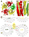
A homology model of GABAARα2β2γ2 was built by threading the primary sequence onto the template of the glutamate-gated chloride channel (GluCl, PDB ID 3RHW, Hibbs and Gouaux, 2011). (A) A view looking down the axis of the ion channel from the extracellular side; α2, β2, and γ2 subunits are rendered as solid ribbons and colored yellow, red, and green, respectively. Several residues mentioned in the text are rendered as space filling surfaces: beta M2 N265 and K274, in pink; M3 M286 and V290, in turquoise; alpha M1 M236, in turquoise; and Loop 2 P52-E59, with carbon, hydrogen, oxygen, and nitrogen in gray, white, red, and blue surfaces. (B) A view from the plane of the lipid bilayer towards the β3 – α1 subunit interface.
Figure 2
Figure 2
Three views of the α1β2γ2L GABAAR showing residues photolabeled by etomidate derivatives. A. View from the extracellular side looking down the ion channel (light blue circle). Residues photolabeled by etomidate derivatives in the transmembrane domain between β3– and α1– subunits have cyan carbons. For reference in the extracellular domain, the agonist site in the same interface is indicated by α1Tyr-205 (~50 Å above the etomidate site) and the benzodiazepine site between the α1– and γ2L subunits is indicated by α1His-202. B. A view from the lipid bilayer towards the β3 – α1 subunit interface. βM3 Met-286, βM3 Val-290 and αM1Met-236 are residues in the etomidate binding site (Li et al., 2006; Chiara et al., 2012). The orientation of residues is suggested by a homology model (Chiara et al., 2013) built on the GLIC crystal structure (Nury et al., 2011). Molecular graphics and analyses were performed with the UCSF Chimera package. Chimera is developed by the Resource for Biocomputing, Visualization, and Informatics at the University of California, San Francisco, with support from the National Institutes of Health (National Center for Research Resources grant 2P41RR001081, National Institute of General Medical Sciences grant 9P41GM103311). C. Helical wheel representation of the GABAAR β/α interface in the trans-membrane domain illustrating the proposed binding site for etomidate (modified from Li et al., 2009) with the residues in αM1 and βM3 photolabeled by [3H]azietomidate (circled residues in green) contributing to a common binding pocket at the β/α interface. Also included in pink is the position in βM2 (N265) that functions as a determinant of etomidate/azietomidate anesthetic potency in vivo(Rudolph & Antkowiak,2004), the residues in αM1 and βM3 identified as sensitivity determinants for direct activation by neurosteroids (boxed residues in yellow [Hosie et al., 2006]), and the positions in αM1 and βM3 that when mutated to Cys can form intersubunit cross-links (red and orange).
Figure 3
Figure 3. Sequence alignment of key alcohol site residues in GABA-R α and β subunits and in GlyR α1 subunits
(a) Sequence alignment of GABA-R “loop A” α subunits with the benzodiazepine-H/R/Q100 (in α6) binding site residues, colored; H (yellow); R (green) and the α6-R100Q alcohol polymorphism (red) (Hanchar et al., 2005). Since this is a highly conserved region all the other amino acid residues shown are identical. (b) Sequence alignment of “Loop2” and “Loop D” comprising two beta sheet structures (β1 and β2, highlighted in yellow) in the acetylcholine binding protein (AChBP, Brejc et al., 2001), verified in the structure of the invertebrate LGIC, GluCl (Hibbs & Gouaux, 2011). Line 1 shows AChBP (with the nicotine binding residue W53 and Q55 boxed) from which structural homology models have been derived (Ernst et al., 2003). Three critical γ “loop D” BZ-site residues in the γ2 subunit (F77, A79, T81) are boxed and colored green (Holden & Czajkowski, 2002). These three residues contribute to the classical BZ binding site at the α(1,2,3,5)+/γ2− subunit interface (abutting the yellow H residue in Loop A shown in (a)). The three β subunits are identical in this region except for residues 66 (boxed, S in β1, A in β2 and Y in β3 (red)). Also boxed in this alignment is residue δH68 that confers diazepam sensitivity to highly EtOH-sensitive α4β3δ receptors when changed to A, the residue present in γ2 at the homologous position (Meera et al.,2010). In addition, we show the GABAR α1 sequence with four residues boxed: α1-D57 is critical for the coupling of agonist binding to channel activation (Kash et al., 2003), and residues D62, F64, and R66 are critical residues for binding of the GABA antagonist Gabazine (Holden and Czajkowski, 2002); F64 is also critical for (affinity labeled by) muscimol binding (Olsen & Li, 2011). Finally, we show Loop 2 alcohol residues (box) from the GABA-R δ, γ2, and the GlyR α1 subunit [blue (Perkins et al., 2009; Crawford et al., 2007)]. These loop 2 residues are flanked not only by ligand binding site residues in loop D (as shown in Fig. 3b), but also by residue Glu-Cl R37 (in loop G) just upstream in GluCl which contributes to glutamate binding sites in the Glu-Cl-glutamate co-crystal structure (see Fig.3b in Hibbs and Gouaux, 2011). Therefore homologous residues could -- depending on the size of the side-chain as well as the size and exact positioning of the ligand in the ligand binding site -- contribute to the ligand binding at receptor subunit interfaces.

References

    1. Arevalo E, Chiara DC, Forman SA, Cohen JB, Miller KW. Gating-enhanced accessibility of hydrophobic sites within the transmembrane region of the nicotinic acetylcholine receptor’s {delta}-subunit. A time-resolved photolabeling study. J Biol Chem. 2005;280:13631–13640. - PubMed
    1. Bali M, Akabas MH. Defining the propofol binding site location on the GABAA receptor. Mol Pharmacol. 2004;65:68–76. - PubMed
    1. Bali M, Jansen M, Akabas MH. GABA-induced inter-subunit conformational movement in the GABAA receptor α1M1-β2M3 transmembrane subunit interface: experimental basis for homology modeling of an intravenous anesthetic binding site. J Neurosci. 2009;29:3083–3092. - PMC - PubMed
    1. Bertaccini EJ, Wallner Br, Trudell JR, Lindahl E. Modeling anesthetic binding sites within the glycine alpha one receptor based on prokaryotic ion channel templates: The problem with TM4. J Chem Inf Model. 2010;50:2248–2255. - PMC - PubMed
    1. Bissantz C, Kuhn B, Stahl M. A medicinal chemist’s guide to molecular interactions. J Med Chem. 2010;53:5061–5084. - PMC - PubMed

Publication types

MeSH terms

Substances

LinkOut - more resources