. 2014 Mar;38(3):694-703.
doi: 10.1111/acer.12286. Epub 2013 Oct 24.

A screen of zebrafish mutants identifies ethanol-sensitive genetic loci

Affiliations

A screen of zebrafish mutants identifies ethanol-sensitive genetic loci

Mary E Swartz et al. Alcohol Clin Exp Res. 2014 Mar.

Abstract

Background: Fetal alcohol spectrum disorders (FASD) are a highly variable set of phenotypes caused by fetal alcohol exposure. Numerous factors influence FASD phenotypes, including genetics. The zebrafish is a powerful vertebrate model system with which to identify these genetic factors. Many zebrafish mutants are housed at the Zebrafish International Resource Center (ZIRC). These mutants are readily accessible and an excellent source to screen for ethanol (EtOH)-sensitive developmental structural mutants.

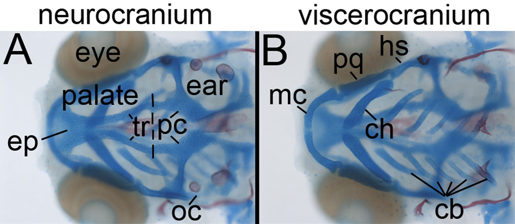
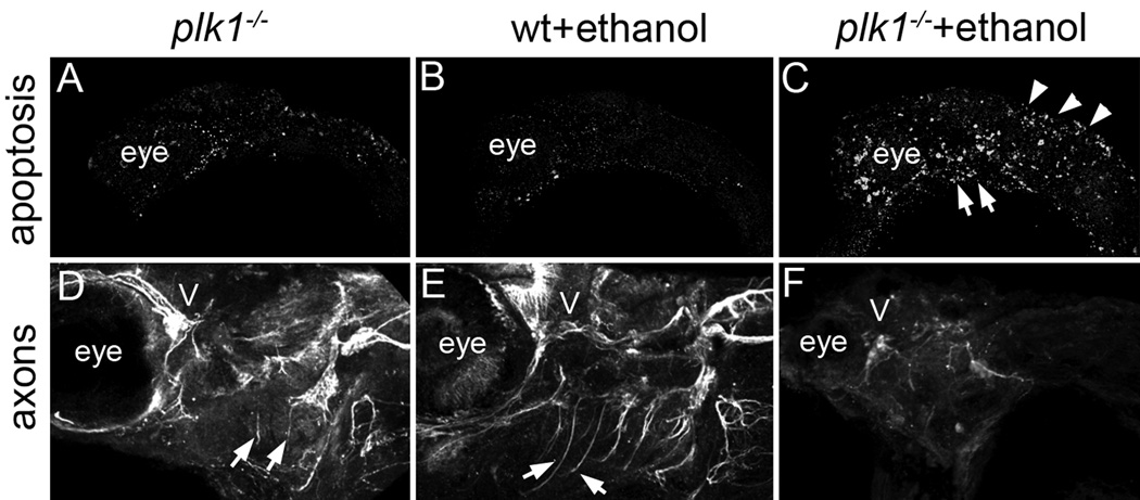
Methods: We screened mutants obtained from ZIRC for sensitivity to EtOH teratogenesis. Embryos were treated with 1% EtOH (41 mM tissue levels) from 6 hours postfertilization onward. Levels of apoptosis were evaluated at 24 hours postfertilization. At 4 days postfertilization, the craniofacial skeleton, peripheral axon projections, and sensory neurons of neuromasts were examined. Fish were genotyped to determine whether there were phenotype/genotype correlations.

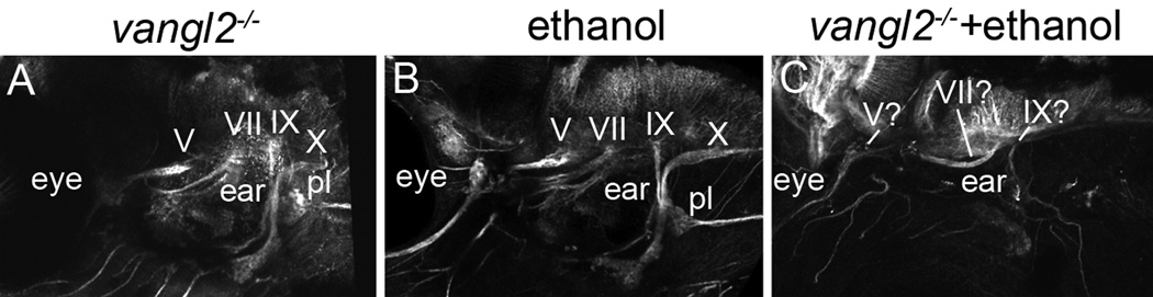
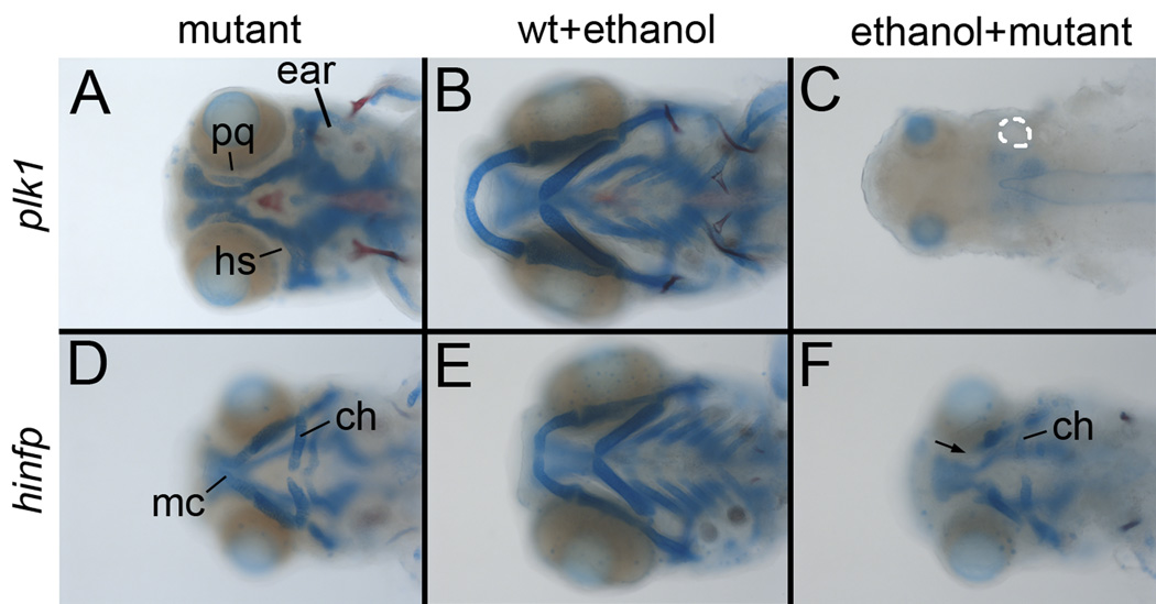
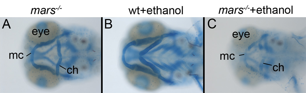
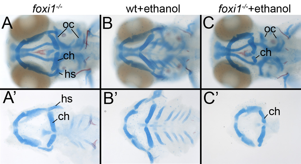
Results: Five of 20 loci interacted with EtOH. Notable among these was that vangl2, involved in convergent extension movements of the embryonic axis, interacted strongly with EtOH. Untreated vangl2 mutants had normal craniofacial morphology, while severe midfacial defects including synophthalmia and narrowing of the palatal skeleton were found in all EtOH-treated mutants and a low percentage of heterozygotes. The cell cycle gene, plk1, also interacted strongly with EtOH. Untreated mutants have slightly elevated levels of apoptosis and loss of ventral craniofacial elements. Exposure to EtOH results in extensive apoptosis along with loss of neural tissue and the entire craniofacial skeleton. Phenotypes of hinfp, mars, and foxi1 mutants were also exacerbated by EtOH.

Conclusions: Our results provide insight into the gene-EtOH interactions that may underlie EtOH teratogenesis. They support previous findings that EtOH disrupts elongation of the embryonic axis. Importantly, these results show that the zebrafish is an efficient model with which to test for gene-EtOH interactions. Understanding these interactions will be crucial to understanding of the FASD variation.

Keywords: Craniofacial; FASD; Genetic Screen; Zebrafish.


Figures

Fig. 1
Fig. 1
Morphology of the zebrafish craniofacial skeleton. (A) Dorsal and (B) ventral views of a 5 dpf zebrafish embryo staining with Alcian Blue/Alizarin Red. (A) The neurocranium can be divided into an anterior, crest-derived portion, and a posterior, presumably mesoderm-derived portion. The crest-derived neurocranium is the zebrafish palatal skeleton and can be further divided into the midline ethmoid plate (ep) and the bilateral trabeculae (tr). The trabeculae fuse to the parachordal cartilages (pc) of the posterior neurocranium. (B) The viscerocranium consists of cartilage elements that are reiterated along the anterior/posterior axis and are generated from pharyngeal arches. Most anteriorly, Meckel’s cartilage (mc) and the palatoquadrate (pq) form the lower and upper jaws, respectively. The ventral ceratohyal (ch) and dorsal hyosymplectic (hs) are the cartilages of the second pharyngeal arch. Pharyngeal arches 3–7 generate the five pairs of ceratobranchial cartilages (cb).
Fig. 2
Fig. 2
Ethanol interacts strongly with vangl2 in craniofacial development. (A–C) Wholemount and (A’–C’) flat mount images of neurocrania. Relative to (A) untreated vangl2 mutants and (B) ethanol-treated wild-type embryos, the eyes of (C) ethanol-treated vangl2 mutants are fused (asterisk). (A’–C’) The fusion of the eyes in ethanol-treated vangl2 mutants is associated with the narrowing of the ethmoid plate (ep) and failure of the trabeculae (tr) to attach to the posterior neurocranium. (D–F) Wholemount and (D’–F’) flatmount images of viscerocranial elements. (F,F’) In ethanol-treated mutants, most of the ceratobranchials are lost. While Meckel’s cartilage and the ceratohyal appear mispositioned in whole mount, flatmounted cartilage elements appear shaped appropriately (n=10/10 genotyped mutants).
Fig. 3
Fig. 3
Ethanol-exposure causes defects to cranial nerves in 5dpf vangl2 mutants. Cranial nerve projections are largely normal in (A) vangl2 mutants and (B) ethanol-treated wild-type embryos. (C) Ethanol-treatment causes severe reductions in cranial nerve projections (n=6/6 genotyped mutants). Cranial nerves are numbered. pl, posterior lateral line ganglion.
Fig. 4
Fig. 4
Interactions with genes regulating the cell cycle. (A–F) Whole mount images focused on the viscerocranium. (A,B) Compared to ethanol-treated wild-type embryos, untreated plk1 mutants have small eyes and loss of most viscerocranial cartilages, with only remnants of the palatoquadrate (pq) and hyosymplectic (hs remaining). While smaller, the neurocranium is present in mutants. (C) The overall size of the entire embryo is greatly reduced and the entire craniofacial skeleton is lost in ethanol-treated plk1 mutants (n=7/7 genotyped mutants). (D,E) Most ceratobranchials are lost in hinfp mutants and the remaining skeletal elements are reduced in size, relative to ethanol-treated wild-type embryos. (F) Ethanol treatment causes a further reduction in the size of the viscerocranium and loss of Meckel’s cartilage (arrow, n=6/6 genotyped mutants).
Fig. 5
Fig. 5
Elevated cell death and axon defects in ethanol-treated plk1 mutants. (A–C) Lateral views of 24 hpf embryos stained with anti-active Caspase antibodies. (A,B) Compared to ethanol-treated wild-type embryos there is an elevation in apoptosis in plk1 mutants. (C) In ethanol-treated plk1 mutants, the levels of apoptosis are dramatically increased and expand throughout the embryo (n=11/11 genotyped mutants). (D–F) Lateral views of 5 dpf embryos stained with anti-acetylated beta-tubulin antibodies. (D,E) while reduced axon projections into the pharyngeal arches are present in untreated plk1 mutants (arrows). (F) Nearly all axon projections are absent in ethanol-treated plk1 mutants. Additionally, only cranial ganglion V is readily identifiable in the ethanol-treated mutants (n=4/4 genotyped mutants).
Fig. 6
Fig. 6
Ethanol interacts with mutants in the biosynthetic Mars enzyme. (A–C) Whole mount images, focused on the viscerocranium. (A,B) Relative to ethanol-treated wild-type embryos, untreated mars mutants have a loss of the ceratobranchials and slight reductions to cartilages derived from the first two pharyngeal arches. (C) Derivatives of the first two pharyngeal arches are greatly reduced in ethanol-treated mars embryos. The neurocranium is also further reduced in ethanol-treated mutants (n=5/5 genotyped mutants). mc; Meckel’s cartilage, ch; ceratohyal.
Fig. 7
Fig. 7
Ethanol treatment exacerbates craniofacial defects in foxi1 mutants. (A–C) Whole mount images show that relative to (A) untreated mutants and (B) ethanol-treated wild-type embryos, (C) ethanol-treated foxi1 mutants have reduction in the size of the otic capsule (oc), surrounding the ear. (A’–C’) Flat mount images show that while (A’) untreated mutants have reductions to the hyosymplectic (hs), ethanol-treated foxi1 mutants show a reduction in the size of the ceratohyal (ch) and a loss of the hyosymplectic (n=12/12 genotyped mutants).
Fig. 8
Fig. 8
Axon projection defects in ethanol-treated foxi1 mutants. (A–C) Lateral views of 5 dpf embryos stained with anti-acetylated beta tubulin antibodies. (A,B) Axon projections into the pharyngeal arches are similar in untreated foxi1 mutants and ethanol-treated wild-type embryos. (C) While axons are present, they do not extend appropriately into the pharyngeal arches of ethanol-treated foxi1 mutants (n=13/13 genotyped mutants).

Similar articles

Cited by

References

    1. Ahlgren SC, Thakur V, Bronner-Fraser M. Sonic hedgehog rescues cranial neural crest from cell death induced by ethanol exposure. Proc Natl Acad Sci U S A. 2002;99(16):10476–10481. - PMC - PubMed
    1. Bilotta J, Barnett JA, Hancock L, Saszik S. Ethanol exposure alters zebrafish development: a novel model of fetal alcohol syndrome. Neurotoxicol Teratol. 2004;26(6):737–743. - PubMed
    1. Blader P, Strahle U. Ethanol impairs migration of the prechordal plate in the zebrafish embryo. Dev Biol. 1998;201(2):185–201. - PubMed
    1. Cartwright MM, Smith SM. Stage-dependent effects of ethanol on cranial neural crest cell development: partial basis for the phenotypic variations observed in fetal alcohol syndrome. Alcohol Clin Exp Res. 1995;19(6):1454–1462. - PubMed
    1. Cavieres MF, Smith SM. Genetic and Developmental Modulation of Cardiac Deficits in Prenatal Alcohol Exposure. Alcoholism: Clinical and Experimental Research. 2000;24(1):102–109. - PubMed

MeSH terms

LinkOut - more resources