. 2014 Mar;90(3):214-23.
doi: 10.3109/09553002.2014.859761. Epub 2014 Feb 6.

Radiation exposure prior to traumatic brain injury induces responses that differ as a function of animal age

Affiliations
Free PMC article

Radiation exposure prior to traumatic brain injury induces responses that differ as a function of animal age

Antiño R Allen et al. Int J Radiat Biol. 2014 Mar.
Free PMC article

Abstract

Purpose: Uncontrolled radiation exposure due to radiological terrorism, industrial accidents or military circumstances is a continuing threat for the civilian population. Age plays a major role in the susceptibility to radiation; younger children are at higher risk of developing cognitive deterioration when compared to adults. Our objective was to determine if an exposure to radiation affected the vulnerability of the juvenile hippocampus to a subsequent moderate traumatic injury.

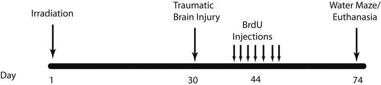
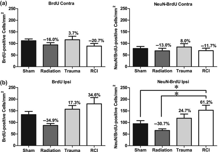
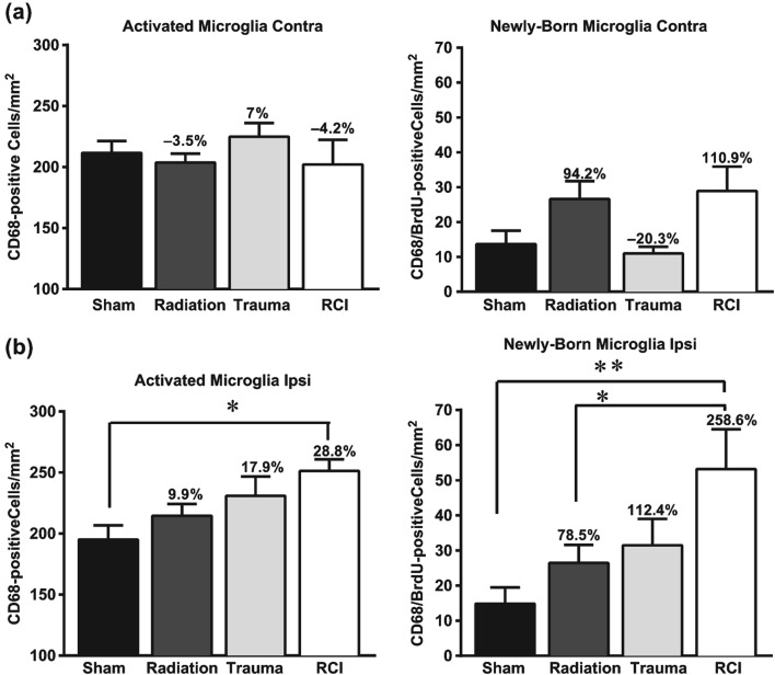
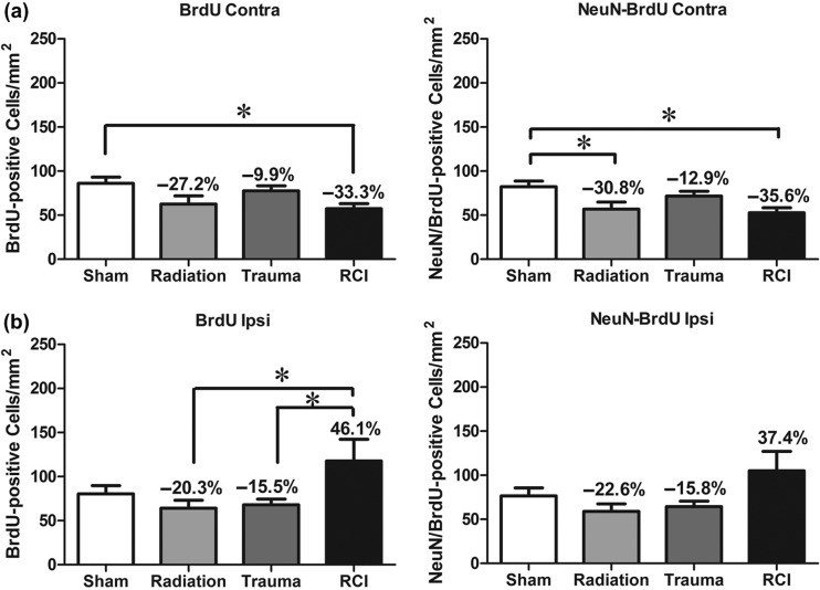
Materials and methods: Three-week-old (juvenile) and eight-week-old young adult C57BL/J6 male mice received whole body cesium-137 ((137)Cs) irradiation with 4 gray (Gy). One month later, unilateral traumatic brain injury was induced using a controlled cortical impact system. Two months post-irradiation, animals were tested for hippocampus-dependent cognitive performance in the Morris water-maze. After cognitive testing, animals were euthanized and their brains frozen for immunohistochemical assessment of activated microglia and neurogenesis in the hippocampal dentate gyrus.

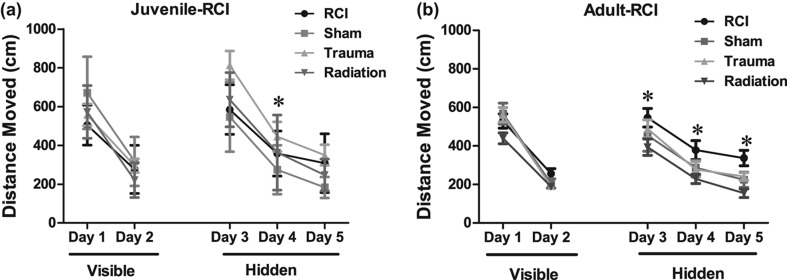
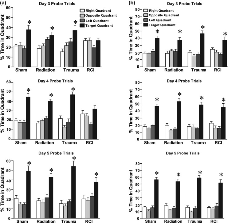
Results: All animals were able to learn the water maze task; however, treatment effects were seen when spatial memory retention was assessed. Animals that received irradiation as juveniles followed by a moderate traumatic brain injury one month later did not show spatial memory retention, i.e., were cognitively impaired. In contrast, all groups of animals that were treated as adults showed spatial memory retention in the probe trials.

Conclusion: Although the mechanisms involved are not clear, our results suggest that irradiation enhanced a young animal's vulnerability to develop cognitive injury following a subsequent traumatic injury.


Figures

Figure 1.
Figure 1.
Schematic diagram showing experimental design. Three-week-old and eight-week-old C57BL/6 mice received whole body irradiation with 4 Gy 137Cs. Four weeks later animals received either focal traumatic brain injury or sham injury. Two weeks later, animals were injected daily for 7 days with BrdU (100 mg/kg). Four weeks after BrdU injections, animals underwent Morris water maze testing for 5 days.
Figure 2.
Figure 2.
Cumulative distance to the target platform during visible and hidden training sessions. (a) During the visible platform training (day 1 and 2), all experimental groups swam similar distances to the platform. All groups showed daily improvements in their abilities to locate during the hidden platform training (day 3–5). However, trauma only (*p < 0.05; day 4, Holms) swam longer escape distances compared to sham mice. (b) All groups showed daily improvements in their abilities to locate during the hidden platform training (day 3–5). Radiation only (*p < 0.05; day 3–5, Holms) swam shorter escape distances compared to sham mice. Each datum point represents the mean of 8–10 mice; error bars are standard error of the mean (SEM).
Figure 3.
Figure 3.
Spatial memory retention in mice during the Morris water maze probe trial following the first day of hidden platform training. (a) Juvenile-RCI mice showed an impairment of hippocampal-dependent spatial memory during the day 3 and day 4 probe trials. (b) All of the adult mice, showed memory retention in the water maze by spending most of their time in target quadrant which contained hidden platform. For all four treatments, when time spent in the target quadrant was compared to all other quadrants there was a significant (p < 0.05) preference. Each bar represents the mean of 8–10 mice; error bars are standard error of the mean (SEM).
Figure 4.
Figure 4.
Total number of BrdU+ cells and BrdU+/NeuN+ cells per mm2 in the dentate subgranular zone of the Juvenile-RCI cohort. (a) In the contralateral hemisphere, there was a no significant group difference for BrdU+ cells (p = 0.242) or BrdU+/NeuN+ (p = 0.697). Percentage change compared with sham-treated animals. (b) In the ipsilateral hemisphere, there was a significant group difference for BrdU+ cells (p = 0.006) and BrdU+/NeuN+ (p = 0.004). RCI significantly increased the numbers of BrdU+ cells and BrdU+/NeuN+ compared to sham-treated and radiation only (p < 0.05). Percentage change compared with sham-treated animals. Each bar represents the mean of 9–10 mice; error bars are standard error of the mean (SEM).
Figure 5.
Figure 5.
Total number of activated microglia (CD68+) and newly born microglia (BrdU+/CD68+) per mm2 in the dentate subgranular zone of the Juvenile-RCI cohort. (a) In the contralateral hemisphere, there was a no significant group difference in the total numbers of activated microglia (p = 0.606). There was a significant group difference for newly born microglia (p = 0.027). There were minor but insignificant increases in total numbers of activated microglia after radiation only and RCI. (b) In the ipsilateral hemisphere, there was a significant group difference for CD68+ cells (p = 0.01) and BrdU+/CD68+ (p = 0.009). RCI significantly increased in the number of activated microglia compared to sham-treated animals. Percentage change compared with sham-treated animals. Each bar represents the mean of 8–10 mice; error bars are standard error of the mean (SEM).
Figure 6.
Figure 6.
Total number of BrdU+ cells and BrdU+/NeuN+ cells in the dentate subgranular zone of the Adult-RCI cohort. (a) In the contralateral hemisphere, there was a significant group difference for BrdU+ cells (p = 0.029) and BrdU+/NeuN+ (p = 0.015). Radiation significantly decreased the numbers BrdU+ cells and BrdU+/NeuN+ neurons in radiation only and RCI mice (p < 0.05). Percentage change compared with sham-treated animals. (b) In the ipsilateral hemisphere, there was a significant group difference for BrdU+ cells (p = 0.027). RCI significantly increased the numbers of BrdU+ cells compared to radiation and trauma only (p < 0.05). In terms of newly born neurons, there was a trend toward a group difference (p = 0.0508). Percentage change compared with sham-treated animals. Each bar represents the mean of 9–10 mice; error bars are standard error of the mean (SEM).

References

    1. Abayomi OK. Pathogenesis of irradiation-induced cognitive dysfunction. Acta Oncol. 1996;35:659–663. - PubMed
    1. American Academy of Pediatrics Committee on Environmental Health. Radiation disasters and children. Pediatrics. 2003;111((6 Pt 1)):1455–1466. - PubMed
    1. Benice TS, Rizk A, Kohama S, Pfankuch T, Raber J. Sex-differences in age-related cognitive decline in C57BL/6J mice associated with increased brain microtubule-associated protein 2 and synaptophysin immunoreactivity. Neuroscience. 2006;137:413–423. - PubMed
    1. Biebl M, Cooper CM, Winkler J, Kuhn HG. Analysis of neurogenesis and programmed cell death reveals a self- renewing capacity in the adult rat brain. Neurosci Lett. 2000;291:17–20. - PubMed
    1. Brody DL, MacDonald C, Kessens CC, Yuede C, Parsadanian M, Spinner M, Kim E, Schwetye KE, Holtzman DM, Bayly PV. Electromagnetic controlled cortical impact device for precise, graded experimental traumatic brain injury. J Neurotrauma. 2007;24:657–673. - PMC - PubMed

Publication types

LinkOut - more resources