. 2014 Jan;34(1):43-56.
doi: 10.1128/MCB.00999-13. Epub 2013 Oct 28.

Crumbs3 is essential for proper epithelial development and viability

Affiliations

Crumbs3 is essential for proper epithelial development and viability

Eileen L Whiteman et al. Mol Cell Biol. 2014 Jan.

Abstract

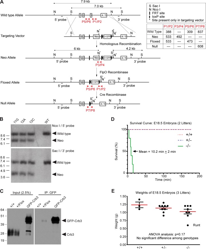
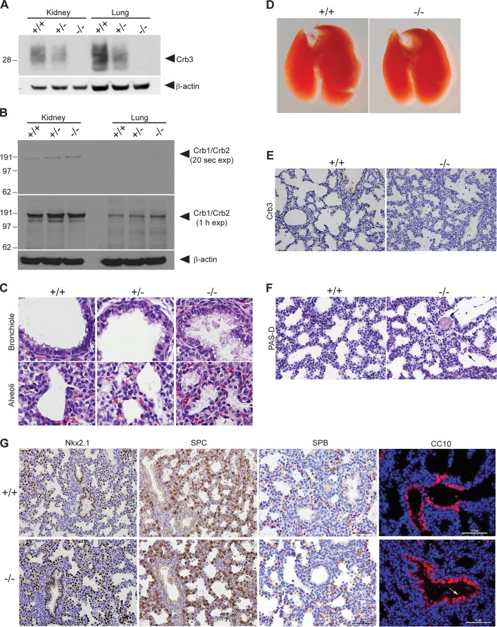
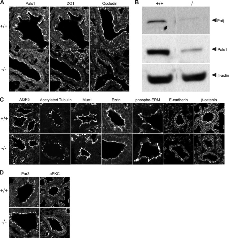
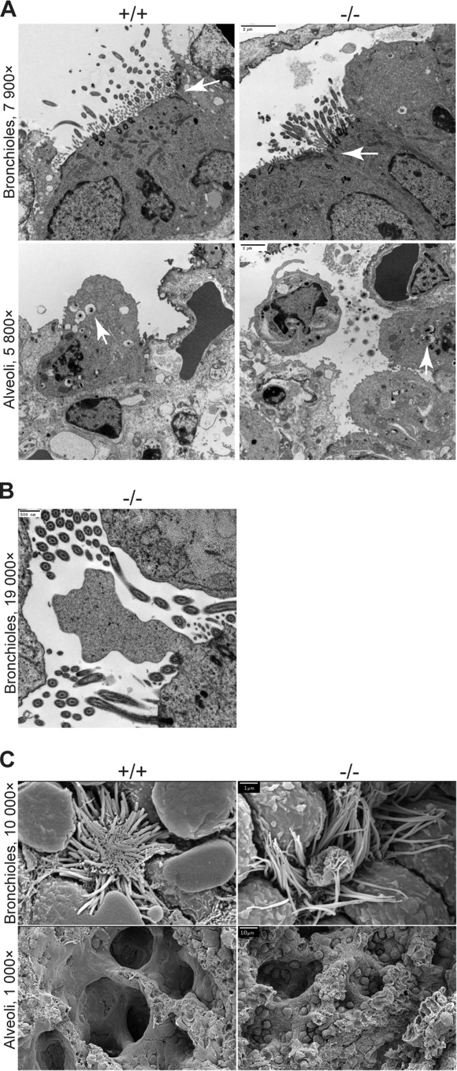
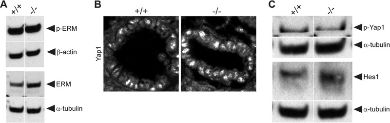
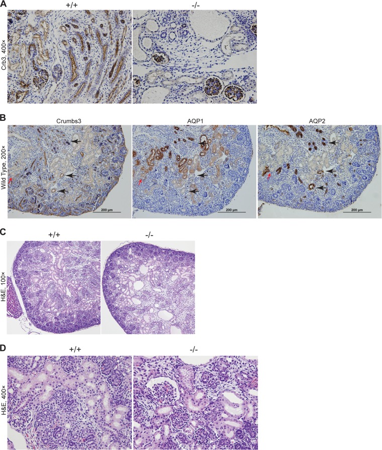
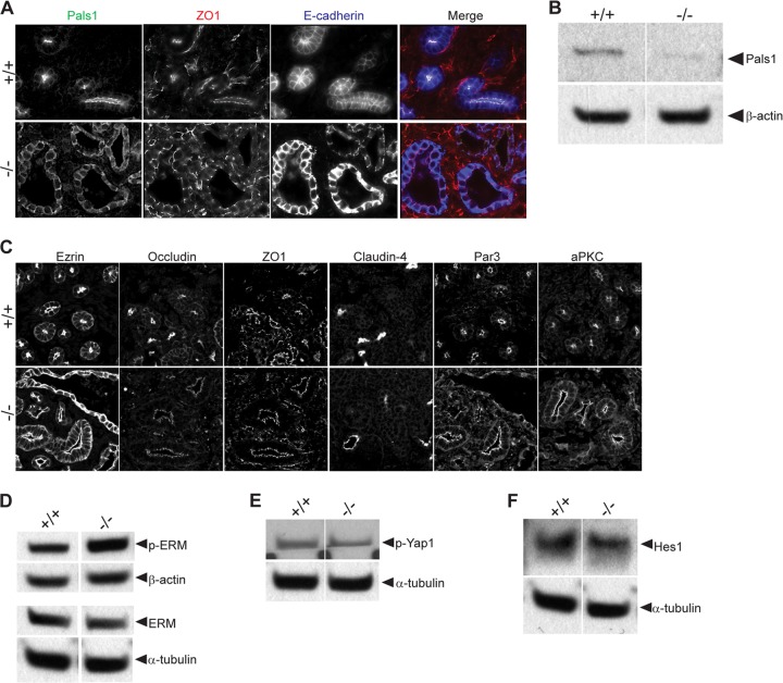
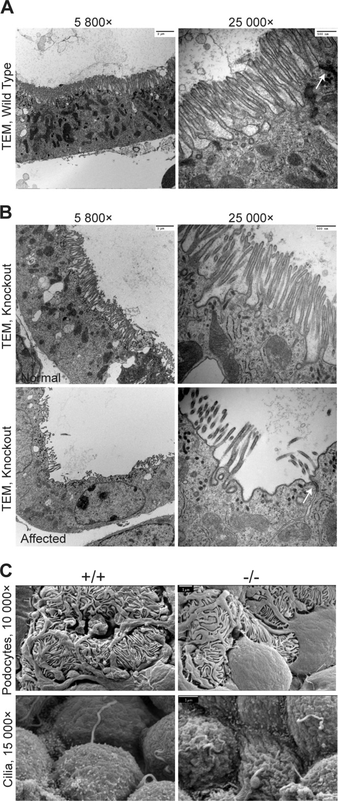
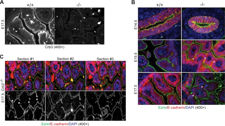
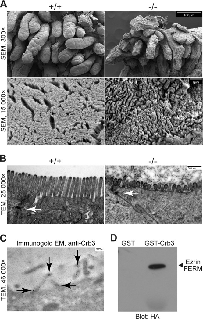
First identified in Drosophila, the Crumbs (Crb) proteins are important in epithelial polarity, apical membrane formation, and tight junction (TJ) assembly. The conserved Crb intracellular region includes a FERM (band 4.1/ezrin/radixin/moesin) binding domain (FBD) whose mammalian binding partners are not well understood and a PDZ binding motif that interacts with mammalian Pals1 (protein associated with lin seven) (also known as MPP5). Pals1 binds Patj (Pals1-associated tight-junction protein), a multi-PDZ-domain protein that associates with many tight junction proteins. The Crb complex also binds the conserved Par3/Par6/atypical protein kinase C (aPKC) polarity cassette that restricts migration of basolateral proteins through phosphorylation. Here, we describe a Crb3 knockout mouse that demonstrates extensive defects in epithelial morphogenesis. The mice die shortly after birth, with cystic kidneys and proteinaceous debris throughout the lungs. The intestines display villus fusion, apical membrane blebs, and disrupted microvilli. These intestinal defects phenocopy those of Ezrin knockout mice, and we demonstrate an interaction between Crumbs3 and ezrin. Taken together, our data indicate that Crumbs3 is crucial for epithelial morphogenesis and plays a role in linking the apical membrane to the underlying ezrin-containing cytoskeleton.


Figures

FIG 1
FIG 1
Generation of Crumbs3-deficient mice. (A) The diagram depicts the gene-targeting strategy and PCR genotyping scheme. The upper box shows the genomic elements; the lower box displays the amplicon sizes for each primer pair (shown in red) which distinguish the alleles. (B) Southern blot analysis of three targeted clones (11G, 12A, and 12C) and wild-type R1 ES cells confirms homologous recombination. (C) Western blot analysis of +/+ and +/flox embryonic lung lysates (embryonic day 18.5 [E18.5]) demonstrates that the GFP-Crb3 protein, a product of the floxed allele, is present in very small quantities in the Crb3+/flox animals. Both panels are taken from the same gel and same exposure time, indicating that GFP-Crb3 is expressed at a tiny fraction of the level of endogenous Crb3 (left panel). The GFP-Crb3 fusion protein is barely detectable following specific immunoprecipitation (IP) of 1 mg lung lysate (right panel). The lanes marked “GFP-Crb3” are positive-control lanes using MDCK cell lysates expressing GFP-Crb3. (D) A survival curve was plotted after videotaping the delivery of E18.5 embryos. The mean lifetime for Crb3−/− pups ± standard error of the mean is indicated. (E) Weights of E18.5 embryos do not appear significantly different among the genotypes by ANOVA.
FIG 2
FIG 2
Analysis of Crumbs3-deficient mice and examination of Crumbs3 knockout lungs. (A) Western blot analysis of kidney and lung lysates (E18.5) confirms Crb3 loss. (B) Upregulation of highly similar proteins Crb1 and Crb2 is not detected in Crb3−/− kidneys or lungs. E18.5 kidney or lung homogenates (100 μg) were resolved by SDS-PAGE, transferred to polyvinylidene difluoride (PVDF), probed using an antibody to the Crumbs3 carboxy-terminal tail, and detected using the very sensitive Bio-Rad Clarity substrate for short and long exposures. We speculate that the positive bands in the 20-s kidney exposure may represent Crb2, whose cross-reactivity with the Crb3 antibody is evident when staining kidney glomeruli (Fig. 6A). We did not observe any enhancement in higher-molecular-mass bands potentially representing Crb1 (>151 kDa) or Crb2 (>137 kDa) in the Crb3−/− animals. (C) Paraffin-embedded lungs from E18.5 pups stimulated to breathe were stained for H&E (×400). (D) Whole-mount images of +/+ and −/− mouse lungs at E18.5. (E) Paraffin sections from E18.5 lungs were stained for Crb3 (brown) and hematoxylin (blue) (×400). (F) Paraffin sections from E18.5 +/+ and −/− mouse lungs were subjected to a PAS-diastase assay. Arrows indicate residual, patchy PAS-diastase-resistant material in the knockout lung alveoli. Scale bar = 50 μm. (G) Paraffin sections from E18.5 +/+ and −/− mouse lungs were stained for Nkx2.1 (brown, ×400), SPC (brown, ×400), SPB (brown; scale bar = 50 μm), and CC10 (red; scale bar = 10 μm [the arrow indicates blebbing of Clara cells]). Nuclei (blue) were stained with hematoxylin or DAPI.
FIG 3
FIG 3
Evaluation of epithelial polarity in Crumbs3 knockout lungs. (A) Frozen sections from E18.5 lungs were stained for Pals1, ZO1, and occludin (×600; zoom, 1.5). (B) Western blot of Crb complex proteins Pals1 and Patj in E18.5 lungs. β-Actin was used as a loading control. Samples were run on nonadjacent lanes of the same gel. (C) Frozen sections from E18.5 +/+ and −/− mouse lungs were stained for AQP5 (alveolar type I cells) (×600; zoom, 2), acetylated tubulin (cilia) (zoom, 1.5), Muc1 (apical surface) (zoom, 1.5), ezrin and phospho-ERM (cytoskeleton) (zoom, 3), E-cadherin and β-catenin (adherens junction/basolateral surface) (zoom, 1.5). (D) Frozen sections from E18.5 +/+ and −/− mouse lungs were stained for Par3 (×600) (zoom, 3) and aPKC (apical polarity complex) (zoom, 1.5).
FIG 4
FIG 4
Electron micrographs of lungs from Crumbs3 knockout mice. (A) Transmission electron micrographs of bronchioles and alveoli from E18.5 lungs (scale bars = 2 μm). Arrows indicate tight junctions (bronchioles) or lamellar bodies (alveoli). (B) Transmission electron micrograph of membrane bleb in Crb3−/− bronchioles from E18.5 lungs (scale bar = 500 nm). (C) Scanning electron micrographs of bronchioles and alveoli from E18.5 lungs (scale bars = 1 μm [top] and 10 μm [bottom]).
FIG 5
FIG 5
Investigation of signaling pathways in Crumbs3 knockout lungs. (A) Western blot of p-ERM and ERM protein levels among +/+ and −/− lungs at E18.5. β-Actin and α-tubulin are shown as loading controls. Samples were run on nonadjacent lanes of the same gel. (B) Frozen sections from E18.5 +/+ and −/− mouse lungs were stained for Yap1 (transcription factor downstream of Hippo pathway) (×600; zoom, 3). (C) Western blot of p-Yap1 (Hippo pathway activation) and Hes1 (Notch pathway activation) among +/+ and −/− lungs at E18.5. α-Tubulin is shown as the loading control. Samples were run on nonadjacent lanes of the same gel.
FIG 6
FIG 6
Examination of Crumbs3 knockout kidney. (A) Paraffin sections from E18.5 kidneys were stained for Crb3 (brown) and hematoxylin (blue). (B) Paraffin-embedded kidneys from wild-type E18.5 mice were stained for Crb3, Aqp1, and Aqp2 (brown) using serial sections. Red arrows indicate areas where weak Crb3-positive tubules were overlaid with Aqp1. Black arrows indicate the overlap of strongly Crb3-positive tubules with Aqp2. Nuclei were counterstained with hematoxylin (blue). (C) Paraffin sections from E18.5 kidneys were stained with H&E. (D) Paraffin-embedded kidneys from E18.5 mice were stained for H&E.
FIG 7
FIG 7
Evaluation of epithelial polarity and signaling pathways in Crumbs3 knockout kidneys. (A) Frozen sections from E17.5 kidneys were stained for Pals1 (green), ZO1 (red), and E-cadherin (blue) (×400 magnification). (B) Western blot of Crb complex protein Pals1 in E18.5 kidneys. β-Actin was used as a loading control. Samples were run on nonadjacent lanes of the same gel. (C) Frozen sections of kidneys from E17.5 to E18.5 mice were stained for ezrin (cytoskeleton), occludin and ZO1 (tight junction), E-cadherin (adherens junction/basolateral surface), claudin-4 (tight junction), Par3, and aPKC (apical polarity complex) (×600; zoom, 1.5). (D) Western blot of p-ERM and ERM protein levels among +/+ and −/− kidneys at E18.5. β-Actin and α-tubulin are shown as loading controls. Samples were run on nonadjacent lanes of the same gel. (E and F) Western blots of p-Yap1 (Hippo pathway activation) and Hes1 (Notch pathway activation) among +/+ and −/− kidneys at E18.5. α-Tubulin is shown as the loading control. Samples were run on nonadjacent lanes of the same gel.
FIG 8
FIG 8
Electron micrographs of kidneys from Crumbs3-deficient mice. (A and B) Transmission electron micrographs of wild-type and knockout proximal tubules with brush borders from E18.5 kidneys (scale bars = 2 μm [left] and 500 nm [right]). Arrows indicate tight junctions. (C) Scanning electron micrographs of podocytes and cilia from E18.5 kidneys (scale bars = 1 μm).
FIG 9
FIG 9
Evaluation of Crumbs3 knockout intestine. (A) Staining of E17.5 duodenum confirms the absence of Crumbs3. Bright spots represent autofluorescent blood cells. (B) Fluorescent staining of paraffin-embedded duodenum sections at different stages of villus development with ezrin (green), E-cadherin (red), and DAPI (blue, to mark nuclei) demonstrates the presence of bridges (marked by asterisks) between villi in Crb3−/− embryos. (C) Staining of Crb3−/− duodenum serial sections with ezrin (green), E-cadherin (red), and DAPI (blue) confirms the presence of bridges (asterisks).
FIG 10
FIG 10
Electron micrographs of intestine from Crumbs3 knockout mice, Crb3 in microvilli (cell culture), and Crb3 interactions with ezrin. (A) Scanning electron micrographs of E18.5 wild-type and Crb3 null intestine (scale bars = 100 μm and 1 μm). (B) Transmission electron micrographs of E18.5 wild-type and Crb3 null intestine (scale bar = 500 nm). Arrows indicate tight junctions. (C) Transmission electron micrograph of immunogold-labeled Crb3 in MDCK cells (scale bar = 100 nm). Arrows indicate immunogold particles and localization of Crb3. (D) GST pulldown experiments assay the interaction between Crb3 intracellular domain and the HA-tagged ezrin FERM domain.

References

    1. Dow LE, Humbert PO. 2007. Polarity regulators and the control of epithelial architecture, cell migration, and tumorigenesis. Int. Rev. Cytol. 262:253–302. 10.1016/S0074-7696(07)62006-3 - DOI - PubMed
    1. Pieczynski J, Margolis B. 2011. Protein complexes that control renal epithelial polarity. Am. J. Physiol. Renal Physiol. 300:F589–F601. 10.1152/ajprenal.00615.2010 - DOI - PMC - PubMed
    1. Datta A, Bryant DM, Mostov KE. 2011. Molecular regulation of lumen morphogenesis. Curr. Biol. 21:R126–R136. 10.1016/j.cub.2010.12.003 - DOI - PMC - PubMed
    1. Tepass U. 2012. The apical polarity protein network in Drosophila epithelial cells: regulation of polarity, junctions, morphogenesis, cell growth, and survival. Annu. Rev. Cell Dev. Biol. 28:655–685. 10.1146/annurev-cellbio-092910-154033 - DOI - PubMed
    1. Bulgakova N, Knust E. 2009. The Crumbs complex: from epithelial-cell polarity to retinal degeneration. J. Cell Sci. 122:2587–2596. 10.1242/jcs.023648 - DOI - PubMed

Publication types

MeSH terms

LinkOut - more resources