. 2014 Jan;34(1):96-109.
doi: 10.1128/MCB.00622-13. Epub 2013 Oct 28.

Identification of the immunoproteasome as a novel regulator of skeletal muscle differentiation

Affiliations

Identification of the immunoproteasome as a novel regulator of skeletal muscle differentiation

Ziyou Cui et al. Mol Cell Biol. 2014 Jan.

Abstract

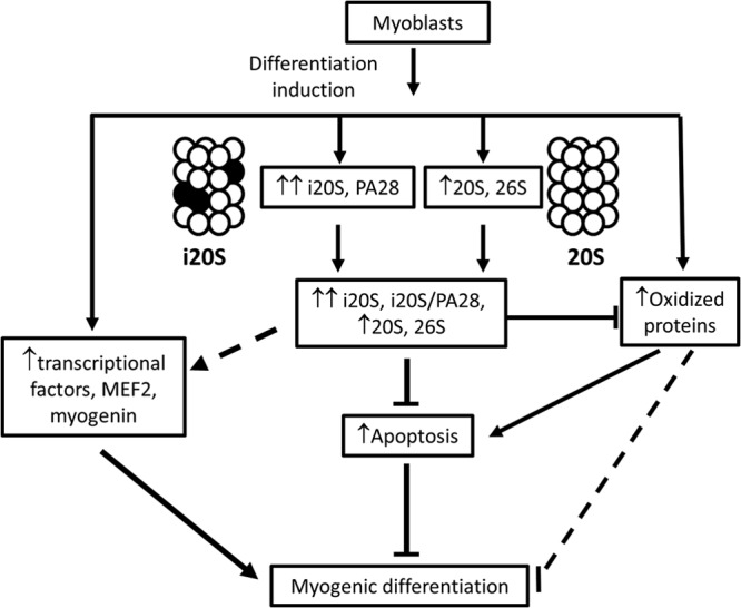
While many of the molecular details of myogenesis have been investigated extensively, the function of immunoproteasomes (i-proteasomes) in myogenic differentiation remains unknown. We show here that the mRNA of i-proteasome subunits, the protein levels of constitutive and inducible proteasome subunits, and the proteolytic activities of the 20S and 26S proteasomes were significantly upregulated during differentiation of skeletal muscle C2C12 cells. Knockdown of the i-proteasome catalytic subunit PSMB9 by short hairpin RNA (shRNA) decreased the expression of both PSMB9 and PSMB8 without affecting other catalytic subunits of the proteasome. PSMB9 knockdown and the use of i-proteasome-specific inhibitors both decreased 26S proteasome activities and prevented C2C12 differentiation. Inhibition of the i-proteasome also impaired human skeletal myoblast differentiation. Suppression of the i-proteasome increased protein oxidation, and these oxidized proteins were found to be more susceptible to degradation by exogenous i-proteasomes. Downregulation of the i-proteasome also increased proapoptotic proteins, including Bax, as well as cleaved caspase 3, cleaved caspase 9, and cleaved poly(ADP-ribose) polymerase (PARP), suggesting that impaired differentiation is likely to occur because of significantly increased apoptosis. These results demonstrate for the first time that i-proteasomes, independent of constitutive proteasomes, are critical for skeletal muscle differentiation of mouse C2C12 cells.


Figures

FIG 1
FIG 1
Dynamic changes of intracellular proteolytic activities during myogenic differentiation and the effects of proteasome inhibition on C2C12 cell differentiation. (A) Bortezomib, the specific proteasome inhibitor, inhibits myogenesis. C2C12 myoblasts (90% confluent) were induced to differentiate in the presence or absence of 10 nM bortezomib for 2 days (D2), 4 days (D4), or 6 days (D6). Images represent three independent experiments. Magnification, ×10. Scale bar, 50 μm. The graph shows the fusion index of C2C12 cells grown in differentiation medium for 6 days after the initiation of differentiation. (B) 26S and 20S proteasome activities increase during myogenic differentiation. Proteasome activities were measured in cell lysates prepared from D0 myoblasts, D1, D2, and D4 differentiating cells, and D6 differentiated cells (myotubes) using fluorescently labeled proteasome substrates. The caspase-like (Z-LLE-AMC, β1), trypsin-like (Boc-LSTR-AMC, β2), and chymotrypsin-like (Suc-LLVY-AMC, β2) proteasome activities were assayed using 25 μg of protein with or without specific proteasome inhibitors. 26S and 20S proteasome activities were determined in the presence of ATP and SDS, respectively. The released free AMC fluorescence was quantified using a 390-nm filter on a Fluoroskan Ascent fluorometer. (C) Calpain activity remains lowered and cathepsin L activity increases during myogenic differentiation. Calpain and cathepsin L activities were measured in D0 to D6 cell lysates using fluorescently labeled substrates. Each value in panels B and C is the mean of at least three measurements (n = 3); error bars denote standard deviations (SD) (*, P < 0.05; **, P < 0.01).
FIG 2
FIG 2
Proteasome subunit gene expression changes during C2C12 cell differentiation. (A) qRT-PCR was utilized to determine the gene expression of constitutive and inducible proteasome subunits as well as proteasome activators in D0 myoblasts, D1, D2, and D4 differentiating cells and D6 differentiated cells (myotubes). The inducible subunits PSMB8, PSMB9, and PSMB10 showed the largest increases in gene expression during C2C12 cell differentiation. Values are means and SD (n = 3) (*, P < 0.05; **, P < 0.01). (B) Agarose gel showing the fragments of RT-PCR products for the inducible subunits and two normalization controls (GUSB and GAPDH).
FIG 3
FIG 3
Localization and dynamic changes of proteasome subunit expression during C2C12 cell differentiation. (A) Protein expression of the constitutive catalytic proteasome subunits PSMB5, PSMB6, and PSMB7, the i-proteasome subunit PSME1, the inducible subunit PSMB8, the 19S subunits PSMC2 and PSMC5, and the 20S subunit PSMA6. Each lane represents a different sample, and three different samples for each experimental condition are included to show the variation between samples at the same time points. Relative expression values are means ± SD (*, P < 0.05; **, P < 0.01). (B) Native gel electrophoresis of D0 and D2 cell lysates. Each lane contains 200 μg of lysate protein which was run on 2 to 5% acrylamide gels and incubated with 50 μM Suc-LLVY-AMC to detect proteasome activity. The clear region on the gel (first image on the left) corresponds to cleaved LLVY-AMC, indicating proteasome activity. The second image on the left is an inverted image of in-gel proteasome activity showing the upregulation of proteasome activity in D2 lysates compared to D0 lysates. After detecting proteasome activity, the gels were immunoblotted with different antibodies to proteasome subunits or stained for total protein with Coomassie blue R-250. (C) The cultured cells (D0) in the chamber slide were immunostained by the rabbit-derived antibody to the inducible subunit PSMB8 and the mouse-derived antibody to the 20S α-type subunit PSMA4. The slide was subsequently incubated with two PLA probes (Duolink II anti-mouse Minus and Duolink II anti-rabbit Plus) followed by the DNA amplification procedure. The panels show cells with DAPI-stained nuclei, immunostained cells, and a merged image. The images were taken using an Zeiss LSM 510 META confocal microscope with 40×/63 objectives. Scale bar, 20 μm.
FIG 4
FIG 4
Effects of PSMB9 suppression on C2C12 cell myogenic differentiation. (A) (Top) C2C12 cells were transfected with a negative control and four different shRNAs to PSMB9 and treated to induce differentiation after puromycin selection. The images were taken at 4 days after the initiation of differentiation. (Bottom) Immunofluorescence analysis of skeletal myosin heavy chain (MyHC) in C2C12 cells grown in differentiation medium for 2 or 4 days in the presence or in the absence of C3 shRNA. The graph shows the fusion indexes of C2C12 cells grown in differentiation medium for 2 days and 4 days after the initiation of differentiation. (B) Expression of MyHC, PSMB9, PSMB8, and myogenin at D2 in C2C12 cells containing the control shRNA (N) or the four PSMB9 shRNAs (C1, C2, C3, and C4). C2 and C3 shRNAs showed the best suppression of PSMB9, PSMB8, and MyHC. (C) Effect of C3 shRNA on MyHC and MyoD protein expression at D0, D1, D2, and D4. (D) qRT-PCR of constitutive and inducible proteolytic proteasome subunits in D2 differentiated cells. Only the RNA expression of the inducible subunits PSMB8 and PSMB9 was suppressed. Values are means and SD (n = 3) (**, P < 0.01). (E) Proteolytic activities (caspase [βi], trypsin [β2], and chymotrypsin [β5] like) of the proteasomes in C2C12 cell lysates from cells containing different shRNAs at D0 and D2. Values are means and SD (n = 3) (*, P < 0.05; **, P < 0.01). (F) Proteasome activity of PSMB9 knockdown cells with C2. (G) Protein expression of constitutive proteolytic proteasome subunits in PSMB9 knockdown cells with different shRNA clones after 2 days of differentiation. The blot shows results for three replicates for each clone.
FIG 5
FIG 5
Effects of inducible subunit inhibitors on C2C12 cell and human skeletal muscle myogenic differentiation. (A) C2C12 myoblasts (90% confluent) were treated to induce differentiation in the presence or absence of i-proteasome inhibitors, UK-101 and LKS01, for up to 6 days (D6). Images show immunofluorescence of MyHC (red) and DAPI (blue). Each image is representative of three independent experiments. The graph shows the fusion index in the presence and absence of i-proteasome inhibitors at D6. (B) Western blots of MyHC and myogenin levels 2 days (D2), 4 days (D4), and 6 days (D6) after differentiation induction in the presence and absence of i-proteasome inhibitors. (C) Western blots of MyHC and GAPDH after two i-proteasome inhibitors (UK-101 and LKS01) were incubated with C2C12 cells for different lengths of time ranging from 2 h to 24 h. Then the medium with inhibitors was replaced with the medium lacking inhibitors, and the cells were left to differentiate for a total of 48 h. (D) Immunofluorescence images of MyHC (red) and DAPI (blue) in myoblasts differentiated for 2 days, after which DMSO or UK-101 and LKS01 were added and the myoblasts allowed to further differentiate for 2 more days. (E) Western blots of MyHC and myogenin to determine the effects of i-proteasome inhibition on differentiation after 2 days of differentiation has already occurred. (F) Human skeletal muscle myoblasts were treated to induce differentiation in the presence or absence of bortezomib (Bor) or i-proteasome inhibitors, UK-101 and LKS01, for 4 days (D4). Images show immunofluorescence of MyHC (red) and DAPI (blue). The graph shows the fusion index of C2C12 myoblasts grown in differentiation medium for 4 days after the initiation of differentiation in the presence of proteasome (bortezomib) or i-proteasome inhibitors compared to control myoblasts. Each image is representative of three independent experiments.
FIG 6
FIG 6
Effects of i-proteasome suppression on oxidized proteins. (A) Oxyblot of C212 cell lysates from D0 myoblasts, D1, D2, and D4 differentiating cells and D6 differentiated cells (myotubes). Carbonyl groups on proteins were first conjugated to DNP; the DNP-bound proteins were then detected using an anti-DNP antibody. Oxidized protein levels increase to a maximum at D2 during C2C12 cell differentiation. (B) Oxyblot of C212 cell lysates from D0 myoblasts and D1 and D2 differentiating C3 and control cells. With C3 shRNA knockdown of PSMB9, protein oxidation further increased in myoblasts and differentiating cells up to D2. Arrows indicate bands which increased in intensity in C3 cells relative to N cells. (C) ROS level in C3 and control cells as measured by flow cytometry. Samples were either myoblasts (D0) or 12-h differentiating C3 and control (N) cells which were harvested, and ROS were measured with the fluorescence probe 2′,7′-dichlorodihydrofluorescein (DCFH). The image is representative of three different experiments. SSC-H, side-scattered light; FL1-H, fluorescent light. (D) Oxyblot of C212 cell lysates from D2 differentiating cells treated with different i-proteasome inhibitors. Carbonyl groups on proteins were first conjugated to DNP; the DNP-bound proteins were then detected using an anti-DNP antibody. The PSMB9-specific inhibitor UK-101 was used at a concentration of 1 μM, and the PSMB8-specific inhibitor LKS01 was used at 0.2 μM. (E) Cytosolic fractions lacking proteasomes from D0 and D2 C2C12 cells were incubated with exogenous 20S i-proteasomes at 37°C for 3 to 6 h. Increases in cleaved proteins were determined by the fluorescent compound fluorescamine, which forms a fluorescent adduct with the N termini of peptides generated by proteasomal cleavage. Results shown are fluorescamine adducts in the cytosolic fractions in the presence of i-proteasomes subtracted from fluorescamine adducts in the absence of exogenous i-proteasomes incubated under the same conditions (n = 4). Higher levels of fluorescent adducts represent greater levels of protein degradation. **, P < 0.001. (F) C2C12 cell D2 homogenates were incubated with constitutive proteasome (c20S), i-proteasome (i20S), or buffer (control) for 3 h at 37°C and then boiled with sample buffer. Western blotting was subsequently carried out to detect oxidized proteins (5 μg protein/lane). Each blot shows results for three replicates for each condition. Although the amount of protein in each lane was 5 μg, samples incubated with c20S and i20S showed decreased total protein staining, suggesting that some of the proteins were being degraded and possibly ran at the dye front (top graph). Quantification of the results showed that i-proteasomes significantly degraded oxidized proteins in D2 myoblasts homogenates relative to constitutive proteasomes (lower graph). n = 3; *, P < 0.05; ** P < 0.001.
FIG 7
FIG 7
Upregulation of apoptosis in i-proteasome-suppressed C2C12 myoblasts. (A) Protein expression of proapoptotic proteins in control (N) and PSMB9 knockdown (C3) myoblasts and myotubes after 0 to 6 days of differentiation. (B) Immunofluorescence analysis of MyHC (red) and DAPI (blue) in C2C12 cells grown in differentiation medium in the presence or in the absence of 20 μM apoptosis activator II for 2 days. (C) Western blots of C2C12 cells for enzymes known to be involved in C2C12 cell differentiation (GSK3β and p38). Total protein staining (Ponceau stain) and GAPDH were used as normalization controls.
FIG 8
FIG 8
Schematic representation of the role of the i-proteasome in C2C12 skeletal muscle cell differentiation showing a possible role of the i-proteasome in C2C12 skeletal muscle cell differentiation.

References

    1. Glickman MH, Ciechanover A. 2002. The ubiquitin-proteasome proteolytic pathway: destruction for the sake of construction. Physiol. Rev. 82:373–428. 10.1152/physrev.00027.2001 - DOI - PubMed
    1. Lecker SH, Goldberg AL, Mitch WE. 2006. Protein degradation by the ubiquitin-proteasome pathway in normal and disease states. J. Am. Soc. Nephrol. 17:1807–1819. 10.1681/ASN.2006010083 - DOI - PubMed
    1. Attaix D, Aurousseau E, Combaret L, Kee A, Larbaud D, Ralliere C, Souweine B, Taillandier D, Tilignac T. 1998. Ubiquitin-proteasome-dependent proteolysis in skeletal muscle. Reprod. Nutrition Dev. 38:153–165. 10.1051/rnd:19980202 - DOI - PubMed
    1. Demartino GN, Gillette TG. 2007. Proteasomes: machines for all reasons. Cell 129:659–662. 10.1016/j.cell.2007.05.007 - DOI - PubMed
    1. Voges D, Zwickl P, Baumeister W. 1999. The 26S proteasome: a molecular machine designed for controlled proteolysis. Annu. Rev. Biochem. 68:1015–1068. 10.1146/annurev.biochem.68.1.1015 - DOI - PubMed

Publication types

MeSH terms