

In silico analysis of the molecular machinery underlying aqueous humor production: potential implications for glaucoma
- PMID: 24165276
- PMCID: PMC3875900
- DOI: 10.1186/2043-9113-3-21
In silico analysis of the molecular machinery underlying aqueous humor production: potential implications for glaucoma
Abstract
Background: The ciliary body epithelia (CBE) of the eye produce the aqueous humor (AH). The equilibrium between the AH production by the CBE and the outflow through the trabecular meshwork ultimately determines the intraocular pressure (IOP). An increased IOP is a major risk factor for primary open angle glaucoma (POAG). This study aims to elucidate the molecular machinery of the most important function of the CBE: the AH production and composition, and aims to find possible new molecular clues for POAG and AH production-lowering drugs.
Methods: We performed a gene expression analysis of the non-pigmented (NPE) and pigmented epithelia (PE) of the human CBE of post mortem eyes. We used 44 k Agilent microarrays against a common reference design. Functional annotations were performed with the Ingenuity knowledge database.
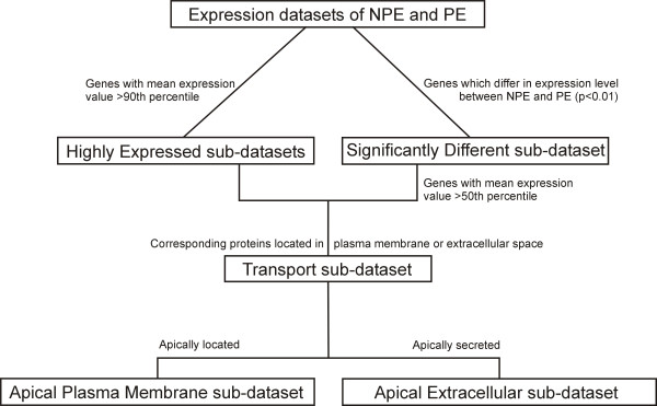
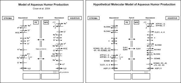
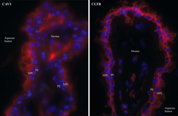
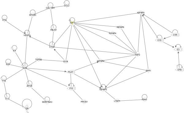
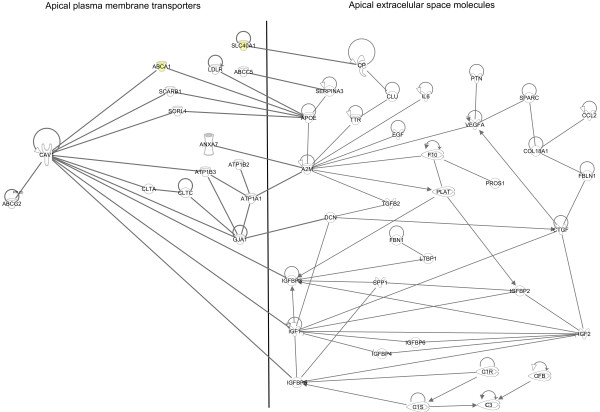
Results: We built a molecular model of AH production by combining previously published physiological data with our current genomic expression data. Next, we investigated molecular CBE transport features which might influence AH composition. These features included caveolin- and clathrin vesicle-mediated transport of large biomolecules, as well as a range of substrate specific transporters. The presence of these transporters implies that, for example, immunoglobins, thyroid hormone, prostaglandins, cholesterol and vitamins can be secreted by the CBE along with the AH. In silico, we predicted some of the molecular apical interactions between the NPE and PE, the side where the two folded epithelia face each other. Finally, we found high expression of seven POAG disease genes in the plasma membrane of extracellular space of the CBE, namely APOE, CAV1, COL8A2, EDNRA, FBN1, RFTN1 and TLR4 and we found possible new targets for AH lowering drugs in the AH.
Conclusions: The CBE expresses many transporters, which account for AH production and/or composition. Some of these entries have also been associated with POAG. We hypothesize that the CBE may play a more prominent role than currently thought in the pathogenesis of POAG, for example by changing the composition of AH.
Figures
Similar articles
-
Gene expression-based comparison of the human secretory neuroepithelia of the brain choroid plexus and the ocular ciliary body: potential implications for glaucoma.Fluids Barriers CNS. 2014 Jan 29;11(1):2. doi: 10.1186/2045-8118-11-2. Fluids Barriers CNS. 2014. PMID: 24472183 Free PMC article.
-
Proteomic analysis of aqueous humor from patients with primary open angle glaucoma.Mol Vis. 2010 Dec 18;16:2839-46. Mol Vis. 2010. PMID: 21203405 Free PMC article.
-
ID1 and ID3 are Negative Regulators of TGFβ2-Induced Ocular Hypertension and Compromised Aqueous Humor Outflow Facility in Mice.Invest Ophthalmol Vis Sci. 2021 May 3;62(6):3. doi: 10.1167/iovs.62.6.3. Invest Ophthalmol Vis Sci. 2021. PMID: 33938911 Free PMC article.
-
Biomarkers and special features of oxidative stress in the anterior segment of the eye linked to lens cataract and the trabecular meshwork injury in primary open-angle glaucoma: challenges of dual combination therapy with N-acetylcarnosine lubricant eye drops and oral formulation of nonhydrolyzed carnosine.Fundam Clin Pharmacol. 2012 Feb;26(1):86-117. doi: 10.1111/j.1472-8206.2011.00969.x. Epub 2011 Aug 24. Fundam Clin Pharmacol. 2012. PMID: 21883446 Review.
-
Role of the Rho GTPase/Rho kinase signaling pathway in pathogenesis and treatment of glaucoma: Bench to bedside research.Exp Eye Res. 2017 May;158:23-32. doi: 10.1016/j.exer.2016.08.023. Epub 2016 Sep 1. Exp Eye Res. 2017. PMID: 27593914 Free PMC article. Review.
Cited by
-
TLR4 deficiency does not alter glaucomatous progression in a mouse model of chronic glaucoma.bioRxiv [Preprint]. 2024 Jun 8:2024.06.07.597951. doi: 10.1101/2024.06.07.597951. bioRxiv. 2024. Update in: Sci Rep. 2025 May 15;15(1):16852. doi: 10.1038/s41598-025-00638-7. PMID: 38895321 Free PMC article. Updated. Preprint.
-
TLR4 deficiency does not alter glaucomatous progression in a mouse model of chronic glaucoma.Sci Rep. 2025 May 15;15(1):16852. doi: 10.1038/s41598-025-00638-7. Sci Rep. 2025. PMID: 40374644 Free PMC article.
-
Gene expression-based comparison of the human secretory neuroepithelia of the brain choroid plexus and the ocular ciliary body: potential implications for glaucoma.Fluids Barriers CNS. 2014 Jan 29;11(1):2. doi: 10.1186/2045-8118-11-2. Fluids Barriers CNS. 2014. PMID: 24472183 Free PMC article.
-
Normal-tension glaucoma: Pathogenesis and genetics.Exp Ther Med. 2019 Jan;17(1):563-574. doi: 10.3892/etm.2018.7011. Epub 2018 Nov 26. Exp Ther Med. 2019. PMID: 30651837 Free PMC article. Review.
-
Prion protein modulates iron transport in the anterior segment: Implications for ocular iron homeostasis and prion transmission.Exp Eye Res. 2018 Oct;175:1-13. doi: 10.1016/j.exer.2018.05.031. Epub 2018 May 31. Exp Eye Res. 2018. PMID: 29859760 Free PMC article.
References
-
- Civan MM. In: Current Topics in Membranes: The eye's aqueous humor. From secretion to glaucoma, Volume 45. Civan MM, editor. San Diego, London, Boston, New York, Sydney, Tokyo, Toronto: Academic Press; 1998. Transport components and net secretion of the aqueous humor and their integrated regulation; pp. 1–24.
LinkOut - more resources
Full Text Sources
Other Literature Sources
Miscellaneous
