

Gastric xanthelasma: an unusual endoscopic finding
- PMID: 24165503
- PMCID: PMC3822080
- DOI: 10.1136/bcr-2013-201017
Gastric xanthelasma: an unusual endoscopic finding
Abstract
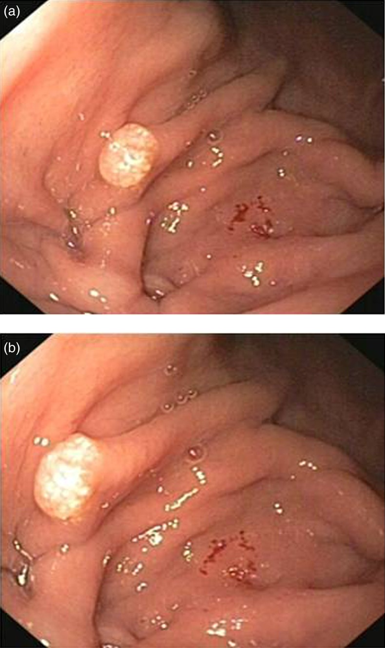
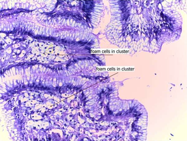
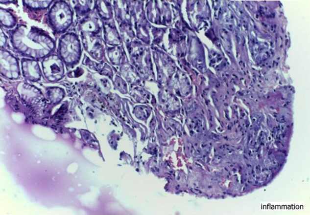
Gastric xanthelasma is a rarely encountered finding in upper gastrointestinal (GI) endoscopy. It is characterised by yellowish-white plaque in the stomach especially in the antrum or the pyloric region. Histologically it consists of foamy macrophages in the lamina propria. It is a benign condition but its appearance mimics malignancy and it is found to be associated with various conditions, some of which are considered premalignant so, histological confirmation is necessary. We present a case of a 44-year-old man who presented to the medicine outpatient department for intermittent pain in epigastrium for the last 2 years. His physical examination was normal. His haematological and biochemical investigations were also normal. His upper GI endoscopy revealed yellowish-white plaque in fundus of the stomach, which was diagnosed as gastric xanthelasma by histological examination with associated chronic gastritis.
Figures
Similar articles
-
An unusual association: gastric xanthelasma presenting with iron deficiency anemia: a case report.J Med Case Rep. 2025 Mar 4;19(1):98. doi: 10.1186/s13256-025-05133-1. J Med Case Rep. 2025. PMID: 40038797 Free PMC article.
-
Xanthomatous hyperplastic polyps of the stomach: clinicopathologic study of 5 patients with polypoid gastric lesions showing combined features of gastric xanthelasma and hyperplastic polyp.Ann Diagn Pathol. 2013 Feb;17(1):72-4. doi: 10.1016/j.anndiagpath.2012.08.001. Epub 2012 Sep 26. Ann Diagn Pathol. 2013. PMID: 23020998
-
Xanthelasma of esophagus and stomach.Indian J Gastroenterol. 2000 Jul-Sep;19(3):135. Indian J Gastroenterol. 2000. PMID: 10918723
-
Gastric Xanthoma Associated with Gastric Cancer Development: An Updated Review.Can J Gastroenterol Hepatol. 2020 Feb 21;2020:3578927. doi: 10.1155/2020/3578927. eCollection 2020. Can J Gastroenterol Hepatol. 2020. PMID: 32149048 Free PMC article. Review.
-
Collagenous gastritis.Dig Endosc. 2013 Sep;25(5):547-9. doi: 10.1111/j.1443-1661.2012.01391.x. Epub 2012 Oct 22. Dig Endosc. 2013. PMID: 23363075 Review.
Cited by
-
Gastric xanthelasma associated with hyperplastic polyp and mucosal erosions: report of an unusual case and literature review.Oxf Med Case Reports. 2018 Aug 11;2018(8):omy051. doi: 10.1093/omcr/omy051. eCollection 2018 Aug. Oxf Med Case Reports. 2018. PMID: 30151218 Free PMC article.
-
Gastric Xanthoma: A Rare Case Report.Iran J Pathol. 2023;18(1):104-107. doi: 10.30699/ijp.2023.551854.2878. Epub 2023 Feb 1. Iran J Pathol. 2023. PMID: 37383159 Free PMC article.
-
Duodenal Xanthoma: From Specks to Obstruction.Cureus. 2019 May 14;11(5):e4661. doi: 10.7759/cureus.4661. Cureus. 2019. PMID: 31328054 Free PMC article.
-
An Unusual Case of a Gastric Xanthoma: A Case Report.Cureus. 2022 May 15;14(5):e25026. doi: 10.7759/cureus.25026. eCollection 2022 May. Cureus. 2022. PMID: 35719823 Free PMC article.
-
A Case of Two Diametrically Opposed Gastric Xanthomas in the Pyloric Antrum: An Unusual and Benign Endoscopic Finding.Cureus. 2024 Jul 24;16(7):e65296. doi: 10.7759/cureus.65296. eCollection 2024 Jul. Cureus. 2024. PMID: 39184778 Free PMC article.
References
-
- Gencosmanoglu R, Sen Oran E, Kurtkaya Yapicier O, et al. Xanthelasmas of the upper gastrointestinal tract. J Gastroenterol 2004;2013:215–19 - PubMed
-
- Gursoy S, Yurci A, Torun E, et al. An uncommon lesion: gastric xanthelasma. Turk J Gastroenterol 2005;2013:167–70 - PubMed
-
- Chen YS, Lin JB, Dai KS, et al. Gastric xanthelasma. Chin Med J 1989;2013:639–43 - PubMed
-
- Ludvikova M, Michal M, Datkova D. Gastric xanthelasma associated with diffuse signet ring carcinoma. A potential diagnostic problem. Histopathology 1994;2013:581–2 - PubMed
-
- Luk IS, Bhuta S, Lewin KJ. Clear cell carcinoid tumor of stomach. A variant mimicking gastric xanthelasma. Arch Pathol Lab Med 1997;2013:1100–3 - PubMed
Publication types
MeSH terms
Substances
LinkOut - more resources
Full Text Sources
Other Literature Sources
Medical