

KCa3.1 mediates activation of fibroblasts in diabetic renal interstitial fibrosis
- PMID: 24166472
- PMCID: PMC3910344
- DOI: 10.1093/ndt/gft431
KCa3.1 mediates activation of fibroblasts in diabetic renal interstitial fibrosis
Abstract
Background: Fibroblast activation plays a critical role in diabetic nephropathy (DN). The Ca2+-activated K+ channel KCa3.1 mediates cellular proliferation of many cell types including fibroblasts. KCa3.1 has been reported to be a potential molecular target for pharmacological intervention in a diverse array of clinical conditions. However, the role of KCa3.1 in the activation of myofibroblasts in DN is unknown. These studies assessed the effect of KCa3.1 blockade on renal injury in experimental diabetes.
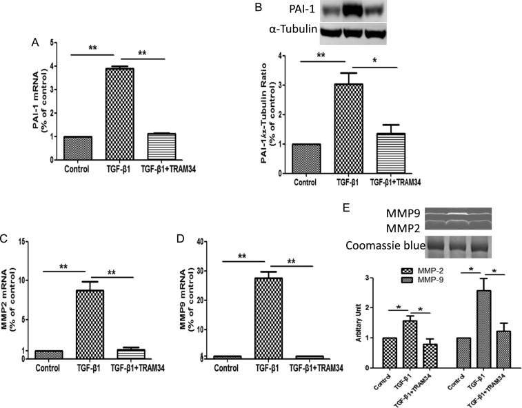
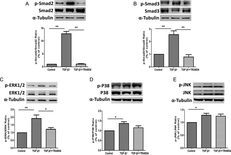
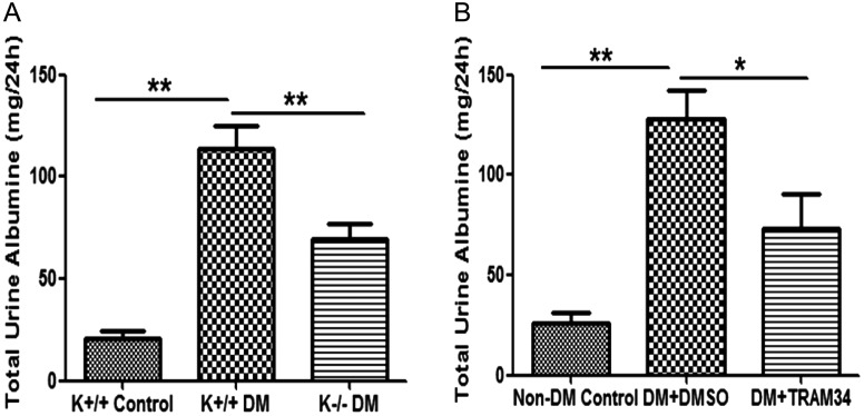
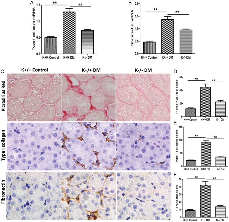
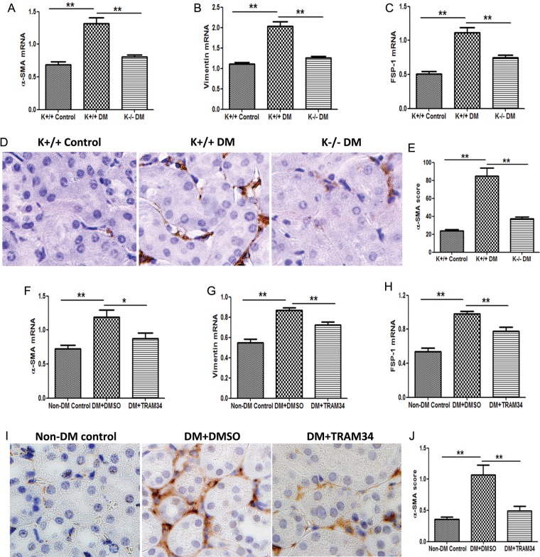
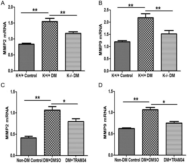
Methods: As TGF-β1 plays a central role in the activation of fibroblasts to myofibroblasts in renal interstitial fibrosis, human primary renal interstitial fibroblasts were incubated with TGF-β1+/- the selective inhibitor of KCa3.1, TRAM34, for 48 h. Two streptozotocin-induced diabetic mouse models were used in this study: wild-type KCa3.1+/+ and KCa3.1-/- mice, and secondly eNOS-/- mice treated with or without a selective inhibitor of KCa3.1 (TRAM34). Then, markers of fibroblast activation and fibrosis were determined.
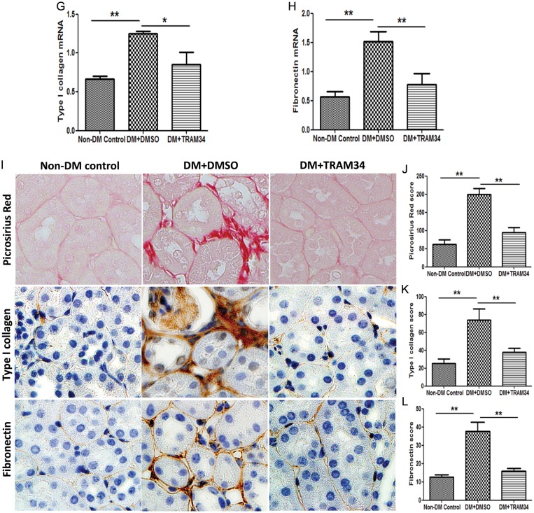
Results: Blockade of KCa3.1 inhibited the upregulation of type I collagen, fibronectin, α-smooth muscle actin, vimentin and fibroblast-specific protein-1 in renal fibroblasts exposed to TGF-β1 and in kidneys from diabetic mice. TRAM34 reduced TGF-β1-induced phosphorylation of Smad2/3 and ERK1/2 but not P38 and JNK MAPK in interstitial fibroblasts.
Conclusions: These results suggest that blockade of KCa3.1 attenuates diabetic renal interstitial fibrogenesis through inhibiting activation of fibroblasts and phosphorylation of Smad2/3 and ERK1/2. Therefore, therapeutic interventions to prevent or ameliorate DN through targeted inhibition of KCa3.1 deserve further consideration.
Keywords: KCa3.1; diabetic nephropathy; fibroblast activation; renal interstitial fibrosis.
Figures

Similar articles
-
Blockade of KCa3.1 ameliorates renal fibrosis through the TGF-β1/Smad pathway in diabetic mice.Diabetes. 2013 Aug;62(8):2923-34. doi: 10.2337/db13-0135. Epub 2013 May 8. Diabetes. 2013. PMID: 23656889 Free PMC article.
-
Inhibition of KCa3.1 suppresses TGF-β1 induced MCP-1 expression in human proximal tubular cells through Smad3, p38 and ERK1/2 signaling pathways.Int J Biochem Cell Biol. 2014 Feb;47:1-10. doi: 10.1016/j.biocel.2013.11.017. Epub 2013 Nov 28. Int J Biochem Cell Biol. 2014. PMID: 24291552
-
Blockade of KCa3.1: A novel target to treat TGF-β1 induced conjunctival fibrosis.Exp Eye Res. 2018 Feb;167:140-144. doi: 10.1016/j.exer.2017.12.003. Epub 2017 Dec 12. Exp Eye Res. 2018. PMID: 29242028 Free PMC article.
-
KCa3.1: a new player in progressive kidney disease.Curr Opin Nephrol Hypertens. 2015 Jan;24(1):61-6. doi: 10.1097/MNH.0000000000000083. Curr Opin Nephrol Hypertens. 2015. PMID: 25415613 Review.
-
Role of the potassium channel KCa3.1 in diabetic nephropathy.Clin Sci (Lond). 2014 Oct;127(7):423-33. doi: 10.1042/CS20140075. Clin Sci (Lond). 2014. PMID: 24963668 Review.
Cited by
-
High glucose induces CCL20 in proximal tubular cells via activation of the KCa3.1 channel.PLoS One. 2014 Apr 14;9(4):e95173. doi: 10.1371/journal.pone.0095173. eCollection 2014. PLoS One. 2014. PMID: 24733189 Free PMC article.
-
KCa3.1 Inhibition Decreases Size and Alters Composition of Atherosclerotic Lesions Induced by Low, Oscillatory Flow.Artery Res. 2021 Jun;27(2):93-100. doi: 10.2991/artres.k.210202.001. Epub 2021 Feb 13. Artery Res. 2021. PMID: 34457083 Free PMC article.
-
Ion channels as a therapeutic target for renal fibrosis.Front Physiol. 2022 Oct 5;13:1019028. doi: 10.3389/fphys.2022.1019028. eCollection 2022. Front Physiol. 2022. PMID: 36277193 Free PMC article. Review.
-
Vascular endothelial growth factor enhances profibrotic activities through modulation of calcium homeostasis in human atrial fibroblasts.Lab Invest. 2020 Feb;100(2):285-296. doi: 10.1038/s41374-019-0341-7. Epub 2019 Nov 20. Lab Invest. 2020. PMID: 31748680
-
[The Role of TGF-β1/SMAD in Diabetic Nephropathy: Mechanisms and Research Development].Sichuan Da Xue Xue Bao Yi Xue Ban. 2023 Nov 20;54(6):1065-1073. doi: 10.12182/20231160108. Sichuan Da Xue Xue Bao Yi Xue Ban. 2023. PMID: 38162063 Free PMC article. Chinese.
References
-
- Zeisberg M, Neilson EG. Mechanisms of tubulointerstitial fibrosis. J Am Soc Nephrol. 2010;21:1819–1834. doi:10.1681/ASN.2010080793. - DOI - PubMed
-
- Desmouliere A, Darby IA, Gabbiani G. Normal and pathologic soft tissue remodeling: role of the myofibroblast, with special emphasis on liver and kidney fibrosis. Lab Invest. 2003;83:1689–1707. doi:10.1097/01.LAB.0000101911.53973.90. - DOI - PubMed
-
- Lan HY. Diverse roles of TGF-beta/Smads in renal fibrosis and inflammation. Int J Biol Sci. 2011;7:1056–1067. doi:10.7150/ijbs.7.1056. - DOI - PMC - PubMed
-
- Wolf G. Growth factors and the development of diabetic nephropathy. Curr Diab Rep. 2003;3:485–490. doi:10.1007/s11892-003-0012-2. - DOI - PubMed
-
- Prud'homme GJ. Pathobiology of transforming growth factor beta in cancer, fibrosis and immunologic disease, and therapeutic considerations. Lab Invest. 2007;87:1077–1091. doi:10.1038/labinvest.3700669. - DOI - PubMed
Publication types
MeSH terms
Substances
LinkOut - more resources
Full Text Sources
Other Literature Sources
Medical
Research Materials
Miscellaneous
