. 2013 Nov 18;210(12):2583-95.
doi: 10.1084/jem.20130761. Epub 2013 Oct 28.

The skin is an important bulwark of acquired immunity against intestinal helminths

Affiliations

The skin is an important bulwark of acquired immunity against intestinal helminths

Kazushige Obata-Ninomiya et al. J Exp Med. .

Abstract

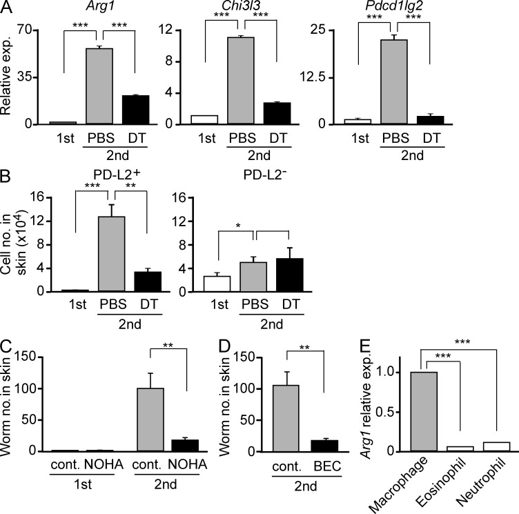
Once animals have experienced a helminthic infection, they often show stronger protective immunity against subsequent infections. Although helminthic infections are well known to elicit Th2-type immune responses, it remains ill-defined where and how acquired protection is executed. Here we show that skin-invading larvae of the intestinal helminth Nippostrongylus brasiliensis are surrounded by skin-infiltrating cells and are prevented from migrating out of infected skin during the second but not the first infection. B cell- or IgE receptor FcεRI-deficient mice showed impaired larval trapping in the skin. Selective ablation of basophils, but not mast cells, abolished the larval trapping, leading to increased worm burden in the lung and hence severe lung injury. Skin-infiltrating basophils produced IL-4 that in turn promoted the generation of M2-type macrophages, leading to the larval trapping in the skin through arginase-1 production. Basophils had no apparent contribution to worm expulsion from the intestine. This study thus reveals a novel mode of acquired antihelminth immunity, in which IgE-armed basophils mediate skin trapping of larvae, thereby limiting lung injury caused by larval migration.


Figures

Figure 1.
Figure 1.
Prominent infiltration of proinflammatory cells in the skin of the larva inoculation site during the second but not first Nb infection. (A) BALB/c mice were left uninfected (left) or infected once (middle) or twice (right) with Nb larvae. Photographs of the subcutaneous tissue of the larva inoculation site were taken 2 d after the first or second inoculation. Bars, 8 mm. (B) Single-cell suspensions were prepared from the skin of the larva inoculation site at the indicated time points after the first and second inoculation and subjected to the flow cytometric analysis to identify the nature of skin-infiltrating cells. The numbers of total and individual cell lineages at each time point are plotted (mean ± SEM; n = 3 each). Data shown in A and B are representative of at least three independent experiments. *, P < 0.05; **, P < 0.01; ***, P < 0.001.
Figure 2.
Figure 2.
Retention of Nb larvae within cellular infiltrates in the infected skin during the second but not first infection. (A and B) The skin of the larva inoculation site was isolated 2 d after the first or second inoculation and subjected to histopathological examination by hematoxylin and eosin staining. Black arrows in A indicate clusters of cells surrounding larvae, and the yellow arrow in B indicates a section of larva surrounded by infiltrating cells. (C) PKH-labeled larvae were inoculated into the skin of naive (left) or previously infected (right) BALB/c mice and subjected to in vivo imaging analysis to examine their retention in the skin of the inoculation site. Photographs were taken 2 d after the larva inoculation. Bars: (A) 500 µm; (B) 200 µm; (C) 1 mm. (D) The skin of the inoculation site was isolated 2 d after the first and second inoculation and subjected to RT-PCR analysis. The relative expression of Nb Actin mRNA is shown (mean ± SEM; n = 3 each), and the level of expression in first infection was set as 1. (E) Larvae were isolated from the skin of the inoculation site at the indicated time points after the first and second inoculation. The number of isolated larvae at each time point is plotted (mean ± SEM; n = 3 each). Data shown in A–E are representative of at least three independent experiments. *, P < 0.05; **, P < 0.01; ***, P < 0.001.
Figure 3.
Figure 3.
Antibodies and FcεRI but not FcγRIII are required for larval retention in the skin. (A–C) WT, μMT, Fcer1g−/−, or Fcgr3−/− C57BL/6 mice (A and B) and WT or Fcer1a−/− BALB/c mice (C) were infected twice with Nb larvae. The skin of the larva inoculation site was isolated on day 2 of the second infection, and the number of larvae was enumerated (mean ± SEM; n = 3 each). Data shown are representative of three independent experiments. **, P < 0.01; ***, P < 0.001.
Figure 4.
Figure 4.
Ablation of basophils but not mast cells abolishes larval retention in the skin. (A) WT and mast cell–deficient KitW-sh/W-sh C57BL/6 mice were infected twice with Nb, and larvae in the skin were enumerated on day 2 of the second infection. (B) The skin of the inoculation site in BALB/c mice was isolated on day 2 of the second infection and subjected to immunohistochemical analysis of tissue sections stained with anti–mMCP-8 (right, brown) or control (left) antibody. Yellow arrows indicate larvae trapped within clusters of cellular infiltrates. Data shown are representative of four independent experiments. Bars, 200 µm. (C and D) Mcpt8DTR BALB/c mice were treated with DT or vehicle (PBS) 1 d before the second inoculation. The skin of the larva inoculation site was isolated 2 d after the second inoculation, and infiltrating cells (C) and larvae (D) were enumerated. (E) Basophils and nonbasophil cells (gray bar) were separately isolated from the bone marrow and spleen of WT C57BL/6 mice 2 d after the second larval inoculation and intraperitoneally transferred into Fcer1g−/− C57BL/6 mice (4 × 10⁴ cells/mouse) that had been infected with larvae 18 d before. On the day of cell transfer, the recipient mice were subjected to the second Nb infection, and 2 d later the skin of the larva inoculation site was isolated and larvae were counted. Data shown in A and C–E are the mean ± SEM (n = 3 each) and are representative of at least three independent experiments. *, P < 0.05; ***, P < 0.001.
Figure 5.
Figure 5.
FcεRI on basophils is essential for larval retention in the skin and their activation in response to Nb antigens. (A) Basophils were isolated from the bone marrow and spleen of WT or Fcer1g−/− C57BL/6 mice 2 d after the second larval inoculation and adoptively transferred into Mcpt8DTR C57BL/6 mice (4 × 10⁴ cells/mouse) that had been infected with larvae 18 d before and treated with DT 1 d before. On the day of cell transfer, the recipient mice were subjected to the second Nb infection, and 2 d later the skin of the larva inoculation site was isolated and larvae were counted (mean ± SEM; n = 3 each). (B) Basophils were enriched from the spleen of uninfected (naive) BALB/c mice or mice infected once with Nb 18 d before (primed) and incubated ex vivo with Nb antigens or control OVA at 37°C for 20 min, followed by flow cytometric analysis for the CD63 expression on their surface. (C) Basophils were enriched from the spleen of primed WT or Fcer1a−/− BALB/c mice and incubated ex vivo with Nb antigens or control OVA at 37°C for 6 h, followed by flow cytometric analysis for intracellular IL-4. In B and C, the frequency (%) of cells positive for CD63 and IL-4 among the basophil population in each group is shown (mean ± SEM; n = 4 each). (D) G4 mice (open histograms) and C57BL/6 mice (shaded histograms) were infected twice with Nb larvae, the skin of the larva inoculation site was isolated on day 2 of the second infection, and GFP expression in the indicated cell types was analyzed by flow cytometry. (E) Mcpt8DTR BALB/c mice were infected with Nb once or twice and treated with DT or vehicle (PBS) 1 d before the second inoculation. The skin of the larva inoculation site was isolated 2 d after the inoculation and subjected to RT-PCR analysis for the Il4 expression (mean ± SEM; n = 4 each). The level of expression in the first infection was set as 1. Data shown in A–E are representative of at least two independent experiments. ***, P < 0.001.
Figure 6.
Figure 6.
Basophils promote the generation of M2-type macrophages expressing Arg1, which contributes to the larval trapping in the skin. (A and B) Mcpt8DTR BALB/c mice were infected with Nb once (white bars) or twice. Mice infected twice were treated with DT or control PBS 1 d before the second inoculation. (A) Gr-1loF4/80+FSChiSSClo macrophages were isolated from the skin of the larva inoculation site 2 d after the final inoculation, and expression of the indicated genes was analyzed by RT-PCR (mean ± SEM; n = 4 each). The level of expression in the first infection was set as 1. (B) PD-L2 expression on Gr-1loF4/80+FSChiSSClo macrophages in the skin lesion was analyzed by flow cytometry, and the numbers of PD-L2+ and PD-L2 macrophages were calculated (mean ± SEM; n = 4 each). (C) BALB/c mice were infected with Nb once or twice and treated twice with nor-NOHA or control PBS (cont.) before and after the final larva inoculation. The skin of the larva inoculation site was isolated 2 d after the final inoculation, and larvae were counted (mean ± SEM; n = 4). (D) BALB/c mice were infected twice with Nb and received drinking water containing BEC or not (cont.) during the second infection. The number of larvae trapped in the skin was determined as in C. (E) BALB/c mice were infected twice with Nb larvae. On day 2 of the second infection, macrophages (Gr-1loF4/80+FSChiSSClo), eosinophils (Gr-1loFSCloSSChi), and neutrophils (Gr-1hi) were separately isolated from the skin of the second inoculation site, and Arg1 expression was analyzed by RT-PCR (mean ± SEM; n = 4 each). The relative expression in macrophages was set as 1. Data shown in A–E are representative of two or three independent experiments. *, P < 0.05; **, P < 0.01; ***, P < 0.001.
Figure 7.
Figure 7.
Ablation of macrophages in the infected skin results in reduced Arg1 expression and impaired larval trapping. (A–C) BALB/c mice were infected twice with Nb and treated with clodronate or control PBS liposomes before the second Nb infection. The skin of the larva inoculation site was isolated 2 d after the second inoculation, and infiltrating cells (A) and larvae (C) were enumerated. (B) Expression of Arg1 was assessed by RT-PCR (mean ± SEM; n = 4). (D–F) WT and Ccr2−/− BALB/c mice were infected twice with Nb larvae. The skin of the larva inoculation site was isolated 2 d after the second inoculation, and infiltrating cells (D) and larvae (F) were enumerated. (E) Arg1 expression was analyzed by RT-PCR (mean ± SEM; n = 3 each). (G) Ccr2−/− BALB/c mice were infected twice with Nb larvae. In the second infection, 3 × 10⁵ monocytes (CD115+Ly6ChiLy6GCD11c) or control neutrophils (Ly6G+Siglec-FCD11c) sorted from bone marrow cells of naive BALB/c mice were adoptively transferred by intradermal injection together with larvae. On day 2 of the second infection, the number of larvae in the skin was counted (mean ± SEM; n = 3 each). (H) BALB/c mice were infected twice with Nb larvae and treated with anti–IL-4 or control rat IgG before the second inoculation. The skin of the larva inoculation site was isolated 2 d after the second inoculation, and larvae were counted (mean ± SEM; n = 3 each). (I) BALB/c mice were left uninfected or infected with Nb by subcutaneous injection of 500 larvae in the back and then percutaneously infected with Nb by laying 100 larvae on shaved abdominal skin for 1 h. The skin of the larva inoculation site was isolated 2 d after the final inoculation, and Nb Actin and mouse Arg1 mRNA expression was analyzed by RT-PCR (mean ± SEM; n = 3 each). The relative expression in the first infection was set as 1. Data shown in A–I are representative of two or three independent experiments. *, P < 0.05; **, P < 0.01; ***, P < 0.001.
Figure 8.
Figure 8.
Basophil ablation in the second Nb infection increases the worm burden in the lung and exacerbates lung injury. (A) BALB/c mice were infected with Nb once or twice, and larvae were isolated from the lung and enumerated at the indicated time points after the first and second larva inoculation (mean ± SEM; n = 3 each). (B–E) Mcpt8DTR BALB/c mice were treated with DT or control PBS 1 d before the second Nb inoculation. (B) The numbers of larvae isolated from the lung 2 d after the second inoculation (mean ± SEM; n = 4 each). (C) Photographs of the left lung lobe isolated 2 d after the second inoculation. Bar, 5 mm. (D) The number of RBCs in the bronchoalveolar lavage fluid (BALF) collected 2 d after the second inoculation (mean ± SEM; n = 4 each). (E) Tissue sections of lung isolated 2 d after the second inoculation and stained with hematoxylin and eosin. Data shown in A–E are representative of at least three independent experiments. *, P < 0.05; **, P < 0.01; ***, P < 0.001.
Figure 9.
Figure 9.
Little or no contribution of basophils to worm expulsion from the small intestine. (A and B) Mcpt8DTR BALB/c mice were infected with Nb once or twice and treated with an intravenous injection of DT or control PBS 1 d before (A) or 2 d after (B) the second larva inoculation. The numbers of worms recovered from the small intestine 5 d after the final inoculation are shown (mean ± SEM; n = 3 each). (C and D) BALB/c mice were left uninfected or infected twice with Nb or Hp. On days 5 and 7 of the second infection, the small intestine was isolated and subjected to immunohistochemical analysis using basophil-specific anti–mMCP-8 mAb or isotype-matched control antibody. Representative images are shown in C. Red arrowheads indicate mMCP-8–expressing basophils (brown), and black arrows indicate Hp larvae in the submucosa. Bars, 500 µm. The number of basophils detected in epithelia, lamina propria, and submucosa per 20 villus crypt units is summarized in D (mean ± SEM; n = 3 each). Data shown are representative of three independent experiments. ***, P < 0.001.

References

    1. Africa C.M. 1931. Studies on the host relations of Nippostrongylus muris with special reference to age resistance and acquired immunity. J. Parasitol. 18:1–13 10.2307/3271737 - DOI
    1. Akuthota P., Weller P.F. 2012. Eosinophilic pneumonias. Clin. Microbiol. Rev. 25:649–660 10.1128/CMR.00025-12 - DOI - PMC - PubMed
    1. Allen J.E., Maizels R.M. 2011. Diversity and dialogue in immunity to helminths. Nat. Rev. Immunol. 11:375–388 10.1038/nri2992 - DOI - PubMed
    1. Anthony R.M., Urban J.F., Jr, Alem F., Hamed H.A., Rozo C.T., Boucher J.L., Van Rooijen N., Gause W.C. 2006. Memory T(H)2 cells induce alternatively activated macrophages to mediate protection against nematode parasites. Nat. Med. 12:955–960 10.1038/nm1451 - DOI - PMC - PubMed
    1. Anthony R.M., Rutitzky L.I., Urban J.F., Jr, Stadecker M.J., Gause W.C. 2007. Protective immune mechanisms in helminth infection. Nat. Rev. Immunol. 7:975–987 10.1038/nri2199 - DOI - PMC - PubMed

Publication types

MeSH terms