. 2013 Nov 18;210(12):2611-25.
doi: 10.1084/jem.20120690. Epub 2013 Oct 28.

On-site education of VEGF-recruited monocytes improves their performance as angiogenic and arteriogenic accessory cells

Affiliations

On-site education of VEGF-recruited monocytes improves their performance as angiogenic and arteriogenic accessory cells

Inbal Avraham-Davidi et al. J Exp Med. .

Abstract

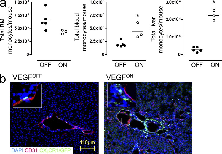
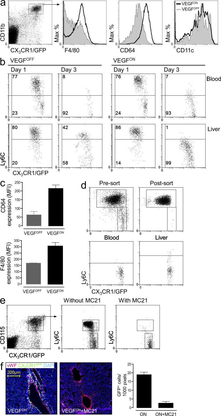
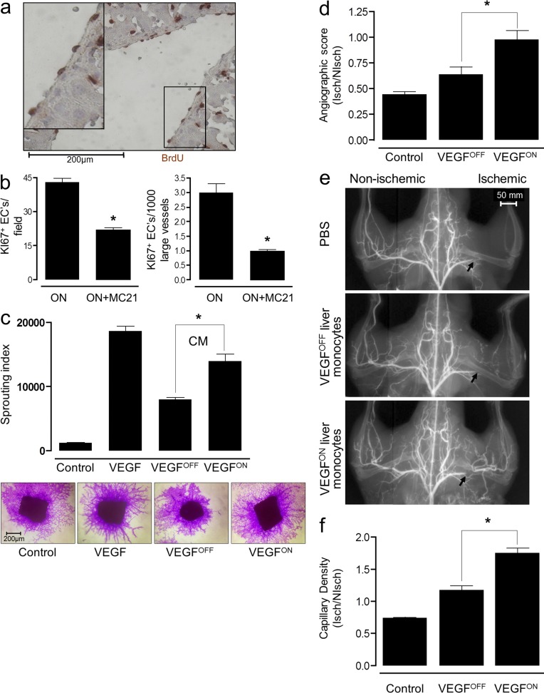
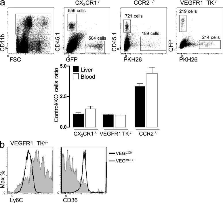
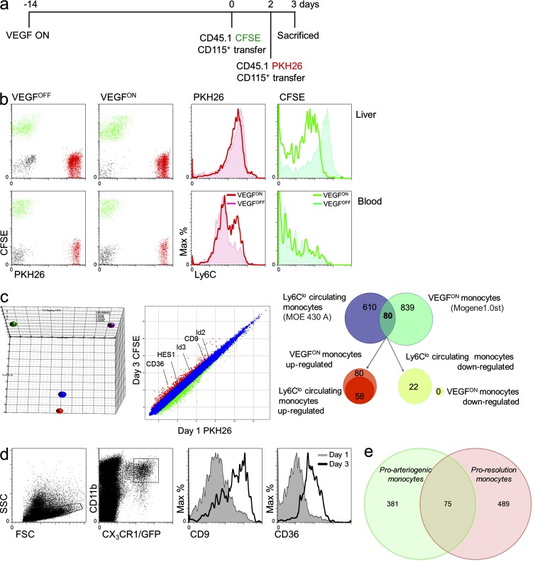
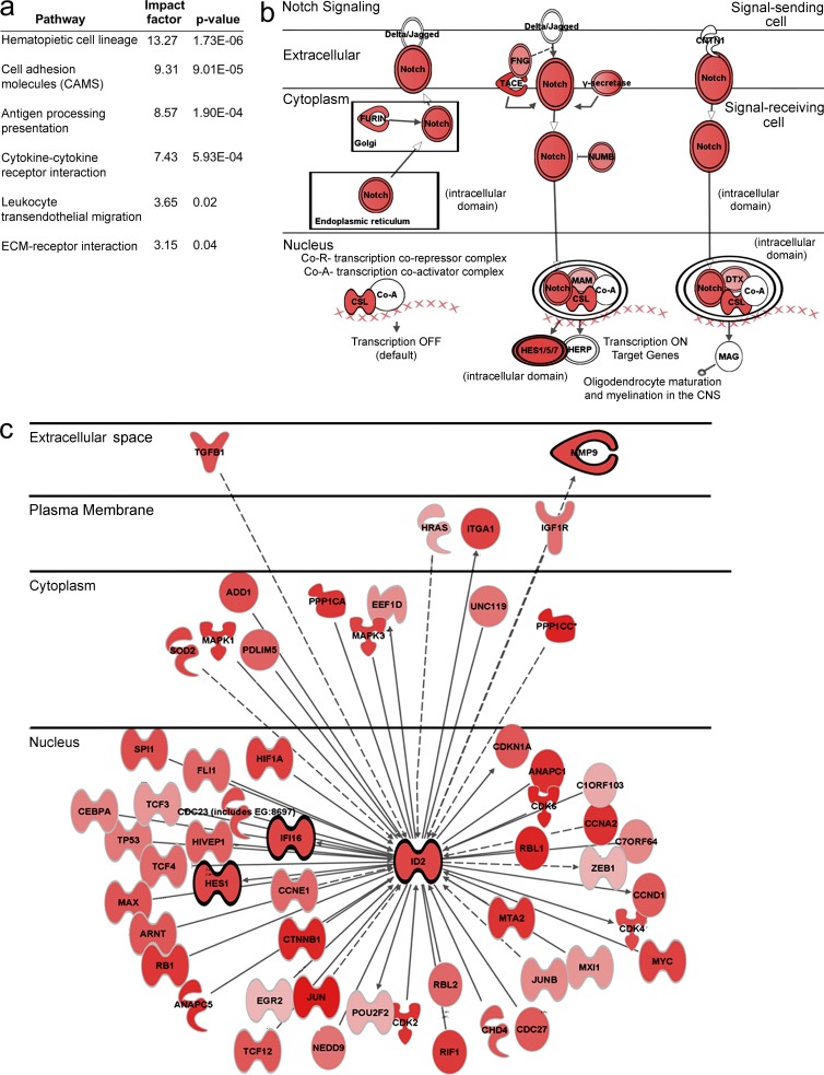
Adult neovascularization relies on the recruitment of monocytes to the target organ or tumor and functioning therein as a paracrine accessory. The exact origins of the recruited monocytes and the mechanisms underlying their plasticity remain unclear. Using a VEGF-based transgenic system in which genetically tagged monocytes are conditionally summoned to the liver as part of a VEGF-initiated angiogenic program, we show that these recruited cells are derived from the abundant pool of circulating Ly6C(hi) monocytes. Remarkably, however, upon arrival at the VEGF-induced organ, but not the naive organ, monocytes undergo multiple phenotypic and functional changes, endowing them with enhanced proangiogenic capabilities and, importantly, with a markedly increased capacity to remodel existing small vessels into larger conduits. Notably, monocytes do not differentiate into long-lived macrophages, but rather appear as transient accessory cells. Results from transfers of presorted subpopulations and a novel tandem transfer strategy ruled out selective recruitment of a dedicated preexisting subpopulation or onsite selection, thereby reinforcing active reprogramming as the underlying mechanism for improved performance. Collectively, this study uncovered a novel function of VEGF, namely, on-site education of recruited "standard" monocytes to become angiogenic and arteriogenic professional cells, a finding that may also lend itself for a better design of angiogenic therapies.


Figures

Figure 1.
Figure 1.
VEGF recruits circulating monocytes to a perivascular site in the angiogenic liver. (a) VEGF expression was induced in the liver of P-LAP-tTA+/tet-VEGF+:CX3CR1GFP/+mice as described in the Materials and methods. BM, circulating, and liver monocytes, identified as CX3CR1GFP/+, CD115+, and CD11b+ triple-positive cells, were analyzed by flow cytometry after 2 wk of VEGF induction. Total numbers of monocytes in the respective organs were compared with those detected in mice maintained in the VEGFOFF mode. Each data point represents an individual animal (n = 3–5), and the experiment was performed twice. *, P < 0.05 versus OFF mice. (b) Immunofluorescence micrographs of livers from VEGFOFF and VEGFON P-LAP-tTA+/tet-VEGF+:CX3CR1GFP/+ mice. Endothelial cells were highlighted by CD31 staining (red), whereas monocytes are identified on the basis of GFP positivity. Enlarged views of the insets further illustrate perivascular positioning of the large number of monocytes recruited by VEGF. Representative image of n = 3 mice per group performed twice.
Figure 2.
Figure 2.
Monocytes recruited by VEGF are Ly6Chi monocytes that dynamically change their surface markers after entrapment in the target organ. (a) Flow cytometry analysis of CX3CR1GFP-positive myeloid cells retrieved as described above from the livers of VEGFON and VEGFOFF mice with respect to expression of the indicated surface markers. Representative of n = 3 mice per group and performed twice. (b) Adoptive transfer of ∼1.5 × 106 CX3CR1GFP/+ BM monocytes to 6–8-wk-old P-LAP-tTA+/tet-VEGF+ mice in which hepatic expression of VEGF was induced 2 wk earlier. Grafted cells from the liver and from the blood of recipient mice were analyzed for their Ly6C expression 1 or 3 d later. Representative dot plot of n = 2–4 mice per group performed three times. (c) Mean expression levels of CD64 and F4/80 on adoptively transferred monocytes retrieved from VEGFOFF and VEGFON livers. Data from three independent experiments with n = 3 mice per group. (d) Ly6Chi CX3CR1GFP/+ BM monocytes were purified to 99% homogeneity through capture on CD115-coated magnetic beads followed by FACS sorting. Isolated cells were analyzed for their purity and adoptively transferred to VEGFON mice. 3 d after the transfer, grafted cells from the blood and the livers of recipient mice were analyzed for Ly6C expression. Representative dot plots of n = 3 mice per group performed twice. (e) Circulating Ly6Chi monocytes were depleted by daily injections of the mAb MC21 for 10 d in P-LAP-tTA+/tet-VEGF+:CX3CR1GFP/+ starting 1 d before the induction of VEGF. Note selective elimination of Ly6Chi, but not of Ly6Clo monocytes from the circulation. Representative dot plots of n = 2–3 mice per group and performed twice. (f) Immunofluorescence micrographs of liver sections from control- and MC21-treated mice with an ongoing VEGF on switch. This reduction in monocyte influx was quantified by counting the mean number of CX3CR1GFP/+ cells per 1,000 pixels of vWF+ blood vessels. Representative image of n = 2–3 mice per group and performed twice.
Figure 3.
Figure 3.
VEGF-recruited monocytes are short-lived. (a) BM CD115+ monocytes were isolated from CX3CR1GFP/+ donor mice and transferred to VEGFON mice. 1, 3, and 7 d after monocyte transfer, grafted cells from the livers of recipient mice were analyzed, and adoptively transferred cells were identified as having CD11b+ and GFP+ expression. Representative dot plots of n = 2 mice per time point per group, performed twice. (b) Monocytes retrieved as described in a at days 1 and 3 after transfer were analyzed by flow cytometry using Annexin V and Sytox and compared with monocytes retrieved from the circulation of the same mice. Representative of three independent individual mice per time point. (c) BM monocytes isolated from CD45.1 donor mice were labeled with the intracellular fluorescent dye CFSE before their transfer to VEGFOFF or VEGFON mice. 3 d after their transfer, CSFE-labeled cells retrieved from the liver or blood of recipient mice were analyzed by flow cytometry for CFSE and Ly6C expression. Note that there was no dilution of CSFE in monocytes from liver relative to monocytes from blood. Representative of n = 3 mice per group and performed twice.
Figure 4.
Figure 4.
VEGF education enhances both angiogenic and arteriogenic performance of recruited monocytes. (a) Endothelial cell proliferation in VEGFON (P-LAP-tTA+/tet-VEGF+) mice was visualized using BrdU immunohistochemistry. Representative n = 3 mice per group, performed twice. (b) Effects of MC21 treatment on VEGF-induced EC proliferation was measured as described above scoring proliferating EC by Ki67 immunostaining. Representative of n = 3 mice per group, performed twice. *, P < 0.05 versus ON mice alone. (c) Mouse aortic segments were embedded in collagen and overlaid with a serum-free control medium, serum-free medium containing 10 ng/ml VEGF, or serum-free medium conditioned by monocytes retrieved from VEGFOFF or VEGFON livers. Equal numbers of monocytes retrieved from VEGFOFF or VEGFON livers were used to obtain the conditioned media. Capillary sprouts were visualized by crystal violet staining and the mean length of capillary sprouts was measured. CM, conditioned medium. Representative of n = 4 mice group, performed three times. *, P < 0.05 versus OFF cells. (d–f) Mice underwent right femoral artery ligation and the effects of intraorbitally injected liver VEGFOFF, VEGFON monocytes (CD11b+ cells), or vehicle, angiogenic (f) and arteriogenic (d and e) responses were compared. *, P < 0.05 versus OFF cells. Representative of n = 6 mice per group, performed three times. (d and e) Arteriogenic response in the proximal region downstream of the ligation site (marked by an arrow in e) of the same animals was evaluated by microangiography. Representative microangiograms are shown in e and angiographic scores are shown in f, n = 6 mice per group, performed three times. *, P < 0.05 versus OFF cells. (f) Angiogenic response in the gastrocnemius muscle was evaluated by counting fibronectin-positive capillaries in both the ligated and contralateral limb. Results are expressed as ischemic to nonischemic microvascular density ratio. Representative of n = 6 mice per group, performed three times.
Figure 5.
Figure 5.
VEGF-instructed recruitment of monocytes nullified for VEGFR1, CX3CR1, and CCR2. (a) The respective mutant monocytes were isolated, mixed with WT monocytes at a 1:1 ratio, and adoptively transferred into mice with an ongoing VEGF switch (VEGFON) as described above. Monocytes were subsequently retrieved 3 d after transfer, and the number of recruited monocytes of each genotype was determined by flow cytometry. To aid distinction between recruited WT and mutant monocytes, the following monocyte mixtures were transferred: CX3CR1+/gfp: (PKH26)-VEGFR1TK−/−, CD45.1: (PKH26)-CCR2−/−, and CD45.1:CX3CR1gfp/gfp. Representative of n = 3 independent mice per group. (b) VEGFR1TK−/− monocytes were adoptively transferred into VEGFON and VEGFOFF mice as described above. 3 d after transfer, cells were retrieved and analyzed for their expression of the membrane markers Ly6C and CD36. Representative of n = 3 independent mice per group.
Figure 6.
Figure 6.
Transcriptomic changes associated with VEGF-instructed monocyte reprogramming. (a) Scheme of the experimental design: to follow the changes occurring in the monocyte-derived populations subsequent to their arrival to the VEGF-induced liver, tandem transfer strategy was designed. In this experiment ,the transfer of CD45.1+ BM monocytes labeled with CFSE (on day 0) was followed by transfer of CD45.1+ BM monocytes labeled with PKH26 (on day 2), and retrieval for analysis occurred a day later (on day 3). (b) Mice were killed on day 3, and the two populations of grafted cells were separated from both blood and liver (left). Analysis of Ly6C expression (right). Representative of n = 4 mice per group, performed twice. (c, left) PCA of naive and VEGF-educated monocytes. PCA analysis of adoptively transferred monocytes comparing input (naive) monocytes (red and blue circles of CFSE- and PKH26-tagged monocytes, respectively) and educated monocytes (purple and green circles representing monocytes retrieved at 72 and 24 h after transfer, respectively. Comparative transcriptomic analysis of CSFE-labeled (veteran/educated) and PKH26-labeled (newly arrived) monocytes highlights the down-regulation of genes between day 1 and 3 of residence in the liver and exposure to the VEGF milieu. (c, right) Differential of the differential. Analysis of the differential between newly arrived (Ly6Chi) and veteran/educated (Ly6Clo) monocytes and the differential between Ly6Chi and Ly6Clo circulating monocytes demonstrated an overlap of only 80 genes. 58 of these genes were up-regulated in both Ly6Clo subsets, whereas 22 genes were specifically up-regulated in the Ly6Clo VEGFON veteran monocytes. Cells were isolated as described in a and b and pooled from seven mice. Experiment was performed in duplicate. (d) Verification of transcriptome analysis, BM CD115+ monocytes were isolated from CX3CR1GFP/+ donors and transferred to VEGFON mice. 1 and 3 d after cell transfer, grafted cells from the livers of recipient mice were analyzed for their expression of CD9 and CD36. Representative of n = 3 mice per group per time point. (e) A Venn diagram comparing the transcriptomes of adoptively transferred Ly6Chi monocytes retrieved from VEGF-induced liver (green) or from a fibrotic liver (pink). Genes subjected for this comparison were all those showing >1.5-fold change (up or down) between 24 and 72 h from transfer. Data on fibrosis proresolution monocytes were obtained from Ramachandran et al. (2012).
Figure 7.
Figure 7.
Transcriptomic changes of adoptively transferred monocytes upon VEGF–instructed reprogramming. Transcriptomes of CSFE-labeled monocytes and PKH26-labeled monocytes obtained as described in Fig. 6 a using the tandem transfer strategy and representing relatively old and new arrivals to the liver, respectively, were compared. (a) GO analysis highlighting some pathways enriched in monocytes retrieved at day 3 relative to monocytes retrieved at day 1. (b and c) Inginuity software-aided analysis of components of the Notch and Id pathways, respectively. Genes surrounded by light ring are genes that were up-regulated between transferred cells and retrieved cells. Genes surrounded by dark ring are genes that were up-regulated between day 1 and day 3. The darker the red the more pronounced the up-regulation.

References

    1. Arnold L., Henry A., Poron F., Baba-Amer Y., van Rooijen N., Plonquet A., Gherardi R.K., Chazaud B. 2007. Inflammatory monocytes recruited after skeletal muscle injury switch into antiinflammatory macrophages to support myogenesis. J. Exp. Med. 204:1057–1069 10.1084/jem.20070075 - DOI - PMC - PubMed
    1. Boring L., Gosling J., Chensue S.W., Kunkel S.L., Farese R.V., Jr, Broxmeyer H.E., Charo I.F. 1997. Impaired monocyte migration and reduced type 1 (Th1) cytokine responses in C-C chemokine receptor 2 knockout mice. J. Clin. Invest. 100:2552–2561 10.1172/JCI119798 - DOI - PMC - PubMed
    1. Cai W., Schaper W. 2008. Mechanisms of arteriogenesis. Acta Biochim. Biophys. Sin. (Shanghai). 40:681–692 - PubMed
    1. De Palma M., Venneri M.A., Roca C., Naldini L. 2003. Targeting exogenous genes to tumor angiogenesis by transplantation of genetically modified hematopoietic stem cells. Nat. Med. 9:789–795 10.1038/nm871 - DOI - PubMed
    1. De Palma M., Venneri M.A., Galli R., Sergi Sergi L., Politi L.S., Sampaolesi M., Naldini L. 2005. Tie2 identifies a hematopoietic lineage of proangiogenic monocytes required for tumor vessel formation and a mesenchymal population of pericyte progenitors. Cancer Cell. 8:211–226 10.1016/j.ccr.2005.08.002 - DOI - PubMed

Publication types

MeSH terms

Substances