. 2013 Nov 12;110(46):18513-8.
doi: 10.1073/pnas.1313491110. Epub 2013 Oct 28.

Keratins as the main component for the mechanical integrity of keratinocytes

Affiliations

Keratins as the main component for the mechanical integrity of keratinocytes

Lena Ramms et al. Proc Natl Acad Sci U S A. .

Abstract

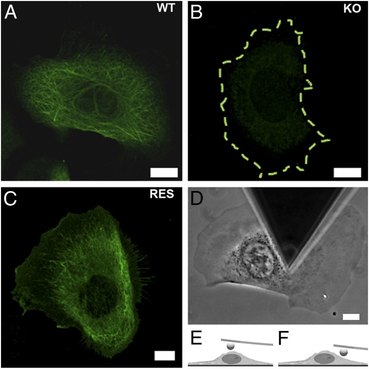
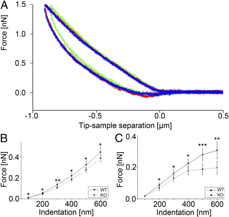
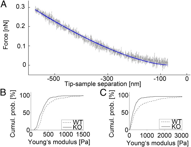
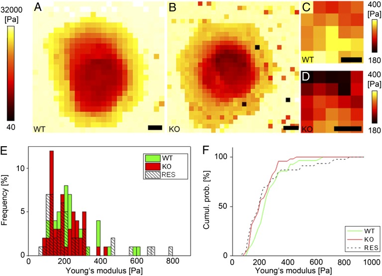
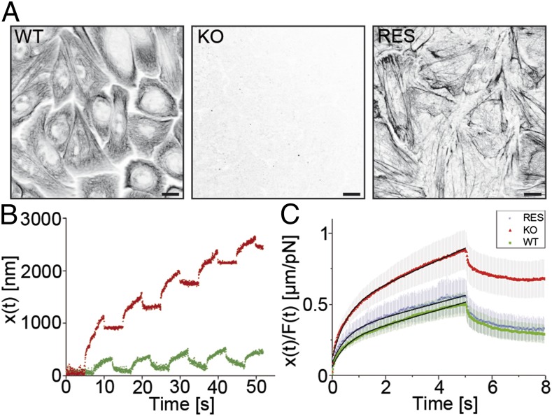
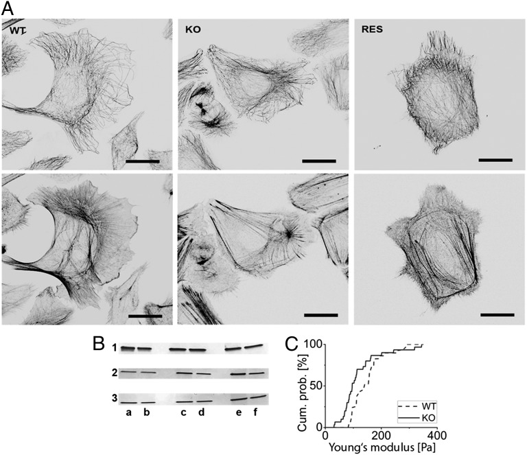
Keratins are major components of the epithelial cytoskeleton and are believed to play a vital role for mechanical integrity at the cellular and tissue level. Keratinocytes as the main cell type of the epidermis express a differentiation-specific set of type I and type II keratins forming a stable network and are major contributors of keratinocyte mechanical properties. However, owing to compensatory keratin expression, the overall contribution of keratins to cell mechanics was difficult to examine in vivo on deletion of single keratin genes. To overcome this problem, we used keratinocytes lacking all keratins. The mechanical properties of these cells were analyzed by atomic force microscopy (AFM) and magnetic tweezers experiments. We found a strong and highly significant softening of keratin-deficient keratinocytes when analyzed by AFM on the cell body and above the nucleus. Magnetic tweezers experiments fully confirmed these results showing, in addition, high viscous contributions to magnetic bead displacement in keratin-lacking cells. Keratin loss neither affected actin or microtubule networks nor their overall protein concentration. Furthermore, depolymerization of actin preserves cell softening in the absence of keratin. On reexpression of the sole basal epidermal keratin pair K5/14, the keratin filament network was reestablished, and mechanical properties were restored almost to WT levels in both experimental setups. The data presented here demonstrate the importance of keratin filaments for mechanical resilience of keratinocytes and indicate that expression of a single keratin pair is sufficient for almost complete reconstitution of their mechanical properties.


Conflict of interest statement

The authors declare no conflict of interest.

Figures

Fig. 1.
Fig. 1.
Keratin network characterization and AFM analysis regions. (A–C) Immunfluorescence micrographs of the keratin network in WT, KtyI−/− KO, and K14 rescue (RES) keratinocytes. The KtyI−/− cell outline is given by a dashed line. (Scale bars, 10 µm.) (D) Top view phase contrast image of an AFM tip-less cantilever aligned with the cell body of a living adherent keratinocyte. (Scale bar, 10 µm.) (E) Side view diagram of a cantilever positioned above the nucleus and (F) the cell body.
Fig. 2.
Fig. 2.
AFM force-distance curves and force-indentation plots. (A) Plot showing the superposition of three AFM force-distance curves (green, red, blue) recorded successively at 5-s intervals on the same position above the nucleus of a WT keratinocyte. (B) Plot showing the average forces needed to reach a certain indentation depth above the nucleus (n = 109–125, depending on indentation depth) and (C) cell body (n = 54–117). Significant differences between WT and KtyI−/− cells (KO) are indicated (*P = 0.05, **P = 0.01, ***P = 0.001); error bars are SEM.
Fig. 3.
Fig. 3.
(A) AFM force distance curve (gray) and data fit to Hertz model (blue). (B) Cumulative histogram of Young's modulus E obtained from indentation experiments performed on the nucleus of WT (n = 110) and KtyI−/− (KO; n = 125) cells. (C) Same plots showing data measured on the cell body (nWT = 117, nKO = 113).
Fig. 4.
Fig. 4.
Stiffness maps and rescue measurements. Stiffness maps of a live WT (A) and KtyI−/− (KO) (B) cell displaying the Young's moduli computed at every position. (Scale bar, 10 µm.) (C and D) Zoom-ins of the nuclear region of A and B, respectively. (Scale bars, 3 µm.) (E) Histogram showing the distribution of Young's moduli obtained from indentation experiments performed on the nucleus of WT (n = 58), KO (n = 48), and RES (n = 44) cells and (F) corresponding cumulative histograms.
Fig. 5.
Fig. 5.
Keratin network localization and magnetic tweezers analyses. (A) Immunfluorescence micrographs of the keratin network of WT, KO (KtyII−/−), and K5 rescue (Res) cells. (Scale bars, 10 µm.) (B) Example of raw magnetic tweezers data: displacement of the superparamagnetic bead incorporated in the cytoplasm of a WT and a KtyII−/− cell (KO) following the application of 5-s force pulses exerted at 10-s intervals. (C) Average overall rescaled peaks recorded on the three phenotypes as a function of time (nWT = 40 for 9 cells; nKO = 39 for 10 cells; nRES = 22 for 6 cells). In black, fits of Eq. 3. Error bars are SEM.
Fig. 6.
Fig. 6.
Independency of biomechanical deficiencies on actin and microtubules. (A) Immunofluorescent staining of microtubules (Upper) and actin (Lower) in WT, KtyI−/− (KO), and rescue cells. (B) Crude protein extract analysis by Western blotting. Protein concentrations were equalized using GAPDH as constitutively expressed marker (1) and subsequently analyzed for tubulin (2) and actin (3) expression. (a and b) Corresponding WTs. (c) KtyI−/−. (d) KtyII−/−. (e) K14 rescue. (f) K5 rescue. (C) Elasticity measurements above the nucleus of WT and KtyI−/− cells by AFM after depolymerization of actin filaments by latrunculin A. n = 30 cells each. (Scale bars, 20 µm.)

Similar articles

Cited by

References

    1. Fuchs E. Keratins and the skin. Annu Rev Cell Dev Biol. 1995;11:123–153. - PubMed
    1. Sun TT, Shih C, Green H. Keratin cytoskeletons in epithelial cells of internal organs. Proc Natl Acad Sci USA. 1979;76(6):2813–2817. - PMC - PubMed
    1. Kasza KE, et al. The cell as a material. Curr Opin Cell Biol. 2007;19(1):101–107. - PubMed
    1. Stricker J, Falzone T, Gardel ML. Mechanics of the F-actin cytoskeleton. J Biomech. 2010;43(1):9–14. - PMC - PubMed
    1. Beil M, et al. Sphingosylphosphorylcholine regulates keratin network architecture and visco-elastic properties of human cancer cells. Nat Cell Biol. 2003;5(9):803–811. - PubMed

Publication types

MeSH terms