. 2013 Nov 12;110(46):18644-9.
doi: 10.1073/pnas.1318257110. Epub 2013 Oct 28.

Regulation of bone remodeling by vasopressin explains the bone loss in hyponatremia

Affiliations

Regulation of bone remodeling by vasopressin explains the bone loss in hyponatremia

Roberto Tamma et al. Proc Natl Acad Sci U S A. .

Erratum in

  • Proc Natl Acad Sci U S A. 2014 Sep 23;111(38):14002

Abstract

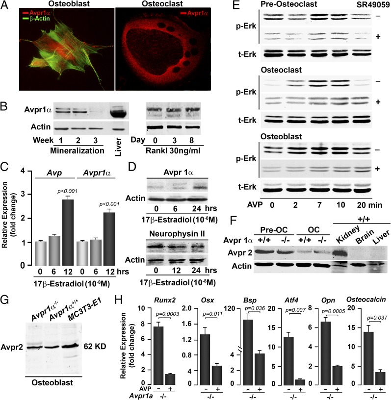
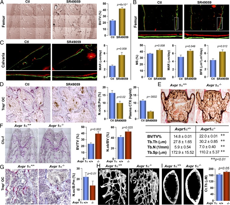
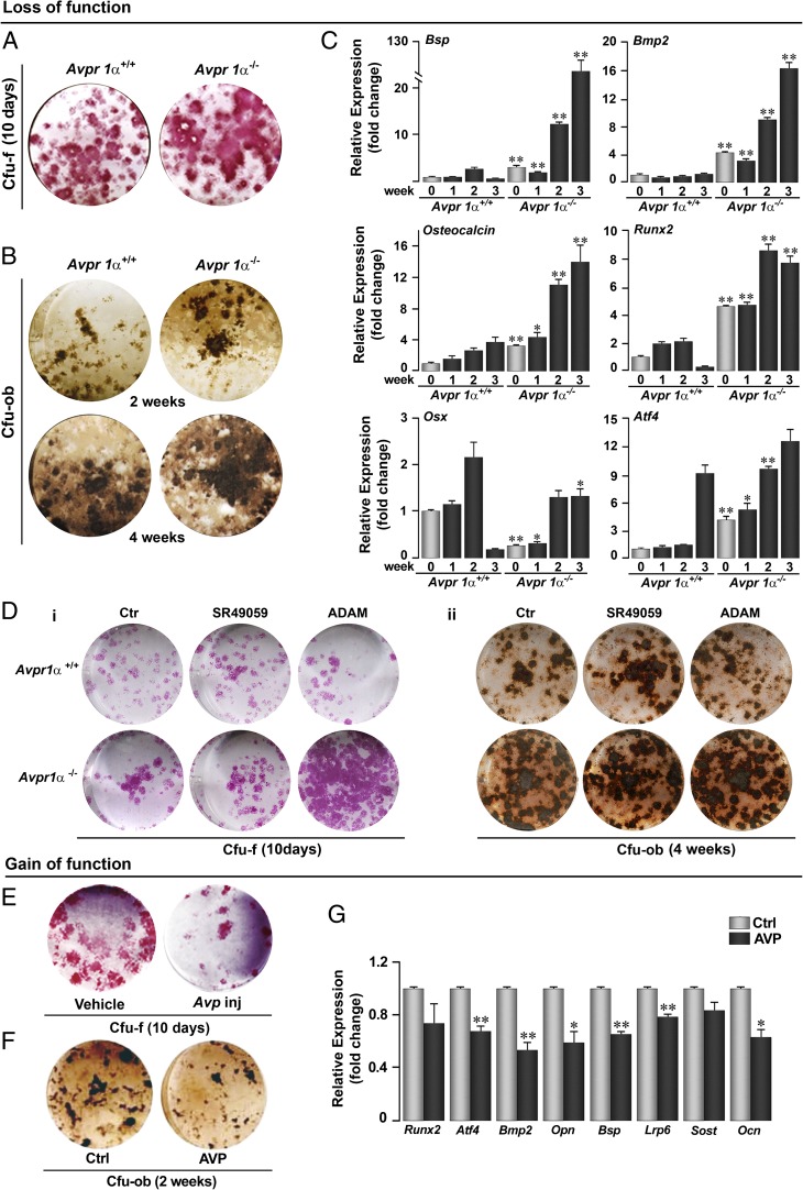
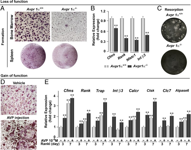
Although hyponatremia is known to be associated with osteoporosis and a high fracture risk, the mechanism through which bone loss ensues has remained unclear. As hyponatremic patients have elevated circulating arginine-vasopressin (AVP) levels, we examined whether AVP can affect the skeleton directly as yet another component of the pituitary-bone axis. Here, we report that the two Avp receptors, Avpr1α and Avpr2, coupled to Erk activation, are expressed in osteoblasts and osteoclasts. AVP injected into wild-type mice enhanced and reduced, respectively, the formation of bone-resorbing osteoclasts and bone-forming osteoblasts. Conversely, the exposure of osteoblast precursors to Avpr1α or Avpr2 antagonists, namely SR49059 or ADAM, increased osteoblastogenesis, as did the genetic deletion of Avpr1α. In contrast, osteoclast formation and bone resorption were both reduced in Avpr1α(-/-) cultures. This process increased bone formation and reduced resorption resulted in a profound enhancement of bone mass in Avpr1α(-/-) mice and in wild-type mice injected with SR49059. Collectively, the data not only establish a primary role for Avp signaling in bone mass regulation, but also call for further studies on the skeletal actions of Avpr inhibitors used commonly in hyponatremic patients.

Keywords: osteopenia; oxytocin; pituitary hormone.


Conflict of interest statement

The authors declare no conflict of interest.

Figures

Fig. 1.
Fig. 1.
Bone cells express Avprs. Immunofluorescence micrographs (A) and Western immunoblotting (B) show the expression of Avpr1α in osteoblasts and osteoclasts, and as a function of osteoblast (mineralization) and osteoclast (with Rankl) differentiation. The expression of Avp (ligand) and Avpr1α (receptor) in osteoblasts is regulated by 17β-estradiol, as determined by quantitative PCR (C) and Western immunoblotting (D). (Magnification: A, 63×.) Because Avp is a small peptide, its precursor neurophysin II is measured. Statistics: Student t test, P values shown compared with 0 h. Stimulation of Erk phosphorylation (p-Erk) as a function of total Erk (t-Erk) by Avp (10−8 M) in osteoclast precursors (preosteoclasts), osteoclasts (OC) and osteoblasts establishes functionality of the Avpr1α in the presence or absence of the receptor inhibitor SR49059 (10−8 M) (E). Western immunoblotting showing the expression of Avpr2 in preosteoclasts, OCs (F), and osteoblasts (G) isolated from Avpr1α−/− mice, as well as in MC3T3.E1 osteoblast precursors (G). Functionality of Avpr2 was confirmed by the demonstration that cells from Avpr1a−/− mice remained responsive to AVP in reducing the expression of osteoblast differentiation genes, namely Runx2, Osx, Bsp, Atf4, Opn, and Osteocalcin (quantitative PCR, P values shown) (H).
Fig. 2.
Fig. 2.
Avprs regulate bone remodeling and bone mass. Pharmacologic inhibition of Avpr1α by the injection of the receptor antagonist SR49059 (or vehicle, Ctl) increases bone mass (unstained sections, quantitated as BV/TV) (A); bone formation (photomicrograph of xylenol/calcein-labeled bones), measured as mineralizing surface, MAR, and bone formation rate in femur epiphyseal bone (B), and MAR in calvarial bone (C); and bone resorption in TRAP-labeled sections measured as osteoclast surfaces (N.oc/B.Pm) and serum C- telopeptide (CTX, by ELISA) (D). Statistics: Unpaired Student t test, P values shown, n = 4–5 mice per group. Consistent with this, Avpr1α−/− mice compared with Avpr1α+/+ littermates (E) showed a profound increase in trabecular bone in the vertebral column (von Kossa stained section; BV/TV, Tb.Th, Tb.N., and Tb.Sp.) and in the femur epiphysis (alkaline phosphatase-stained section, BV/TV); this is accompanied by an increase and decrease, respectively, in Cfu-f and N.ob./BPm (F), and osteoclast numbers (Trap-stained surface, N.oc/B.Pm) (G). Microtomography (H) confirms the increase in bone mass in both trabecular [femur metaphysis and cortical bone (Ct.Th)] (I). Unpaired Student t test, P values shown, n = 5 mice per group. (Magnification: AC and F, 10×; D, 40×; E, 2×; G, 20×; H, 4×.)
Fig. 3.
Fig. 3.
Avpr1α and Avpr2 regulate osteoblastogenesis. Genetic deletion of Avpr1α in Avpr1α−/− mice causes a dramatic increase in osteoblastogenesis, measured by alkaline phosphatase-positive Cfu-f (10 d) (A) and von Kossa-positive Cfu-ob (2 and 4 wk) (B). For Cfu-f and Cfu-ob assays, bone marrow stromal cells were grown in α-MEM and 15% (vol/vol) FCS with acorbate-2-phosphate (1 mM) (Materials and Methods). (C) The enhanced osteoblastogenesis was confirmed by a strong up-regulation of differentiation genes (quantitative PCR), namely Bsp, Bmp2, Osteocalcin, Runx2, Osx, and Atf4. (D) Effect of inhibitors of Avpr1α (SR49059) or Avpr2 (ADAM) on Cfu-f (i, 10 d) and Cfu-ob (ii, 4 wk) formation. (E) Ex vivo Cfu-f formation (10 d) in mice injected with AVP (4 µg per mouse, twice, 6-wk-old, killed 12 h after second injection). Cfu-ob formation (F) and expression of Runx2, Atf4, Bmp2, Opn, Bsp, Lrp6, Sost, and Ocn (G) (at 2 wk) in the presence of AVP in vitro. Statistics: Unpaired Student t test, corrected by Bonferroni’s; *P < 0.05; **P < 0.01, comparison between Avpr1α+/+ and Avpr1α−/− cells or between Ctl and AVP. (Magnification: all stained images, 1×.)
Fig. 4.
Fig. 4.
Avpr1α regulates osteoclast formation and function. Genetic deletion of Avpr1α in Avpr1α−/− mice causes a dramatic reduction in osteoclastogenesis in bone marrow and spleen cell cultures (A), which is accompanied by a reduction in the expression (quantitative PCR) of differentiation genes, namely Cfms, Rank, Nfatc1, and Int β3 (B), and by a reduction in bone resorption by mature osteoclasts plated on dentine slices (von Kossa-stained) (C). Gain-of-function experiments evaluating the stimulatory effect of AVP injections (4 µg per mouse, twice, 6-wk-old mice killed 12 h after second injection) on TRAP-positive osteoclast formation in bone marrow cell cultures treated with Rankl (30 ng/mL) (D), and on osteoclast genes, namely Cfms, Rank, Trap, Int β3, Calcr, Ctsk, Clc7, and Atpase6 (E). Statistics by Unpaired Student t test; **P < 0.01 vs. vehicle (−, no AVP) treated at 3 and 7 d of culture. (Magnification: A, Upper, and D, 10×; A, Lower, and C, 2×.)

References

    1. Abe E, et al. TSH is a negative regulator of skeletal remodeling. Cell. 2003;115(2):151–162. - PubMed
    1. Sun L, et al. FSH directly regulates bone mass. Cell. 2006;125(2):247–260. - PubMed
    1. Zaidi M, et al. ACTH protects against glucocorticoid-induced osteonecrosis of bone. Proc Natl Acad Sci USA. 2010;107(19):8782–8787. - PMC - PubMed
    1. Tamma R, et al. Oxytocin is an anabolic bone hormone. Proc Natl Acad Sci USA. 2009;106(17):7149–7154. - PMC - PubMed
    1. Zaidi M, et al. Primer on the Metabolic Bone Disease and Disorders of Mineral Metabolism. 2013. The pituitary-bone connection. 8th ed. eds Rosen CJ, Bouillon R, Compston JE, Rosen V (Wiley-Blackwell, Hoboken, NJ), pp 969–977.

Publication types

Substances