

Keratins significantly contribute to cell stiffness and impact invasive behavior
- PMID: 24167274
- PMCID: PMC3832002
- DOI: 10.1073/pnas.1310493110
Keratins significantly contribute to cell stiffness and impact invasive behavior
Abstract
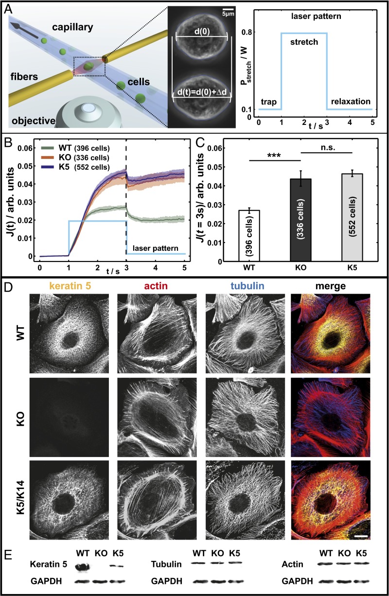
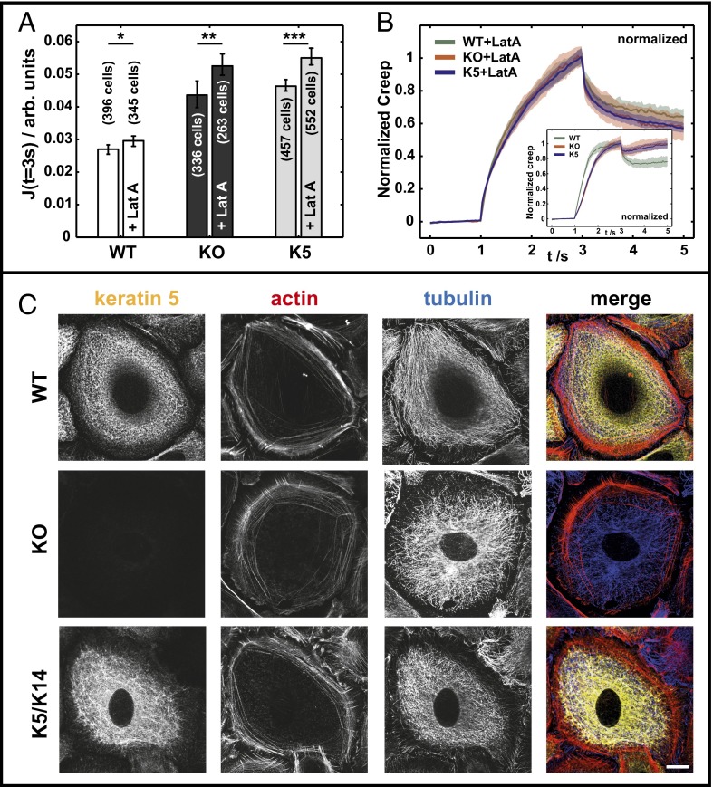
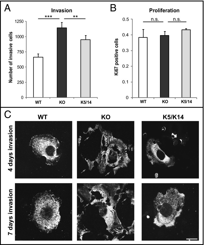
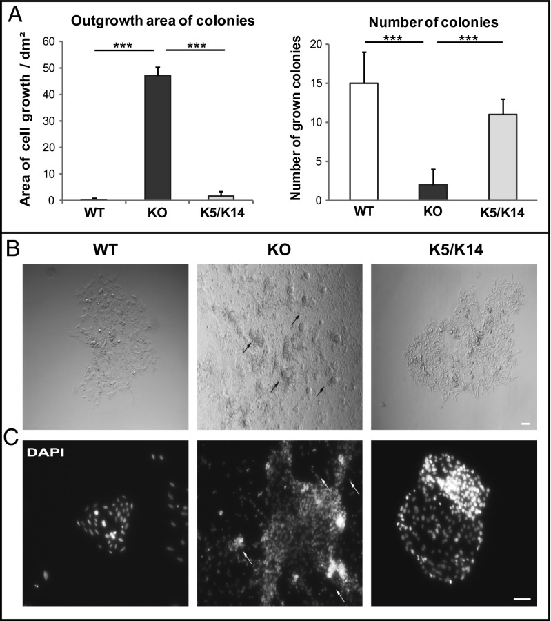
Cell motility and cell shape adaptations are crucial during wound healing, inflammation, and malignant progression. These processes require the remodeling of the keratin cytoskeleton to facilitate cell-cell and cell-matrix adhesion. However, the role of keratins for biomechanical properties and invasion of epithelial cells is only partially understood. In this study, we address this issue in murine keratinocytes lacking all keratins on genome engineering. In contrast to predictions, keratin-free cells show about 60% higher cell deformability even for small deformations. This response is compared with the less pronounced softening effects for actin depolymerization induced via latrunculin A. To relate these findings with functional consequences, we use invasion and 3D growth assays. These experiments reveal higher invasiveness of keratin-free cells. Reexpression of a small amount of the keratin pair K5/K14 in keratin-free cells reverses the above phenotype for the invasion but does not with respect to cell deformability. Our data show a unique role of keratins as major players of cell stiffness, influencing invasion with implications for epidermal homeostasis and pathogenesis. This study supports the view that down-regulation of keratins observed during epithelial-mesenchymal transition directly contributes to the migratory and invasive behavior of tumor cells.
Keywords: Boyden chamber; cell mechanics; intermediate filaments; optical stretcher.
Conflict of interest statement
The authors declare no conflict of interest.
Figures
Similar articles
-
Keratins as the main component for the mechanical integrity of keratinocytes.Proc Natl Acad Sci U S A. 2013 Nov 12;110(46):18513-8. doi: 10.1073/pnas.1313491110. Epub 2013 Oct 28. Proc Natl Acad Sci U S A. 2013. PMID: 24167246 Free PMC article.
-
Keratins mediate localization of hemidesmosomes and repress cell motility.J Invest Dermatol. 2013 Jan;133(1):181-90. doi: 10.1038/jid.2012.256. Epub 2012 Aug 16. J Invest Dermatol. 2013. PMID: 22895363 Free PMC article.
-
Expression of complete keratin filaments in mouse L cells augments cell migration and invasion.Proc Natl Acad Sci U S A. 1993 May 1;90(9):4261-5. doi: 10.1073/pnas.90.9.4261. Proc Natl Acad Sci U S A. 1993. PMID: 7683431 Free PMC article.
-
Keratin expression by corneal and limbal stem cells during development.Exp Eye Res. 2020 Nov;200:108206. doi: 10.1016/j.exer.2020.108206. Epub 2020 Aug 31. Exp Eye Res. 2020. PMID: 32882212 Review.
-
Keratin intermediate filaments: intermediaries of epithelial cell migration.Essays Biochem. 2019 Oct 31;63(5):521-533. doi: 10.1042/EBC20190017. Essays Biochem. 2019. PMID: 31652439 Review.
Cited by
-
A Drosophila Model of Epidermolysis Bullosa Simplex.J Invest Dermatol. 2015 Aug;135(8):2031-2039. doi: 10.1038/jid.2015.129. Epub 2015 Apr 1. J Invest Dermatol. 2015. PMID: 25830653 Free PMC article.
-
How cancer invasion takes shape.Nature. 2020 Sep;585(7825):355-356. doi: 10.1038/d41586-020-02490-3. Nature. 2020. PMID: 32879478 No abstract available.
-
Roadmap to Local Tumour Growth: Insights from Cervical Cancer.Sci Rep. 2019 Sep 4;9(1):12768. doi: 10.1038/s41598-019-49182-1. Sci Rep. 2019. PMID: 31484955 Free PMC article. Clinical Trial.
-
Interaction of plectin with keratins 5 and 14: dependence on several plectin domains and keratin quaternary structure.J Invest Dermatol. 2014 Nov;134(11):2776-2783. doi: 10.1038/jid.2014.255. Epub 2014 Jun 18. J Invest Dermatol. 2014. PMID: 24940650
-
Directed expression of a chimeric type II keratin partially rescues keratin 5-null mice.J Biol Chem. 2014 Jul 11;289(28):19435-47. doi: 10.1074/jbc.M114.553867. Epub 2014 May 27. J Biol Chem. 2014. PMID: 24867950 Free PMC article.
References
-
- Fuchs E, Cleveland DW. A structural scaffolding of intermediate filaments in health and disease. Science. 1998;279(5350):514–519. - PubMed
-
- Herrmann H, Hesse M, Reichenzeller M, Aebi U, Magin TM. Functional complexity of intermediate filament cytoskeletons: From structure to assembly to gene ablation. Int Rev Cytol. 2003;223:83–175. - PubMed
-
- Fuchs E, Green H. Changes in keratin gene expression during terminal differentiation of the keratinocyte. Cell. 1980;19(4):1033–1042. - PubMed
Publication types
MeSH terms
Substances
LinkOut - more resources
Full Text Sources
Other Literature Sources
Research Materials
