. 2013 Nov 12;110(46):E4355-61.
doi: 10.1073/pnas.1307088110. Epub 2013 Oct 28.

A neuropeptide speeds circadian entrainment by reducing intercellular synchrony

Affiliations

A neuropeptide speeds circadian entrainment by reducing intercellular synchrony

Sungwon An et al. Proc Natl Acad Sci U S A. .

Abstract

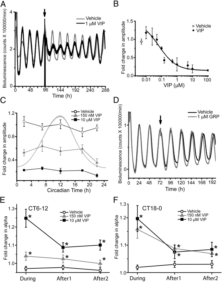
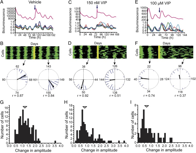
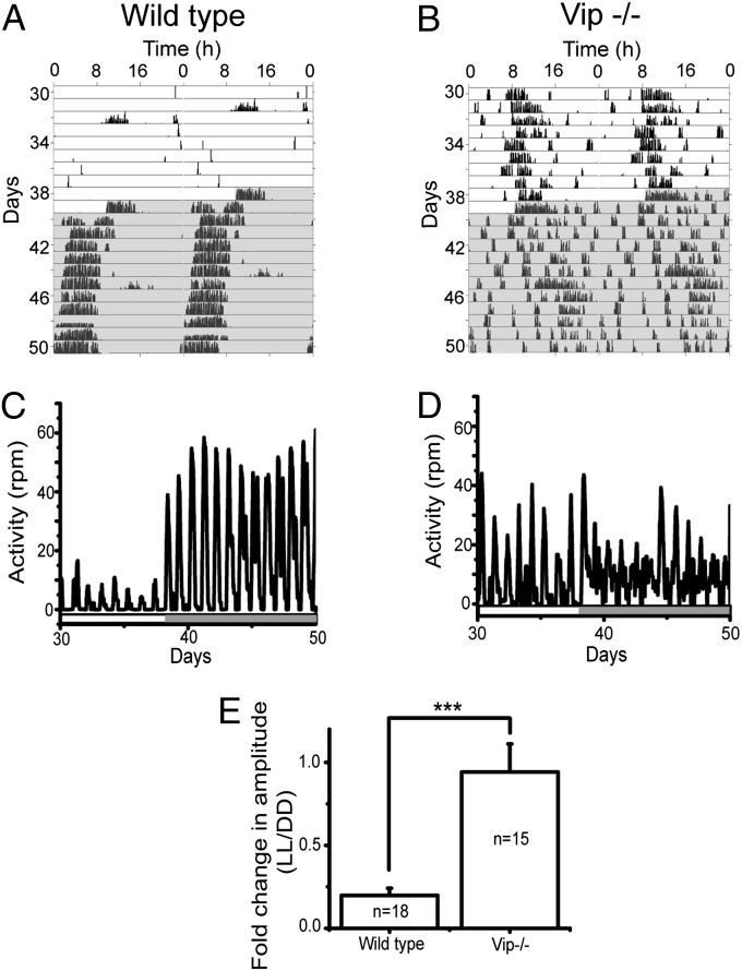
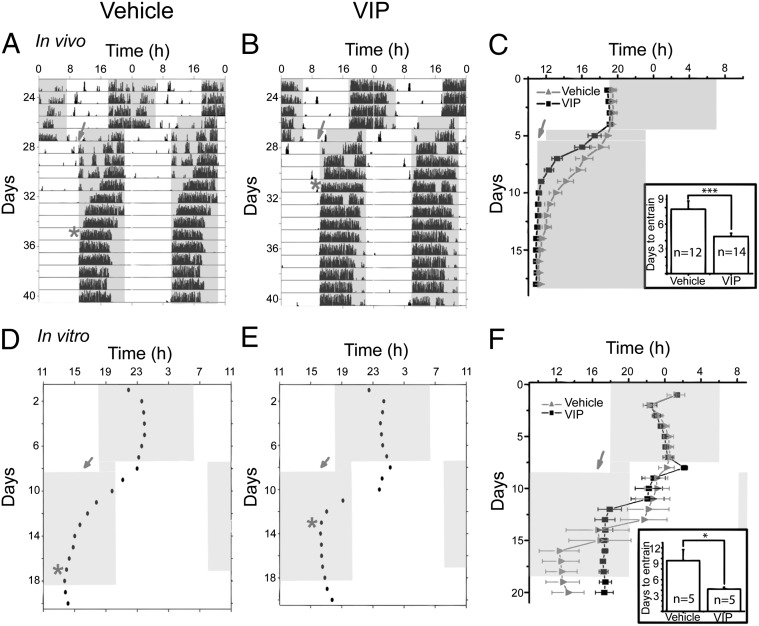
Shift work or transmeridian travel can desynchronize the body's circadian rhythms from local light-dark cycles. The mammalian suprachiasmatic nucleus (SCN) generates and entrains daily rhythms in physiology and behavior. Paradoxically, we found that vasoactive intestinal polypeptide (VIP), a neuropeptide implicated in synchrony among SCN cells, can also desynchronize them. The degree and duration of desynchronization among SCN neurons depended on both the phase and the dose of VIP. A model of the SCN consisting of coupled stochastic cells predicted both the phase- and the dose-dependent response to VIP and that the transient phase desynchronization, or "phase tumbling", could arise from intrinsic, stochastic noise in small populations of key molecules (notably, Period mRNA near its daily minimum). The model also predicted that phase tumbling following brief VIP treatment would accelerate entrainment to shifted environmental cycles. We tested this using a prepulse of VIP during the day before a shift in either a light cycle in vivo or a temperature cycle in vitro. Although VIP during the day does not shift circadian rhythms, the VIP pretreatment approximately halved the time required for mice to reentrain to an 8-h shifted light schedule and for SCN cultures to reentrain to a 10-h shifted temperature cycle. We conclude that VIP below 100 nM synchronizes SCN cells and above 100 nM reduces synchrony in the SCN. We show that exploiting these mechanisms that transiently reduce cellular synchrony before a large shift in the schedule of daily environmental cues has the potential to reduce jet lag.

Keywords: biological clock; circadian oscillator; period gene; vasoactive intestinal peptide; vasopressin.


Conflict of interest statement

The authors declare no conflict of interest.

Figures

Fig. 1.
Fig. 1.
VIP dose-dependently reduces the amplitude of circadian rhythms in the SCN. (A) Representative detrended bioluminescence traces from PER2::LUC SCN explants were treated with 1 μM VIP (solid line) or vehicle (shaded line) delivered near the peak of PER2 expression (CT12, arrow). Note that the amplitude of the VIP-treated SCN decreased and, then, gradually recovered. Each trace was normalized to the peak before treatment. (B) The dose-dependent amplitude decrease (mean ± SEM; n = 3–5 cultures at each dose) by VIP application at CT12. Between 150 nM and 10 μM VIP, the amplitude decreased linearly with logarithmic increases in VIP concentration. Data were fitted with a logistic function (solid line). Amplitude was measured as the trough-to-peak magnitude 48 h after VIP application. (C) The amplitude reduction of PER2 cycling (mean ± SEM) was greater following 10 µM VIP (squares; n = 20) than 150 nM VIP (triangles; n = 16) at all times (P < 0.00001, F6,66 = 38.53, n = 74; two-way ANOVA with a Scheffé post hoc). Notably, 10 µM VIP delivery at CT22 had a larger effect on amplitude than at other times (P < 0.03, F4,14 = 3.87, n = 19; one-way ANOVA with a Scheffé post hoc). Vehicle (open circles) did not reduce the amplitude at any time. The shaded line corresponds to a PER2-expression rhythm peaking at CT12. (D) Representative bioluminescence traces from SCN explants treated at the peak of PER2 expression (arrow) showing that another neuropeptide, 1 μM gastrin-releasing peptide (GRP) (solid line), did not reduce amplitude compared with vehicle (shaded line) applied at CT12. (E) VIP application transiently broadens the waveform of PER2 expression. The fold change (mean ± SEM) in the duration (α) of PER2 expression is plotted relative to the PER2 duration on the day before treatment. When applied near the peak of PER2 (E), VIP dose-dependently increased the width of PER2 expression on the day of treatment and for the 2 d after (10 μM VIP, squares, n = 3; 150 nM VIP, triangles, n = 8; vehicle, circles, n = 8; P < 0.000003, F2,18 = 32.5, two-way ANOVA with a Scheffé post hoc). The peak broadening effect of VIP decreased with days after treatment (P < 0.05, F2,18 = 2.8, Two-way ANOVA with a Scheffé post hoc). Similarly, when applied near the trough of PER2 (F), 10 μM VIP (n = 12) and 150 nM VIP (n = 7) increased the width of daily PER2 expression compared with vehicle (n = 8; P < 0.0005, F2,26 = 8.3, two-way ANOVA). This effect on α persisted for the 2 d after VIP application (P = 0.006, F2,26 = 5.5, two-way ANOVA).
Fig. 2.
Fig. 2.
VIP dose-dependently reduces circadian synchrony among SCN cells. (A) PER2::LUC bioluminescence traces of five randomly selected cells treated with vehicle (blue arrow). Note that the cells retained their phase relationships and amplitudes so that their summed expression (purple trace) shows a circadian rhythm with sustained amplitude. (B) A raster plot shows the daily increase (green) and decrease (black) in PER2 expression from 20 representative cells in the same SCN slice treated with vehicle (yellow bar). Two Rayleigh plots show distribution of phases among cells (n = 140) in this SCN on the day before and 1 d after vehicle administration. Each dot represents the time of daily peak PER2 expression for one cell. Note that the length of the mean vector (r) did not change following the treatment, indicating that the cells remained synchronized. In contrast, treatment with VIP reduced synchrony among SCN cells depending on the concentration of VIP as illustrated by (C and E) representative PER2::LUC traces from 5 cells and (D and F) raster plots from 20 representative cells and Rayleigh plots before and after VIP administration. Note that, compared with vehicle (G), VIP-treated cells in each of these representative cultures remained rhythmic with modest effects on their peak-to-trough amplitude (H and I), but with reduced synchrony.
Fig. 3.
Fig. 3.
VIP mediates the amplitude reduction of locomotor rhythms by constant light (LL). (A and B) Representative actograms of a wild-type and a VIP-knockout (Vip−/−) mouse kept in LL for 39 d and then constant darkness (DD) for 11 d. Each line shows wheel-running activity in 6-min bins over 48 h with the last 24 h of data replotted on the line below to illustrate free-running circadian periodicity. Cage changes on days 32, 39, and 45 induced locomotor activity, showing that the mice were capable of running on their wheel. (C and D) Time series plots reveal the rapid switch from low-amplitude rhythms in LL to high-amplitude rhythms in DD of the wild-type mouse from A compared with the weak circadian rhythms in LL and DD of the Vip−/− mouse from B. The bar at the bottom of each plot shows the times of lights on (open) and off (shaded). (E) The fold change in the peak-to-trough amplitude of daily locomotion in wild-type animals was reduced dramatically in LL compared with DD, but did not change in Vip−/− mice (mean ± SEM, n indicates the number of mice; ***P < 0.0005, Student’s two-tailed t test).
Fig. 4.
Fig. 4.
VIP accelerates circadian synchronization to an advanced schedule in vivo and in vitro. (A and B) Representative actograms of two mice exposed to an 8-h advance in their light schedule on recording day 27. Mice received either 20 or 200 pmol VIP (B) or vehicle (A) at ZT3 before the shift (shaded arrow) and stably entrained (*) after 4 d (B) or 8 d (A) in the new light schedule. (C) The daily activity onset of all vehicle- or VIP-injected animals (mean ± SEM) and days required to entrain (mean ± SEM; Inset). (D and E) Representative actograms of two SCN cultures in a temperature cycle (shaded, 35 °C; open, 36.5 °C). Points show the daily peaks of PER2 expression before and after application of either vehicle (D) or 10 µM VIP (E) at CT3 (arrow). Note that the vehicle-treated SCN required 8 d to entrain to the new temperature cycle (*) whereas VIP-treated SCN synchronized within 5 d. (F) The daily peak of PER2 expression (mean ± SEM) of all vehicle- (n = 5) and VIP-treated SCN (n = 5). Note that the vehicle-treated SCN had greater variability in their phases at the end of the temperature cycle. Inset shows that cultures that received VIP entrained significantly faster than controls (mean ± SEM; *P < 0.05, Student’s two-tailed t test).

References

    1. Cho K. Chronic ‘jet lag’ produces temporal lobe atrophy and spatial cognitive deficits. Nat Neurosci. 2001;4(6):567–568. - PubMed
    1. Davidson AJ, et al. Chronic jet-lag increases mortality in aged mice. Curr Biol. 2006;16(21):R914–R916. - PMC - PubMed
    1. Herzog ED. Neurons and networks in daily rhythms. Nat Rev Neurosci. 2007;8(10):790–802. - PubMed
    1. Vosko AM, Schroeder A, Loh DH, Colwell CS. Vasoactive intestinal peptide and the mammalian circadian system. Gen Comp Endocrinol. 2007;152(2–3):165–175. - PMC - PubMed
    1. Shinohara K, Honma S, Katsuno Y, Abe H, Honma KI. Circadian rhythms in the release of vasoactive intestinal polypeptide and arginine-vasopressin in organotypic slice culture of rat suprachiasmatic nucleus. Neurosci Lett. 1994;170(1):183–186. - PubMed

Publication types

Substances