. 2013 Nov 12;110(46):18692-7.
doi: 10.1073/pnas.1312902110. Epub 2013 Oct 28.

Mind wandering away from pain dynamically engages antinociceptive and default mode brain networks

Affiliations

Mind wandering away from pain dynamically engages antinociceptive and default mode brain networks

Aaron Kucyi et al. Proc Natl Acad Sci U S A. .

Abstract

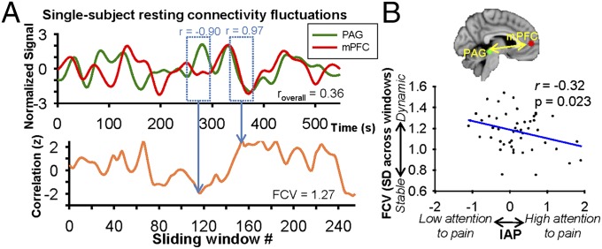
Human minds often wander away from their immediate sensory environment. It remains unknown whether such mind wandering is unsystematic or whether it lawfully relates to an individual's tendency to attend to salient stimuli such as pain and their associated brain structure/function. Studies of pain-cognition interactions typically examine explicit manipulation of attention rather than spontaneous mind wandering. Here we sought to better represent natural fluctuations in pain in daily life, so we assessed behavioral and neural aspects of spontaneous disengagement of attention from pain. We found that an individual's tendency to attend to pain related to the disruptive effect of pain on his or her cognitive task performance. Next, we linked behavioral findings to neural networks with strikingly convergent evidence from functional magnetic resonance imaging during pain coupled with thought probes of mind wandering, dynamic resting state activity fluctuations, and diffusion MRI. We found that (i) pain-induced default mode network (DMN) deactivations were attenuated during mind wandering away from pain; (ii) functional connectivity fluctuations between the DMN and periaqueductal gray (PAG) dynamically tracked spontaneous attention away from pain; and (iii) across individuals, stronger PAG-DMN structural connectivity and more dynamic resting state PAG-DMN functional connectivity were associated with the tendency to mind wander away from pain. These data demonstrate that individual tendencies to mind wander away from pain, in the absence of explicit manipulation, are subserved by functional and structural connectivity within and between default mode and antinociceptive descending modulation networks.

Keywords: experience sampling; pain modulation; salience network; stimulus-independent thought; ventral attention network.


Conflict of interest statement

The authors declare no conflict of interest.

Figures

Fig. 1.
Fig. 1.
Experience sampling during painful stimulation reveals frequency and sensory/cognitive aspects of attentional fluctuations away from pain. (A) Task trial design (Upper) and example of fluctuations in attentional state during the psychophysics session (Lower). (B) Distribution of the incidence of trials in session 1 (n = 51) (Upper) and session 2 (n = 50) (Lower) in which subjects experienced pain and/or something else. (C) Group averages (±SD) for ratings of something else being MW, TRIs, and EDs for session 1 and session 2. (D) Consistency of responses between session 1 and session 2 in IAP scores (ICC = 0.83; P = 4.7 × 10⁻¹⁰). (E) IAP scores from session 1 positively correlated with pain catastrophizing scale scores (r = 0.30; P = 0.03). ISI, interstimulus interval.
Fig. 2.
Fig. 2.
Tendency to attend to pain relates to the disruptive effect of pain on cognitive task performance. (A) The task required subjects to choose the box with the greatest number of digits (highest count). Green-outlined boxes show the correct response in this example. Subjects performed the task with pain (P) and without pain (NP). Subjects with mean reaction time (RT) for P faster than for NP trials were classified as A type (attention dominates). Subjects with slower RTs for P compared with NP trials were P-type (pain dominates) (3, 22). (B) Positive correlation between ΔRT [P − NP] in the cognitive interference task and IAP from experience sampling (n = 48) (r = 0.42; P = 0.003). Quadrants show classification of subjects of A/P type and low/high IAP.
Fig. 3.
Fig. 3.
Salience network and DMN activations relate to fluctuations in attention to pain (n = 32). (A) Regions with greater activation during periods preceding reports of attention to pain compared with attention to something else. Bar graphs show mean % signal change (±SD), extracted from 3-mm-radius spheres at peak coordinates. (B) Regions with greater activation during periods preceding reports of attention to something else compared with attention to pain. Bar graphs show mean % signal change (±SD), extracted from 3-mm-radius spheres at peak coordinates. Statistical images are thresholded at FWE-corrected Z > 2.3; cluster P < 0.05.
Fig. 4.
Fig. 4.
Mean change of activation within the DMN core, defined as the medial prefrontal cortex and posterior cingulate cortex/precuneus, between attention to something else vs. attention to pain [ΔDMN activation (Else > Pain)], correlated with postscan ratings of the degree to which Something Else reports were due to external sensory distractions (ρ = −0.61, P = 0.0002) (Left), task-related interferences (ρ = −0.24, P = 0.19) (Center), and mind-wandering (ρ = −0.45, P = 0.011) (Right).
Fig. 5.
Fig. 5.
(A) Functional connectivity of the PAG relates to fluctuations in attention to pain (n = 32). Statistical image shows greater functional connectivity of the PAG with areas of the DMN during periods preceding reports of attention to something else compared with attention to pain (FWE-corrected Z > 2.3; cluster P < 0.05). Bar graph shows % mean change (±SD) of functional connectivity between the PAG and mPFC for the two contrasted conditions, extracted from a 6-mm-radius sphere surrounding peak mPFC coordinates. (B) Structural connectivity between PAG and mPFC relates to individual differences in IAP. Image shows the across-subject aligned white matter mPFC–PAG skeleton (yellow) overlaid on the common pathway between PAG and mPFC identified with probabilistic tractography (Methods). Plot shows a negative correlation of IAP with mean FA in the mPFC–PAG skeleton (n = 51) (r = −0.36, P = 0.009).
Fig. 6.
Fig. 6.
Dynamic resting state functional connectivity between the PAG and mPFC relates to individual differences in IAP. (A) Single-subject example of PAG and mPFC signals during a resting state scan (Upper) and fluctuations in functional connectivity between PAG and mPFC across 40-s sliding windows (each window progressively sliding every 2 s) (Lower). (B) Group-level significant negative correlation between IAP and mPFC–PAG functional connectivity variability (SD of correlation values across sliding time windows) (r = −0.32, P = 0.023).

Comment in

References

    1. Killingsworth MA, Gilbert DT. A wandering mind is an unhappy mind. Science. 2010;330(6006):932. - PubMed
    1. Kane MJ, et al. For whom the mind wanders, and when: An experience-sampling study of working memory and executive control in daily life. Psychol Sci. 2007;18(7):614–621. - PubMed
    1. Seminowicz DA, Mikulis DJ, Davis KD. Cognitive modulation of pain-related brain responses depends on behavioral strategy. Pain. 2004;112(1–2):48–58. - PubMed
    1. Sprenger C, et al. Attention modulates spinal cord responses to pain. Curr Biol. 2012;22(11):1019–1022. - PubMed
    1. Valet M, et al. Distraction modulates connectivity of the cingulo-frontal cortex and the midbrain during pain—An fMRI analysis. Pain. 2004;109(3):399–408. - PubMed

Publication types