. 2013 Sep:111:93-118.

The spectrum of optic disc ischemia in patients younger than 50 years (an Amercian Ophthalmological Society thesis)

Affiliations

The spectrum of optic disc ischemia in patients younger than 50 years (an Amercian Ophthalmological Society thesis)

Anthony C Arnold et al. Trans Am Ophthalmol Soc. 2013 Sep.

Abstract

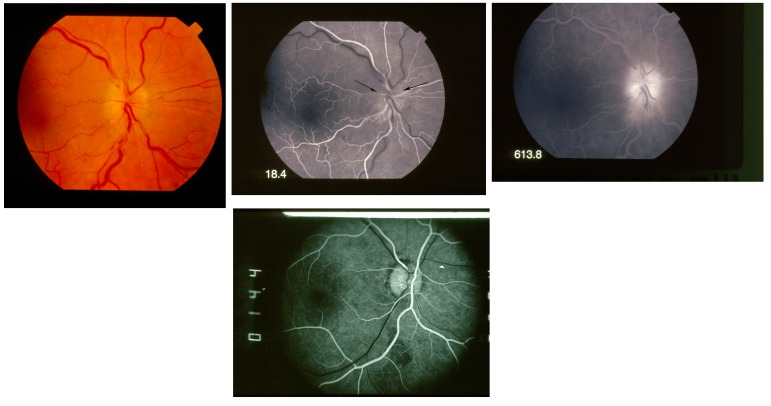
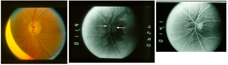
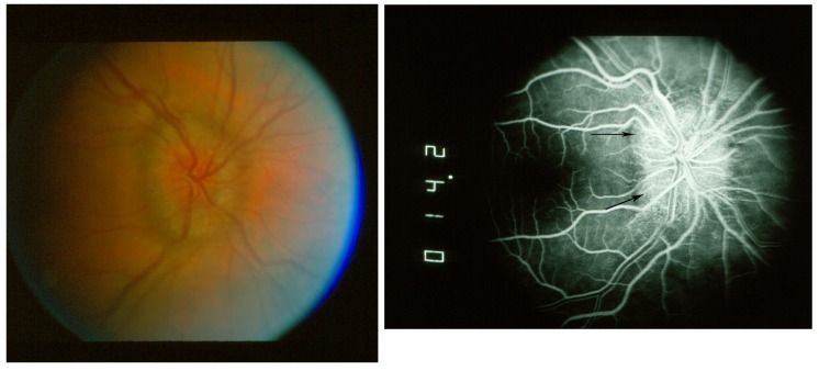
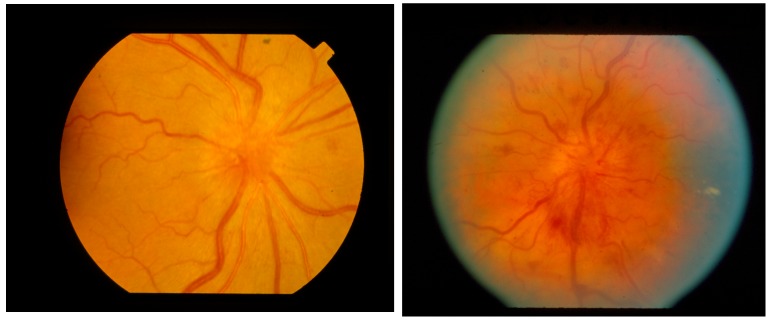
Purpose: To identify the spectrum of clinical and fluorescein angiographic features of optic disc ischemia in patients younger than 50 years.

Methods: This retrospective comparative case series from a university consultative neuro-ophthalmology practice consisted of two phases. The first compared 108 cases of nonarteritic anterior ischemic optic neuropathy in patients younger than 50 years (NAIONy) to a cohort of 108 cases in patients 50 years or older (NAIONo). Predisposing risk factors, fluorescein angiographic features, and clinical course were compared. In the second phase, 12 cases of diabetic papillopathy under age 50 were assessed by fluorescein angiographic criteria for evidence of optic disc ischemia and compared to patients with NAIONy.

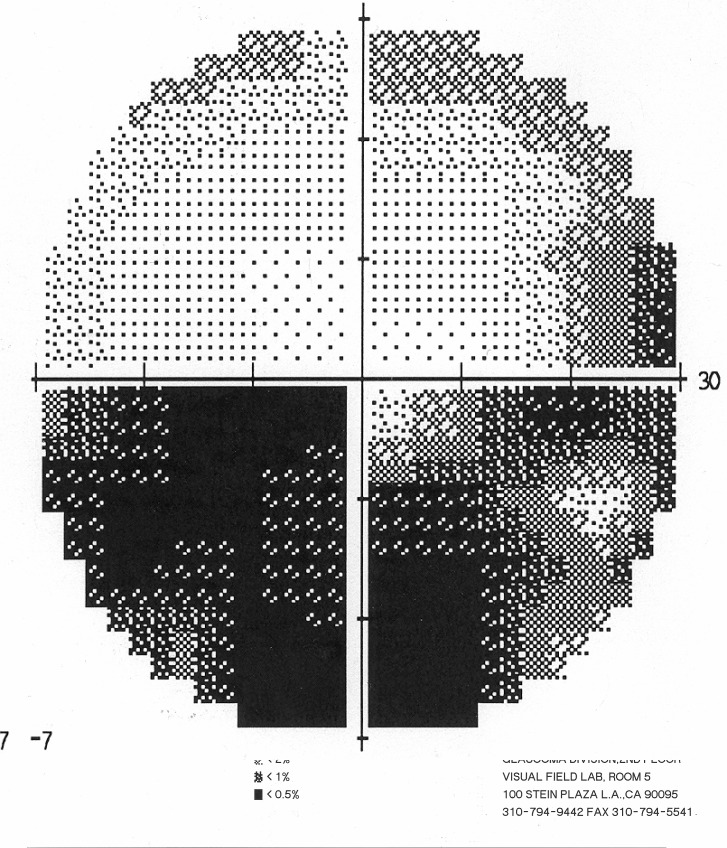
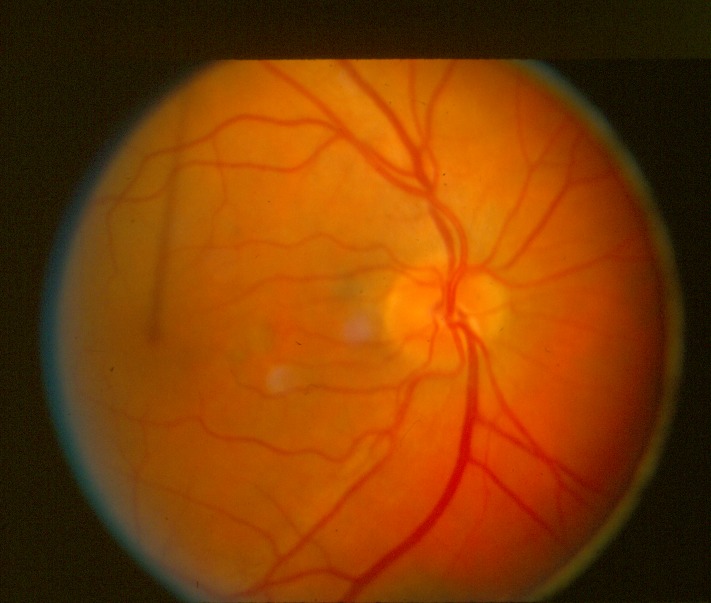
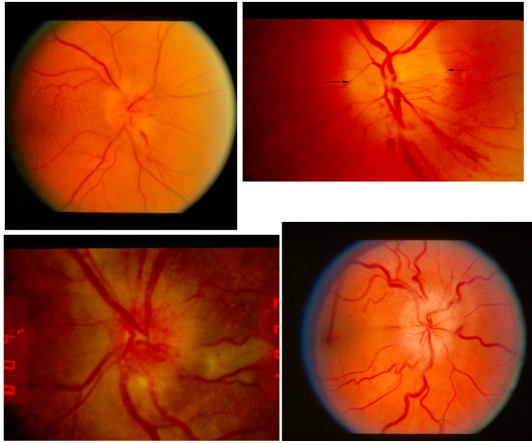
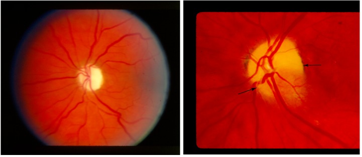
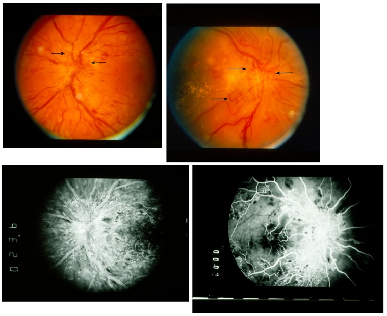
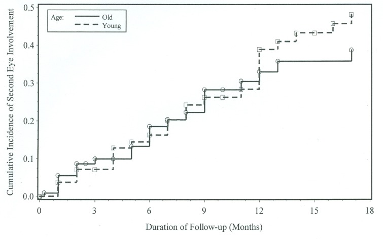
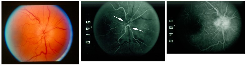
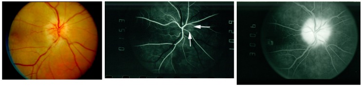
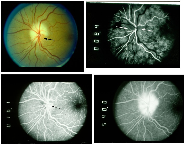
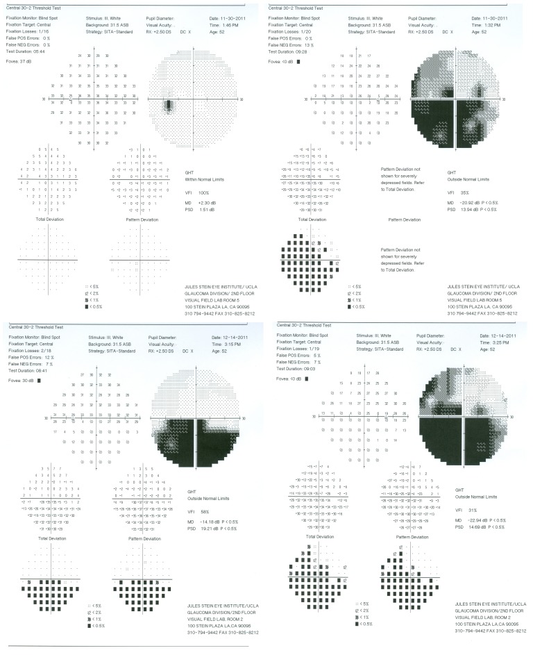
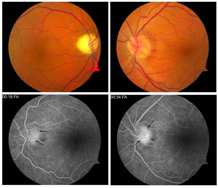
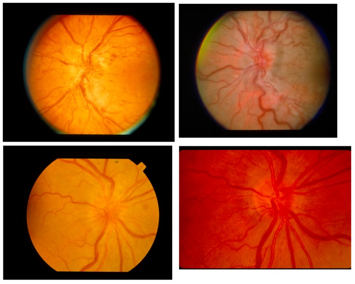
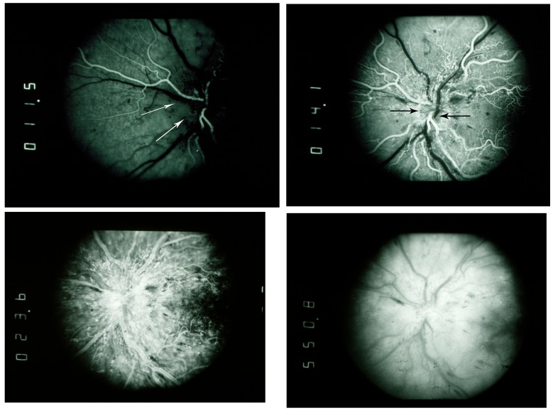
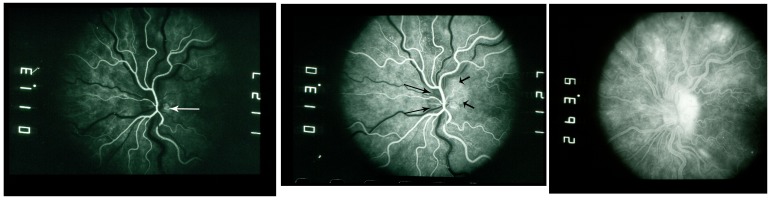
Results: NAIONy comprised 108 (12.7%) of 848 NAION cases reviewed. Chronic renal failure with dialysis and migraine were more common in NAIONy. Fellow eye involvement rate was significantly higher for NAIONy patients (46/108, 42.6%) than for NAIONo patients (32/108, 29.6%). Fluorescein angiographic features of ischemia were documented in 44 (81.5%) of 54 eyes studied. In one case, these features were documented in pre-NAION edema. Diabetic papillopathy demonstrated delayed filling consistent with ischemia in 7 of 10 (70.0%), without significant visual field loss.

Conclusions: Ischemic optic neuropathy in patients younger than 50 years is not rare. Fellow eye involvement is more frequent in younger patients. Fluorescein angiography confirmation of impaired perfusion in multiple syndromes of optic neuropathy corroborates a spectrum of optic disc ischemia ranging from perfusion delay without visual loss to severely impaired perfusion and visual loss and incorporates optic neuropathies previously considered nonischemic.


Figures

FIGURE 1
FIGURE 1
Visual field in nonarteritic anterior ischemic optic neuropathy in older patients (NAIONo). Quantitative perimetry shows inferior altitudinal visual field defect in right eye.
FIGURE 2
FIGURE 2
Fundus photograph showing fellow eye in nonarteritic anterior ischemic optic neuropathy in older patients (NAIONo). Optic disc is small in diameter, with cup-disc ratio <0.2.
FIGURE 3
FIGURE 3
Fundus photograph showing disc edema in nonarteritic anterior ischemic optic neuropathy in older patients (NAIONo). Upper left, Optic disc with diffuse edema and adjacent flame hemorrhage. Upper right, Optic disc with segmental edema worse inferiorly below arrows. Lower left, Optic disc with dilated surface vessels. Lower right, Optic disc with pale edema.
FIGURE 4
FIGURE 4
Fundus photograph showing optic atrophy in nonarteritic anterior ischemic optic neuropathy. Left, Optic disc with diffuse atrophy. Right, Optic disc with segmental atrophy above arrows.
FIGURE 5
FIGURE 5
Fluorescein angiography in nonarteritic anterior ischemic optic neuropathy diffuse. Upper left, Fundus photograph showing diffuse optic disc edema. Upper middle, Fluorescein angiogram, early arteriovenous phase, showing diffusely poor filling of optic disc (arrows) at 18.4 seconds. Upper right, Fluorescein angiogram, late phase, showing late leakage from optic disc at 613.8 seconds. Lower, Fluorescein angiography, early arteriovenous phase in normal eye, showing normal diffuse filling of the optic disc at 14.4 seconds
FIGURE 6
FIGURE 6
Fluorescein angiography in nonarteritic anterior ischemic optic neuropathy segmental. Left, Fundus photograph showing diffuse optic disc edema. Middle, Fluorescein angiogram, early phase, showing diffusely poor filling of optic disc at 11.9 seconds (arrows). Right, Fluorescein angiogram, early arteriovenous phase, showing poorly filling optic disc (short arrows, left) with relatively spared segmental region of early filling at 14.1 seconds (long arrows, right) .
FIGURE 7
FIGURE 7
Fluorescein angiography in nonischemic optic disc edema. Left, Fundus photograph showing diffuse optic disc edema. Right, Fluorescein angiogram, early arteriovenous phase, showing complete filling of optic disc (arrows) at 14.2 seconds.
FIGURE 8
FIGURE 8
Fundus photograph showing diabetic papillopathy. Left, Optic disc with diffuse edema. Right, Optic disc with diffuse edema and prominent surface vascular dilation.
FIGURE 9
FIGURE 9
Fluorescein angiography, diabetic papillopathy vs optic disc neovascularization. Upper left, Diabetic papillopathy, fundus photograph, optic disc edema with prominent surface vascular dilation. Vessels (arrows) predominantly are radial and extend minimally past disc margin. Upper right, Diabetic papillopathy and optic disc neovascularization, fundus photograph, optic disc edema with optic disc neovascularization. Vessels (arrows) are nonradial, irregular, and extend far past disc margin. Lower left, Diabetic papillopathy, fluorescein angiogram, mid phase, optic disc edema with prominent surface vascular dilation; leakage is primarily from radial disc surface vessels extending adjacent to disc. Lower right, Diabetic papillopathy and optic disc neovascularization, fluorescein angiogram, mid phase, optic disc edema with optic disc neovascularization, leakage from disc is relatively mild compared with extensive irregular vascular abnormality and leakage throughout posterior pole.
FIGURE 10
FIGURE 10
Kaplan-Meier curve of time to fellow eye involvement (up to 18 months) among 213 nonarteritic anterior ischemic optic neuropathy (NAION) patients without bilateral involvement at onset. Log-rank test P = .49 for comparing Kaplan-Meier curves between patients with NAION under age 50 (NAIONy, 43 of 105 patients [41%] with fellow eye involvement) and patients with NAION aged 50 and over (NAIONo, 32 of 108 patients [30%] with fellow eye involvement).
FIGURE 11
FIGURE 11
Fluorescein angiography in nonarteritic anterior ischemic optic neuropathy in young patients (NAIONy) diffuse. Left, Fundus photograph showing diffuse optic disc edema. Middle, Fluorescein angiogram, early arteriovenous phase, showing poor filling of optic disc (arrows) at 19.5 seconds. Right, Fluorescein angiogram, late phase, showing late leakage from optic disc at 48.8 seconds.
FIGURE 12
FIGURE 12
Fluorescein angiography in nonarteritic anterior ischemic optic neuropathy in young patients (NAIONy) segmental. Left, Fundus photograph showing diffuse optic disc edema. Middle, Fluorescein angiogram, early arteriovenous phase, showing poor filling of optic disc with relatively spared segmental region of early filling (arrows) at 15.3 seconds. Right, Fluorescein angiogram, late phase, showing late leakage from optic disc at 300.6 seconds.
FIGURE 13
FIGURE 13
Fluorescein angiography in nonarteritic anterior ischemic optic neuropathy in young patients (NAIONy), chronic renal failure (CRF). Upper left, Fundus photograph showing segmental, pale optic disc edema. Inferotemporal segment (arrow) is relatively spared. Upper right, Fluorescein angiogram, early arteriovenous phase, showing diffusely poor filling of the optic disc (arrows) at 8.4 seconds. Lower left, Fluorescein angiogram, early arteriovenous phase, showing diffusely poor filling of the optic disc with inferotemporal segment of relatively intact filling (arrow) at 16.1 seconds. Lower right, Fluorescein angiogram, late phase, showing late leakage from optic disc at 540.0 seconds.
FIGURE 14
FIGURE 14
Visual fields in pre-nonarteritic anterior ischemic optic neuropathy optic disc edema. Upper left, Quantitative perimetry showing normal visual field, left eye. Upper right, Quantitative perimetry showing inferior altitudinal visual field defect, right eye. Lower left, Quantitative perimetry showing inferior altitudinal visual field defect, left eye. Lower right, Quantitative perimetry showing inferior altitudinal visual field defect, right eye.
FIGURE 15
FIGURE 15
Fluorescein angiography in pre-nonarteritic anterior ischemic optic neuropathy optic disc edema. Upper left, Fundus photograph, right eye, showing optic atrophy from prior NAION. Upper right, Fundus photograph, left eye, showing diffuse optic disc edema. Lower left, Fluorescein angiogram, arteriovenous phase, showing poor filling of optic disc with relatively spared segment temporally (arrows) at 19.0 seconds. Fluorescein angiogram, mid phase, showing continued poor filling of optic disc with progressive hyperfluorescence at relatively spared segment temporally (arrows) at 34.0 seconds.
FIGURE 16
FIGURE 16
Fundus photograph in diabetic papillopathy. Upper left, Optic disc left eye with diffuse edema and prominent surface vascular dilation. Upper right, Optic disc left eye with diffuse nonspecific edema. Lower left, Optic disc right eye with diffuse nonspecific edema at initial presentation. Lower right, Optic disc right eye after resolution of edema (3 months), showing normal appearance.
FIGURE 17
FIGURE 17
Fluorescein angiography in diabetic papillopathy with prominent surface vascular dilation. Upper left, Fluorescein angiogram, early arteriovenous phase, showing absent optic disc filling (arrows) at 11.5 seconds. Upper right, Fluorescein angiogram, early arteriovenous phase, showing poor optic disc filling (arrows) and very early leakage from adjacent surface vessels at 14.1 seconds. Lower left, Fluorescein angiogram, mid phase, showing diffuse early leakage from dilated surface vessels at 23.6 seconds. Prelaminar layer of disc filling is obscured by dilated leaking surface vasculature. Lower right, Fluorescein angiogram, late phase, showing late leakage from optic disc at 550.8 seconds.
FIGURE 18
FIGURE 18
Fluorescein angiography in diabetic papillopathy with nonspecific diffuse edema. Left, Fluorescein angiogram, early arteriovenous phase, showing nearly absent filling of the optic disc with temporal segment of relatively intact filling (arrow) at 11.3 seconds. Middle, Fluorescein angiogram, early arteriovenous phase, showing poor filling of the optic disc nasally (long arrows), with temporal segment of progressive hyperfluorescence at relatively spared segment temporally (short arrows) at 13.0 seconds. Right, Fluorescein angiogram, late phase, showing late leakage from optic disc at 263.9 seconds.

References

    1. Arnold AC. Ischemic optic neuropathy. In: Miller NR, Newman NJ, editors. Walsh & Hoyt’s Clinical Neuro-Ophthalmology. 6th ed. Vol. 1. Philadelphia, PA: Lippincott Williams & Wilkins; 2005. pp. 349–384.
    1. Spencer WH, Hoyt WF. A fatal case of giant-cell arteritis (temporal or cranial arteritis) with ocular involvement. Arch Ophthalmol. 1960;64(6):862–867.
    1. MacFaul PA. Ciliary artery involvement in giant cell arteritis. Br J Ophthalmol. 1967;51(8):505–512. - PMC - PubMed
    1. Henkind P, Charles NC, Pearson J. Histopathology of ischemic optic neuropathy. Am J Ophthalmol. 1970;69(1):78–90. - PubMed
    1. Knox DL, Duke JR. Slowly progressive ischemic optic neuropathy. A clinicopathologic case report. Trans Am Acad Ophthalmol Otolaryngol. 1971;75(5):1065–1068. - PubMed

Publication types