. 2013 Oct 22;8(10):e77558.
doi: 10.1371/journal.pone.0077558. eCollection 2013.

Isozyme-specific ligands for O-acetylserine sulfhydrylase, a novel antibiotic target

Affiliations

Isozyme-specific ligands for O-acetylserine sulfhydrylase, a novel antibiotic target

Francesca Spyrakis et al. PLoS One. .

Abstract

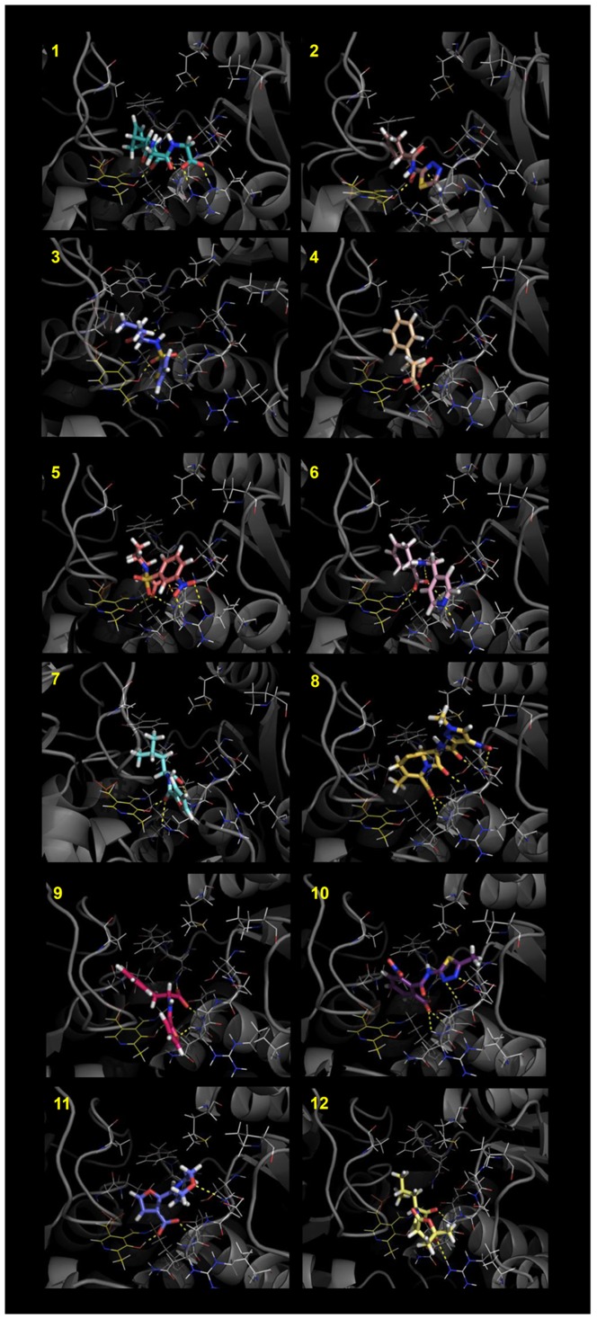
The last step of cysteine biosynthesis in bacteria and plants is catalyzed by O-acetylserine sulfhydrylase. In bacteria, two isozymes, O-acetylserine sulfhydrylase-A and O-acetylserine sulfhydrylase-B, have been identified that share similar binding sites, although the respective specific functions are still debated. O-acetylserine sulfhydrylase plays a key role in the adaptation of bacteria to the host environment, in the defense mechanisms to oxidative stress and in antibiotic resistance. Because mammals synthesize cysteine from methionine and lack O-acetylserine sulfhydrylase, the enzyme is a potential target for antimicrobials. With this aim, we first identified potential inhibitors of the two isozymes via a ligand- and structure-based in silico screening of a subset of the ZINC library using FLAP. The binding affinities of the most promising candidates were measured in vitro on purified O-acetylserine sulfhydrylase-A and O-acetylserine sulfhydrylase-B from Salmonella typhimurium by a direct method that exploits the change in the cofactor fluorescence. Two molecules were identified with dissociation constants of 3.7 and 33 µM for O-acetylserine sulfhydrylase-A and O-acetylserine sulfhydrylase-B, respectively. Because GRID analysis of the two isoenzymes indicates the presence of a few common pharmacophoric features, cross binding titrations were carried out. It was found that the best binder for O-acetylserine sulfhydrylase-B exhibits a dissociation constant of 29 µM for O-acetylserine sulfhydrylase-A, thus displaying a limited selectivity, whereas the best binder for O-acetylserine sulfhydrylase-A exhibits a dissociation constant of 50 µM for O-acetylserine sulfhydrylase-B and is thus 8-fold selective towards the former isozyme. Therefore, isoform-specific and isoform-independent ligands allow to either selectively target the isozyme that predominantly supports bacteria during infection and long-term survival or to completely block bacterial cysteine biosynthesis.


Conflict of interest statement

Competing Interests: Paolo Benedetti, is a consultant of Molecular Discovery Ltd, as clearly stated into his affiliation, but the authors do guarantee that this does not alter their adherence to all the PLOS ONE policies on sharing data and materials.

Figures

Figure 1
Figure 1. Cysteine biosynthesis.
Upper panel: Intermediates of cysteine biosynthesis in mammals and bacteria. The red arrows indicate the biosynthetic pathway in mammals and the yellow arrows the biosynthetic pathway in bacteria. Lower panel: Sulfur assimilation in bacteria. Sulfate and thiosulfate are the most abundant forms of extracellular sulfur, the latter being predominant under less oxidizing conditions. Inorganic sulfur enters the cells through specific transporters. In contrast to OASS-A, OASS-B can directly use thiosulfate for cysteine biosynthesis. The product S-sulfo-L-cysteine is reduced by glutaredoxins to cysteine and sulfide that enters in the last step of the sulfate reduction pathway , .
Figure 2
Figure 2. Structural comparison of OASS-A and OASS-B.
Panel A: Structure-based amino acid sequence alignment of OASS-A and OASS-B from Salmonella typhimurium. The alignment, carried out on the PDB entries 1OAS and 2JC3 using the Flexible structure AlignmenT (FATCAT) method , gave an overall identity of 40.32% and a similarity of 56.51%. Identical residues have a red background and residues with similar physicochemical properties are shown in red. Similarity scores were calculated by the ESPript program using the Blosum62 matrix set at global score of 0.2. Residues of the first active site shell are indicated by dark circles below the alignment. Panel B: Active site of OASS-A. Residues of the first active site shell and PLP are shown in ball and stick style, colored pink and yellow, respectively. Panel C: Active site of OASS-B. Residues of the first active site shell and PLP are shown in ball and stick style, colored cyan and yellow, respectively.
Figure 3
Figure 3. LigPlot of the wild type tetrapeptide ligand in the active site of Haemophilus influenzae OASS.
The interactions between the Asn-Leu-Asn-Ile tetrapeptide and the active site residues of H. influenzae OASS-A (PDB code: 1Y7L) are reported. The figure was drawn with LigPlot program version 4.5.3 .
Figure 4
Figure 4. Compounds selected by SBVS/LBVS-docking procedures for OASS-A and OASS-B.
Figure 5
Figure 5. Best HINT scored conformations of the compounds selected by the SBVS/LBVS-docking procedures for OASS-A.
The images were prepared with PyMOL (The PyMOL Molecular Graphics System, Version 1.5.0.4 Schrödinger, LLC.)
Figure 6
Figure 6. Best HINT scored conformations of the compounds selected by the LBVS/docking procedures for OASS-B.
The images were prepared with PyMOL (The PyMOL Molecular Graphics System, Version 1.5.0.4 Schrödinger, LLC.)
Figure 7
Figure 7. Binding of ligands to StOASS.
Panel A. Fluorescence emission spectra upon excitation at 412 nm (slitex =  6 nm, slitem =  6 nm) of a solution containing 50 nM StOASS-A and increasing concentrations of Compound 1 in 100 mM Hepes buffer, pH 7.0, at 20°C. Inset: Dependence of the fluorescence emission intensity at 500 nm on the ligand concentration. The line drawn through data points is the fit to a binding isotherm with Kd  =  3.7 ± 0.4 µM. Panel B. Fluorescence emission spectra upon excitation at 412 nm (slitex =  4 nm, slitem =  4 nm) of a solution containing 1 µM StOASS-B and increasing concentrations of Compound 13 in 100 mM Hepes buffer, pH 7.0, at 20 °C. Inset: Dependence of the fluorescence emission intensity at 500 nm on the ligand concentration. The line drawn through data points is the fit to a binding isotherm with Kd  =  33 ± 2 µM.
Figure 8
Figure 8. Docking pose of best binders to the two isozymes placed into the active sites.
Panel A: Docking pose of 1 in the OASS-A binding pocket. Red and green contours identify the hydrogen bond acceptor and hydrophobic GRID MIFs. Hydrogen bond donor hot spots have not been shown for clarity. Panel B: Docking pose of compound 13 in the OASS-B binding pocket. Red and green contours identify the hydrogen bond acceptor and hydrophobic GRID MIFs. Hydrogen bond donor hot spots have not been shown for clarity.
Figure 9
Figure 9. GRID MIFs calculated for OASS-A and OASS-B.
Red, blue and green contours identify the hydrogen bond acceptor, hydrogen bond donor and hydrophobic MIFs, respectively, calculated for OASS-A (pink cartoons) towards OASS-B (cyan cartoons). In Panel B compounds 1 and 13 are shown in ball and stick.

References

    1. Kessler D (2006) Enzymatic activation of sulfur for incorporation into biomolecules in prokaryotes. FEMS Microbiol Rev 30: 825–840. - PubMed
    1. Beinert H (2000) A tribute to sulfur. Eur J Biochem 267: 5657–5664. - PubMed
    1. Westrop GD, Goodall G, Mottram JC, Coombs GH (2006) Cysteine biosynthesis in Trichomonas vaginalis involves cysteine synthase utilizing O-phosphoserine. J Biol Chem 281: 25062–25075. - PMC - PubMed
    1. Bhave DP, Muse WB (2007) Drug targets in mycobacterial sulfur metabolism. Infect Disord Drug Targets 7: 140–158. - PMC - PubMed
    1. Muller S, Liebau E, Walter RD, Krauth-Siegel RL (2003) Thiol-based redox metabolism of protozoan parasites. Trends Parasitol 19: 320–328. - PubMed

MeSH terms