. 2013 Jul;3(3):152-66.
doi: 10.1177/1941874412466090.

Neurological complications of solid organ transplantation

Affiliations

Neurological complications of solid organ transplantation

Amy A Pruitt et al. Neurohospitalist. 2013 Jul.

Abstract

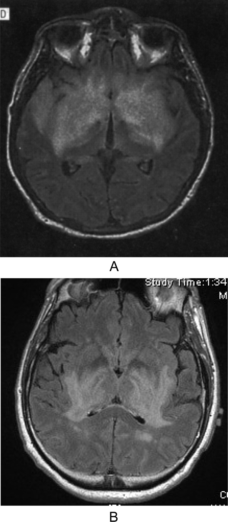
Solid organ transplantation (SOT) is the preferred treatment for an expanding range of conditions whose successful therapy has produced a growing population of chronically immunosuppressed patients with potential neurological problems. While the spectrum of neurological complications varies with the type of organ transplanted, the indication for the procedure, and the intensity of long-term required immunosuppression, major neurological complications occur with all SOT types. The second part of this 2-part article on transplantation neurology reviews central and peripheral nervous system problems associated with SOT with clinical and neuroimaging examples from the authors' institutional experience. Particular emphasis is given to conditions acquired from the donated organ or tissue, problems specific to types of organs transplanted and drug therapy-related complications likely to be encountered by hospitalists. Neurologically important syndromes such as immune reconstitution inflammatory syndrome (IRIS), posterior reversible encephalopathy syndrome (PRES), and posttransplantation lymphoproliferative disorder (PTLD) are readdressed in the context of SOT.

Keywords: hepatic encephalopathy; immune reconstitution inflammatory syndrome; organ transplantation; posterior reversible encephalopathy syndrome; posttransplantation lymphoproliferative disorder; progressive multifocal leukoencephalopathy; toxoplasmosis.


Conflict of interest statement

Declaration of Conflicting Interests: The authors declared no potential conflicts of interest with respect to the research, authorship, and/or publication of this article.

Figures

Figure 1.
Figure 1.
Liver transplant–related encephalopathies. Patient 1 (A and B) with hyperammonemic encephalopathy has distinct FLAIR (A) and DWI (B) cortical abnormalities sparing occipital cortex. Patient 2 (C and D) was imaged 2 weeks after liver transplant for alcoholic cirrhosis. Patient 2 had a slight tremor pretransplant that became disabling on tacrolimus. The MRI shows hyperintense lesions on FLAIR (C) and DWI (D) involving the globus pallidus. These were seen on a preoperative CT (not shown) and were thought to represent hepatocerebral degeneration. FLAIR indicates fluid attenuated inversion recovery; DWI, diffusion-weighted imaging; CT, computed tomography.
Figure 2.
Figure 2.
FLAIR MRI (A) of patient 2 weeks after liver transplant with evidence of PRES and also subacute infarction on diffusion-weighted imaging (B) with diffuse narrowing of proximal vertebral and basilar arteries indicative of vasculopathy on MRA (C). FLAIR indicates fluid attenuated inversion recovery; MRI, magnetic resonance imaging; MRA, magnetic resonance angiography.
Figure 3.
Figure 3.
Donor-transmitted infections. A, FLAIR MRI shows large areas of signal abnormality in the basal ganglia and temporal lobes in a patient who contracted rabies encephalitis from organ donor. Reprinted with permission from Arch Neurol (2005;62:873-882). B, FLAIR MRI of a patient with characteristic Flavivirus (West Nile virus) pattern of basal ganglia signal abnormality that also was seen in brain stem (not shown). FLAIR indicates fluid attenuated inversion recovery; MRI, magnetic resonance imaging.
Figure 4.
Figure 4.
Progressive hydrocephalus due to cryptococcosis and IRIS. This case illustrates the difficulty of interpreting host response to a fungal infection in the setting of changing immunosuppressive regimen and organ rejection. Progressive hydrocephalus demonstrated on FLAIR MRI at presentation with headache (A), and 3 months later (B), in a heavily immunosuppressed liver transplant patient with recurrent hepatitis C and ongoing organ rejection. Gadolinium-enhanced MRI (not shown) showed fluctuating leptomeningeal enhancement. The CSF was abnormal with 84 lymphocytes, glucose 19 mg/dL and protein 165 to 258 mg /dL. The indolent course, CSF, and negative PCRs and cultures suggested possible lymphoma or neurosarcoidosis. Multiple negative cytologies and meningeal biopsy failed to make a diagnosis until the third lumbar puncture finally revealed cryptococcal meningitis. Antifungal therapy cleared cryptococcal meningitis without worsening what appears to be an example of infection- and rejection-precipitated IRIS. C, Unrelated OLT patient whose gadolinium-enhanced MRI shows multifocal nodular enhancement initially interpreted as recurrent cryptococcal meningitis but ultimately found to be IRIS. Figure 4C reprinted with permission from Liver Transpl (2008;14:1671-1674). FLAIR indicates fluid attenuated inversion recovery; MRI, magnetic resonance imaging; CSF, cerebrospinal fluid; PCR, polymerase chain reaction; IRIS, immune reconstitution inflammatory syndrome; OLT, orthotopic liver transplantation.
Figure 5.
Figure 5.
A multifactorial problem. These MRIs demonstrate the diagnostic complexity of a patient 2 years after liver transplant, who presented with diarrhea due to Clostridium difficile and then sudden paraplegia due to extensive transverse myelitis seen on sagittal (A) and axial (B) T2 MRI. The patient also had multiple DWI abnormalities on brain MRI (C) and evidence of intraventricular hemorrhage on gradient echo MRI (D). Tacrolimus level was high on presentation and the patient was found to have CMV in an acellular CSF. Acute myelopathy was ascribed to possible CMV-associated vasculitis, although linking all of the neuroimaging findings pathophysiologically remained difficult. There was no improvement in foscarnet and ganciclovir therapy and the patient ultimately succumbed to Klebsiella sepsis. FLAIR indicates fluid attenuated inversion recovery; MRI, magnetic resonance imaging; DWI, diffusion-weighted imaging; CSF, cerebrospinal fluid; CMV, cytomegalovirus.
Figure 6.
Figure 6.
Osmotic demyelination. A, FLAIR MRI of a patient post liver transplant with clinical signs of dysarthria, dysphagia, and abducens palsies after rapid correction of hyponatremia. B, DWI MRI of a renal failure patient pretransplantation with osmotic demyelination demonstrating that extrapontine sites can be involved, often after recent hemodialysis. FLAIR indicates fluid attenuated inversion recovery; DWI, diffusion-weighted imaging; MRI, magnetic resonance imaging.

References

    1. Organ Procurement and Transplantation Network. http://optn.transplant.hrsa.gov Accessed March 24, 2012
    1. Senzolo M, Ferronato C, Burra P. Neurologic complications after solid organ transplantation. Transpl Int. 2009;22(3):269–278 - PubMed
    1. Dhar R, Human T. Central nervous system complications after transplantation. Neurol Clin, 2011;29(4):943–972 - PubMed
    1. Pustavoitau A, Bhardwaj A, Stevens R. Neurological complications of transplantation. J Intensive Care Med. 2011;26(4):209–222 - PubMed
    1. Sharma P, Eesa M, Scott JN. Toxic and acquired metabolic encephalopathies: MRI appearance. AJR Am J Roentgenol 2009;193(3):879–886 - PubMed

LinkOut - more resources