. 2013 Jul;4(7-8):261-72.
doi: 10.1177/1947601913499020.

Extracellular Vesicle-Mediated Transfer of a Novel Long Noncoding RNA TUC339: A Mechanism of Intercellular Signaling in Human Hepatocellular Cancer

Affiliations

Extracellular Vesicle-Mediated Transfer of a Novel Long Noncoding RNA TUC339: A Mechanism of Intercellular Signaling in Human Hepatocellular Cancer

Takayuki Kogure et al. Genes Cancer. 2013 Jul.

Abstract

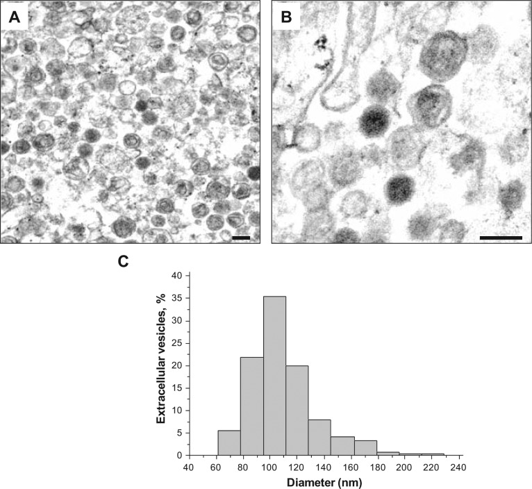
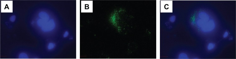
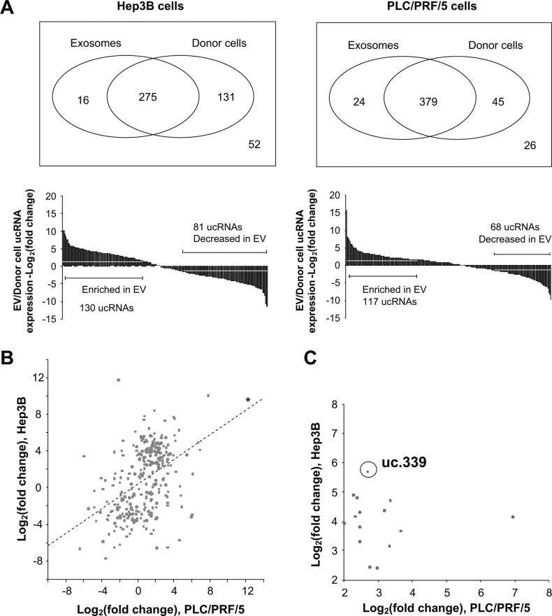
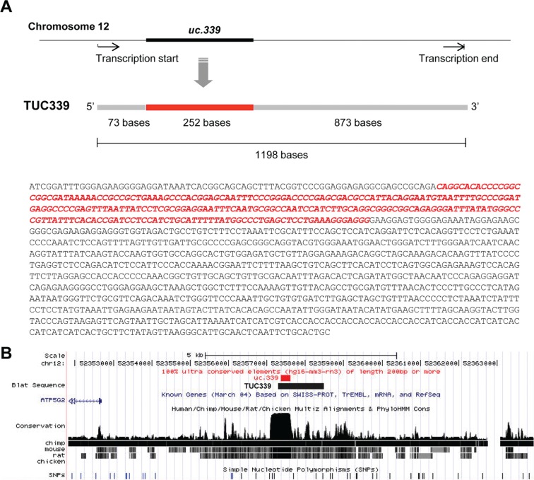
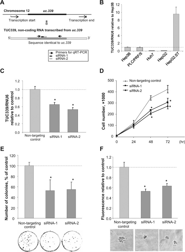
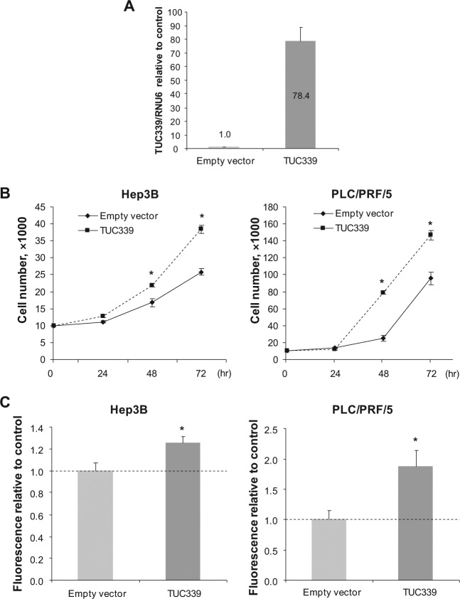
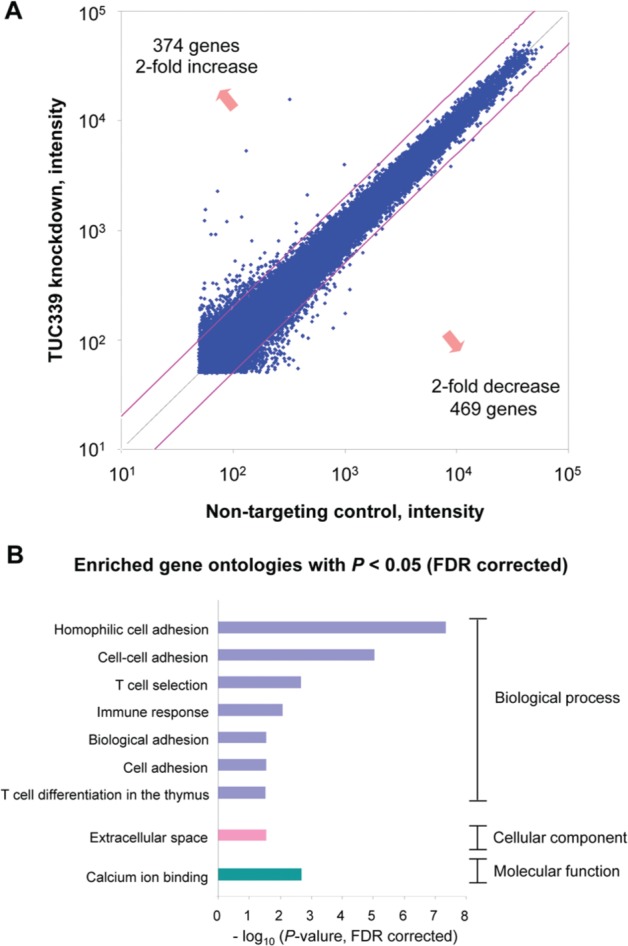
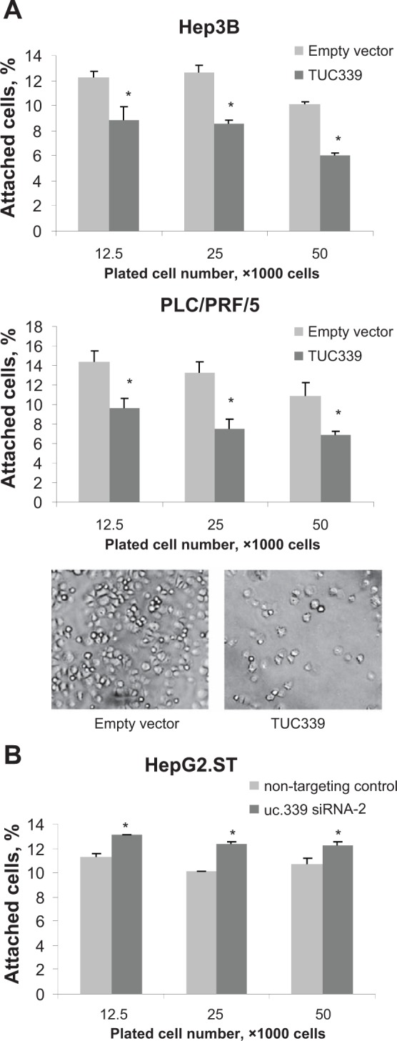
Although the expression of long noncoding RNA (lncRNA) is altered in hepatocellular cancer (HCC), their biological effects are poorly defined. We have identified lncRNA with highly conserved sequences, ultraconserved lncRNA (ucRNA) that are transcribed and altered in expression in HCC. Extracellular vesicles, such as exosomes and microvesicles, are released from tumor cells and can transfer biologically active proteins and RNA across cells. We sought to identify the role of vesicle-mediated transfer of ucRNA as a mechanism by which these novel lncRNA could influence intercellular signaling with potential for environmental modulation of tumor cell behavior. HCC-derived extracellular vesicles could be isolated from cells in culture and taken up by adjacent cells. The expression of several ucRNA was dramatically altered within extracellular vesicles compared to that in donor cells. The most highly significantly expressed ucRNA in HCC cell-derived extracellular vesicles was cloned and identified as a 1,198-bp ucRNA, termed TUC339. TUC339 was functionally implicated in modulating tumor cell growth and adhesion. Suppression of TUC339 by siRNA reduced HCC cell proliferation, clonogenic growth, and growth in soft agar. Thus, intercellular transfer of TUC339 represents a unique signaling mechanism by which tumor cells can promote HCC growth and spread. These findings expand the potential roles of ucRNA in HCC, support the existence of selective mechanisms for lncRNA export from cells, and implicate extracellular vesicle-mediated transfer of lncRNA as a mechanism by which tumor cells can modulate their local cellular environment. Intercellular transfer of functionally active RNA molecules by extracellular vesicles provides a mechanism that enables cells to exert genetic influences on other cells within the microenvironment.

Keywords: RNA genes; extracellular vesicles; gene expression; liver cancer; paracrine signaling.


Conflict of interest statement

Declaration of Conflicting Interests: The author(s) declared no potential conflicts of interest with respect to the research, authorship, and/or publication of this article.

Figures

Figure 1.
Figure 1.
Transmission electron microscopy (TEM) of extracellular vesicles isolated from HCC cells. Morphology of PLC/PRF/5-derived extracellular vesicles was examined by ultrathin section TEM using an EM208S Electron Microscope (Philips). (A) Low magnification. (B) High magnification; scale bar, 100 nm. (C) The sizes of vesicles were analyzed using NIH Image J software (National Institutes of Health). The histogram shows the distribution of the size of vesicles.
Figure 2.
Figure 2.
Internalization of Hep3B-derived extracellular vesicles into other cells. HepG2 cells in culture were incubated with Hep3B-derived extracellular vesicles labeled with PKH67 green dye for 24 hours. Cells are fixed with methanol at −20°C and mounted with ProLong Gold Antifade Reagent with DAPI (Molecular Probes). Images were obtained using a fluorescent microscope (Nikon Eclipse 80i, Nikon Instruments Inc.). (A) DAPI (blue), (B) PKH67 (green), and (C) Merge. Hep3B-derived extracellular vesicles are shown to be internalized into the cytoplasm of HepG2 cells.
Figure 3.
Figure 3.
ucRNA expression in extracellular vesicles (EV) and their donor cells. Profiling of ucRNAs was performed using quantitative RT-PCR, and the expression level of each ucRNA in extracellular vesicles was normalized using the median threshold cycle (CT) value and expressed relative to their donor cells. (A) The mean values of fold change of expression of ucRNA detected in extracellular vesicles relative to that in donor cells is shown (n = 4) and the numbers of ucRNA that were exclusively detected in either donor cells or extracellular vesicles are depicted. A total of 291 ucRNAs were identified in Hep3B-derived extracellular vesicles, and 403 ucRNAs in PLC/PRF/5-derived extracellular vesicles. (B) Correlation of ucRNA expression in Hep3B-derived extracellular vesicles and in PLC/PRF/5-derived extracellular vesicles are shown. A total of 279 ucRNAs were detected in extracellular vesicles from both Hep3B and PLC/PRF/5. *, coefficient, .434; P < 0.0001. (C) ucRNAs with fold change >4 and P < 0.05 in both Hep3B and PLC/PRF/5 are plotted. uc.339 was identified to be one of highly expressed ucRNAs in extracellular vesicles secreted from both Hep3B and PLC/PRF/5.
Figure 4.
Figure 4.
Cloning of TUC339. (A) Schematic representation of TUC339, the ultraconserved RNA transcribed from the region including uc.339. RACE cloning was performed using SMARTer RACE cloning kit (Clontech) to identify the sequence of full-length TUC339. The complete sequence of TUC339 is shown with the uc.339 sequence identified by Bejerano et al., shown in red. (B) Location of uc.339 and TUC339 in human genome in UCSC Genome Browser (http://genome.ucsc.edu/). No confirmed protein coding transcripts overlapping with TUC339 were reported.
Figure 5.
Figure 5.
TUC339 knockdown decreases HCC cell proliferation. (A) Schematic representation of uc.339, TUC339, primers, and siRNAs. The location of the primers used for quantitative RT-PCR (qRT-PCR) and siRNAs are shown. (B) Basal expression level of TUC339 in human HCC cell lines. RNA was extracted from HCC cell lines using TRIzol reagent and qRT-PCR with SYBR green was performed. Expression of TUC339 was normalized using the expression of RNU6B and standardized to Hep3B. Bars express mean ± SE. (C) Knockdown efficiency of siRNAs against TUC339. HepG2.ST cells were transfected with siRNAs against TUC339 or nontargeting control (Dharmacon) using Lipofectamine 2000 (Invitrogen). After 48 hours cells were collected for qRT-PCR to detect TUC339 (C), or used for the following experiments (D-F). Bars represent the mean ± SEM. *, P < 0.05. (D) Growth curve assay. Cells were plated on 24-well plates at 1,000 cells per well and the number of viable cells in each well was counted using hemocytometer with trypan blue staining. Each plot represents mean ± SEM of 3 separate determinants. *, P < 0.05. (E) Clonogenic assay. Cells were plated on 6-well plates at 200 cells per well. After 7 days colonies were fixed and stained using neutralized buffered formalin containing 0.5% crystal violet. Images were taken and the number of the colonies were counted using NIH Image J software and expressed as a percentage of control. Data represent mean ± SEM of 3 determinants. *, P < 0.05. (F) Soft agar assay. Cells were plated in a 96-well plate with cell culture medium containing 0.4% agar at 600 cells per well over a base agar layer consisting of culture medium containing 0.6% agar. After incubation of 7 days, the number of colonies was evaluated fluorometrically. Bars represent the mean ± SEM of 6 separate determinations. *, P < 0.05.
Figure 6.
Figure 6.
Enforced expression of TUC339 enhances cell growth. Cells were transfected with plasmids expressing full-length TUC339 or empty vector control using electroporation (Amaxa Nucleofector V kit, Lonza), and after 48 hours cells were used for anchorage-dependent and anchorage-independent growth assays. (A) Quantitative RT-PCR for TUC339. RNA was extracted from PLC/PRF/5 cells at 48 hours after transfection and qRT-PCR was performed. The expression of TUC339 was expressed relative to that with empty vector control. Bars represent mean ± SEM of 3 determinants. (B) Growth curve assay. Cells were plated on 24-well plates at 1,000 cells per well and the number of viable cells in each well was counted using hemocytometer with trypan blue staining. Each plot represents mean ± SEM of 3 separate determinants. *, P < 0.05. (C) Soft agar assay. Cells were plated in a 96-well plate with cell culture medium containing 0.4% agar at 600 cells per well over a base agar layer consist of culture medium containing 0.6% agar. After incubation of 7 days, the number of colonies was evaluated fluorometrically. Bars represent the mean ± SEM of 6 separate determinations. *, P < 0.05.
Figure 7.
Figure 7.
TUC339 knockdown alters gene expression. (A) A genome-wide expression analysis was performed by comparing the expression of genes in HepG2.ST cells with TUC339 knockdown to that with nontargeting control using NimbleGen gene expression 12 × 135K arrays (Roche NimbleGen). Differential expressions of genes are plotted (axis, intensity). (B) Enriched gene ontologies of genes with altered expression by TUC339 knockdown. Total 843 genes with fold change >2 or <2 were analyzed using DAVID v6.7 program. Enriched gene ontologies with P < 0.05 (false discovery rate corrected) are shown. FDR = false discovery rate.
Figure 8.
Figure 8.
Enforced TUC339 expression modulates cell attachment. Hep3B and PLC/PRF/5 cells transfected with TUC339 expression vector or empty vector control and HepG2.ST cells transfected with siRNA against TUC339 or nontargeting control cells were labeled with green fluorescent dye (CellTrackerGreen CMFDA, Invitrogen), seeded on a collagen I-coated 96-well plate and incubated at 37°C for 30 minutes. Cells were washed with serum-free medium and fluorescence was measured before and after washing. The number of cells attached to the plate was estimated based on the change in fluorescence before and after the wash. (A) Hep3B and PLC/PRF/5 with representative images of PLC/PRF/5 cells after washing at 50,000 cells per well. Bars express mean ± SEM of 3 studies, *, P < 0.05. (B) HepG2.ST. Bars express mean ± SEM of 3 studies, *, P < 0.05.

References

    1. Hui EE, Bhatia SN. Micromechanical control of cell-cell interactions. Proc Natl Acad Sci U S A. 2007;104:5722-6 - PMC - PubMed
    1. van Niel G, Porto-Carreiro I, Simoes S, Raposo G. Exosomes: a common pathway for a specialized function. J Biochem. 2006;140:13-21 - PubMed
    1. Valadi H, Ekstrom K, Bossios A, Sjostrand M, Lee JJ, Lotvall JO. Exosome-mediated transfer of mRNAs and microRNAs is a novel mechanism of genetic exchange between cells. Nat Cell Biol. 2007;9:654-9 - PubMed
    1. Kogure T, Lin WL, Yan IK, Braconi C, Patel T. Intercellular nanovesicle-mediated microRNA transfer: a mechanism of environmental modulation of hepatocellular cancer cell growth. Hepatology. 2011;54:1237-48 - PMC - PubMed
    1. Braconi C, Valeri N, Kogure T, et al. Expression and functional role of a transcribed noncoding RNA with an ultraconserved element in hepatocellular carcinoma. Proc Natl Acad Sci U S A. 2011;108:786-91 - PMC - PubMed